AlphaFold vs. Experimental Structures: When to Use Predicted Models
By Dr. Zubair Khalid, DVM, MS, PhD ·

An AlphaFold model is a computational prediction of how a protein chain most likely folds, while an experimental structure is a physical measurement of one actual conformation that the protein adopted in a sample. The rule of thumb for choosing between them is simple. Reach for the experimental structure whenever your answer depends on a specific conformational state, a bound ligand, an interface between chains, or precise side-chain geometry. Reach for AlphaFold when the protein has no experimental structure at all, when you need a credible fold quickly as a starting point, or when you need thousands of folds at once for a proteome-scale analysis.
This article explains the difference between AlphaFold and experimental structures in operational terms, gives a side-by-side comparison, walks through real research scenarios where the choice matters, and covers the mistakes that waste the most time. By the end, you should be able to answer the question "alphafold or experimental structures?" for your own project in a few minutes rather than a few days.
What an Experimental Structure Actually Is
An experimental structure is a three-dimensional model derived from physical measurements on a purified sample. Three methods dominate structural biology.
X-ray crystallography measures how protein crystals diffract X-rays. The result is an electron density map, into which an atomic model is built and refined. The deposited coordinates in the Protein Data Bank (PDB) come with a resolution value, expressed in angstroms. A 1.8 Å structure resolves individual atoms well, while a 3.5 Å structure may leave side-chain positions and even loop tracing ambiguous. Crystal structures carry two systematic caveats. First, crystallography captures one conformation out of many the protein samples in solution, often the one stabilized by the crystal lattice. Second, crystal packing contacts can distort surface loops, and disordered regions simply produce no density, so they appear as gaps in the model.
Cryo-electron microscopy (cryo-EM) images individual protein particles frozen in a thin film of vitreous ice. Modern cryo-EM routinely reaches near-atomic resolution for large, rigid assemblies and is the method of choice for membrane proteins and large complexes. Like crystallography, it reports a population-averaged structure, and flexible regions blur out.
Nuclear magnetic resonance (NMR) spectroscopy determines structures of small proteins in solution, typically under about 30 kDa. NMR uniquely reports an ensemble of models rather than a single structure, which makes it valuable for proteins that sample multiple conformations.
Every experimental structure is a hypothesis constrained by data. The coordinates carry quality indicators such as resolution, R-free (a cross-validation statistic for crystal structures), and B-factors (per-atom thermal motion and disorder). Validation reports from the PDB flag clashes, geometry outliers, and fit-to-density problems. None of this makes an experimental structure wrong, but it means an experimental structure is a measured structure of a specific construct, under specific conditions, in a specific state. A ligand-bound crystal structure of a kinase tells you about that inhibitor-bound state, and it may differ substantially from the apo state or from an AlphaFold prediction of the same sequence.
What an AlphaFold Model Actually Is
AlphaFold is a deep learning system that predicts protein structure from amino acid sequence. AlphaFold 2 demonstrated a step-change in accuracy at the CASP14 assessment, and AlphaFold 3 extended the approach to complexes with proteins, nucleic acids, and small molecules. The AlphaFold Database, hosted by EMBL-EBI, provides precomputed AlphaFold 2 predictions for cataloged proteins across many species, which means that for most single proteins you never need to run the model yourself.
Two numbers decide whether an AlphaFold model is trustworthy, and you should never open a model without looking at them.
pLDDT (predicted local distance difference test) is a per-residue confidence score from 0 to 100, stored in the B-factor column of the deposited model files. Very high pLDDT (above 90) indicates a well-determined backbone and side-chain positions. Scores from about 70 to 90 usually mean the backbone is reliable while side chains may vary. Scores from about 50 to 70 indicate low confidence, and scores below 50 mean the region is very likely disordered in solution. AlphaFold models of intrinsically disordered regions come out as extended, unstructured tangles precisely because the model has learned that these sequences do not adopt a single fold, and structure-classification work on AlphaFold models has documented the correlation between low confidence and intrinsic disorder [1].
PAE (predicted aligned error) describes the expected positional error between every pair of residues. It is the number to check for multidomain proteins, because a model can have excellent pLDDT within each domain while the relative orientation of the domains is uncertain. The PAE matrix, downloadable as JSON alongside every AlphaFold Database entry, shows this as blocks of high error between domains.
Three properties of AlphaFold models follow directly from how they are built, and they drive most of the alphafold vs. experimental structures decision:
- AlphaFold predicts a single, most-likely conformation. It does not sample alternative states. For proteins that switch between active and inactive conformations, the model usually resembles the dominant state seen in the PDB training data.
- Standard AlphaFold Database models contain no ligands, cofactors, post-translational modifications, or membrane environment. Binding pockets are predicted as empty cavities, and residues that change conformation upon ligand binding are predicted in whatever state is most common across homologs.
- Prediction accuracy is not uniform across the sequence. Well-ordered domains are often near-experimental accuracy. Flexible loops, ligand-binding pockets, and interdomain hinges are where predictions deviate.
A systematic comparison of AlphaFold 2 predictions against experimental structures across the nuclear receptor family made these limitations quantitative. AlphaFold 2 achieved high accuracy for stable conformations with proper stereochemistry, but it systematically underestimated ligand-binding pocket volumes, captured only single conformational states in homodimeric receptors where experimental structures show functionally important asymmetry, and showed markedly higher variability in ligand-binding domains (coefficient of variation 29.3%) than in DNA-binding domains (17.7%) [2].
Side-by-Side Comparison
| Feature | Experimental structure (X-ray, cryo-EM, NMR) | AlphaFold model |
|---|---|---|
| Origin | Physical measurement on purified sample | Neural network prediction from sequence |
| Coverage | Limited to proteins someone has crystallized, imaged, or assigned | Nearly complete for cataloged proteomes in the AlphaFold Database |
| Confidence measures | Resolution, R-free, B-factors, validation reports | pLDDT (per residue) and PAE (per residue pair) |
| Conformational states | One measured state per structure, multiple states if several structures exist | Single most-likely state, usually matching dominant PDB conformations |
| Ligands and cofactors | Present if co-crystallized or co-complexed, with real binding geometry | Absent in standard database models, pockets predicted empty |
| Complexes and assemblies | Real interfaces, quaternary structure, symmetry as measured | Chain-level models in the main database, complex predictions require AlphaFold-Multimer or AlphaFold 3 |
| Flexible and disordered regions | Missing density, gaps, or blurred maps | Low pLDDT extended chains, no meaningful single conformation |
| Error structure | Measurable against the data, locally validated | Estimated statistically, can be confidently wrong in rare cases |
| Cost and turnaround | Weeks to years, specialized facilities, variable cost per construct | Minutes to hours of compute, or instant download from the database |
| Best for | Ligand binding, drug design, precise interfaces, mechanism, catalytic geometry | Fold assignment, starting models, proteome-scale surveys, constructs, search models |
| Weak point | Conformational bias from sample preparation, incomplete coverage | Ligand pockets, flexible regions, alternative states, complex stoichiometry |
The table compresses the difference between AlphaFold and experimental structures into one idea. Experimental structures are sparse but grounded. AlphaFold models are dense but generic. Almost every practical decision follows from that asymmetry.
When to Use AlphaFold
The question of when to use alphafold has a clear pattern across the literature. AlphaFold wins whenever the task needs a credible fold, broad coverage, or a consistent set of models across many proteins, and loses whenever the task needs a specific functional state.
Molecular Replacement and Experimental Phasing
If you are solving a crystal structure, an AlphaFold model of your target or a close homolog is now the search model of first resort for molecular replacement (MR), the technique of placing a known model into your diffraction data to solve the phase problem. MrParse, a pipeline built for this decision, consolidates the search by scanning both the PDB for experimental homologs and the EBI AlphaFold database, ranking candidates with eLLG (an expected log-likelihood-gain score that estimates whether a search model will solve the structure), and displaying predicted secondary structure, coiled coils, and transmembrane regions to guide the protocol choice [3]. The practical workflow is to trim the AlphaFold model to well-ordered domains using pLDDT, remove long flexible loops, and submit the trimmed fragments to MR. Even an imperfect predicted fold often carries enough correct signal to solve a structure that would otherwise sit unsolved.
Family-Wide and Proteome-Wide Structural Comparison
Before AlphaFold, comparing structures across a whole protein family meant whatever handful of experimental structures existed. Now you can classify entire families. A study of fungal hydrophobins, small self-assembling surface proteins, first benchmarked AlphaFold against the available X-ray and NMR structures, then used AlphaFold models to analyze more than 7,000 class I and class II hydrophobins in UniProt and classify them into six distinct structural clades, work that would have been impossible from the few solved structures alone [4].
The same pattern scales up to search and classification infrastructure. AlphaFind is a web search engine that accepts a UniProt ID, PDB ID, or gene symbol and returns the most structurally similar chains across the entire AlphaFold Database, with 3D superpositions and similarity metrics [5]. 3D-AF-Surfer provides real-time structure-based search over AlphaFold models using 3D Zernike descriptors, a rotationally invariant mathematical representation of shape, and reports structure classifications across the modeled proteomes [1]. For any question of the form "which other proteins look like this one?", the AlphaFold Database plus these search tools is usually the right answer, because experimental coverage is nowhere near dense enough to answer it.
Drug Discovery Triage and Computational Docking
Structure-based drug discovery (SBDD), the design of molecules against a three-dimensional model of the target, was historically restricted to the fraction of disease-relevant proteins with solved structures. A recent review of AlphaFold in medicinal chemistry concludes that predicted structures have substantially expanded the reachable target space for target identification, binding-site prediction, molecular docking, virtual screening, and lead optimization, while stressing that prediction confidence and accuracy vary by target and by region within a target [6]. The operational translation is that AlphaFold models are excellent for early triage, for identifying that a pocket exists and is druggable, and for docking campaigns intended to rank chemotypes, but experimental structures become necessary as candidates advance toward optimization and structure-activity relationship (SAR) work.
Real pipelines reflect this split. A molecular docking dataset comparing human and zebrafish orthologs of the aryl hydrocarbon receptor and Keap1 drew receptor structures from AlphaFold predictions, X-ray crystallography, and cryo-EM as available, prepared and refined them, and docked reference ligands and pollutants with AutoDock Vina [7]. A mechanistic study of PFAS alternatives acting on coagulation factor XII used both FXII crystal structures and AlphaFold structures for molecular dynamics and virtual alanine scanning, exploiting the experimental structures where they existed and the predicted models for domain coverage the crystals did not provide [8]. In both cases the choice was not alphafold versus experimental structures but a deliberate mix, weighted toward experiment wherever a measured structure was on hand.
Proteome-Scale Pipelines
When a computation must run across thousands of proteins, AlphaFold models are usually the only feasible input. AlphaCross-XL automates the mapping of cross-linked peptides from crosslinking mass spectrometry (XL-MS) onto 3D structures at proteome scale, retrieving structures from the AlphaFold Protein Structure Database rather than requiring manual selection of PDB entries, because no curated experimental set can support that throughput [9]. Similarly, PypKa, a server for pKa prediction and structure preparation, has precomputed results for over 200,000 structures from both the PDB and the AlphaFold Database, letting researchers retrieve pH-dependent protonation states for either source without recalculation [10]. Structure preparation from AlphaFold models is now routine enough that preparation servers treat the two databases as parallel sources.
Bulk access matters here. The AlphaFoldDB Structure Extractor is a web server and API that downloads AlphaFold structures in PDB, mmCIF, bCIF, or PAE JSON formats using inputs as varied as NCBI Taxonomy IDs, RefSeq accessions, locus tags, or UniProt accessions, tested up to 5,000 accessions per run, and it generates an ID mapping file so outputs trace back to standard accessions [11]. This fills a real gap, since the standard AlphaFold Database interface is organized around single UniProt entries and a limited list of proteomes.
Construct Design and Reagent Planning
Deciding which fragment of a protein to express is a daily problem in antibody discovery, recombinant reagent production, and binder design. OpenAntigens, a structure-aware database covering 5,328 human secreted, GPI-anchored, single-pass, and multipass proteins, integrates AlphaFold pLDDT and PAE, PDB precedent, domain annotations, and ortholog sequence similarity to produce 55,305 construct suggestions, with an interactive designer linking sequence, structure, and confidence so users can adjust boundaries before ordering DNA [12]. The logic is instructive. Well-predicted, well-ordered regions are good expression and targeting candidates, while low-confidence segments are candidates to avoid or treat explicitly, and experimental PDB structures take precedence wherever they exist.
Interpreting Solution Data
AlphaFold models also serve as reference points against which other data are interpreted. A careful examination of small-angle X-ray scattering (SAXS) data from the Small-Angle Scattering Biological Data Bank compared scattering profiles computed from AlphaFold predictions against the experimental curves. In several cases the single predicted structure could not account for the data, while ensemble models built by treating the high-confidence predicted domains as rigid bodies connected by flexible linkers agreed well. The authors generated representative conformer pools with Monte Carlo sampling of backbone dihedral angles in the flexible regions and used fast ensemble modeling methods [13]. The lesson is constructive rather than negative. The predicted structured domains were accurate enough to anchor the analysis, and the failure was confined to the flexible linkers, which SAXS correctly revealed as conformationally heterogeneous.
When to Use an Experimental Structure
The inverse question, when an experimental structure is the only defensible choice, has equally clear patterns.
Ligand-binding pockets and binding-site geometry. AlphaFold 2 systematically underestimated ligand-binding pocket volumes in nuclear receptors, the family where this has been examined most thoroughly [2]. Pocket volume, pocket shape, and the conformation of pocket-lining side chains determine docking poses and virtual screening outcomes. If a crystal or cryo-EM structure of your target, ideally ligand-bound, exists, use it for docking and for any SAR interpretation. Reserve AlphaFold structures for targets with no experimental coverage or for domains the experimental construct did not include.
Alternative and asymmetric states. Experimental structures capture the state the sample adopted. For homodimeric nuclear receptors, experimental structures reveal functionally important asymmetry between the two chains that AlphaFold 2 collapses into a single symmetric prediction [2]. Any question about allostery, cooperativity, or state-specific conformations requires experimental structures of those states, or at minimum experimental validation that the predicted state is the relevant one.
Complexes with peptides, antibodies, and flexible partners. Predicting the bound structure of a nanobody with a short peptide epitope using AlphaFold succeeded in just over half of tested cases, 4 of 6 pairs, and even models that succeeded in isolation failed to translate to more complex experimental contexts, pointing to an underappreciated dependence of accuracy on the size and complexity of the modeling inputs [14]. Antibody-antigen interfaces, transient complexes, and peptide-bound states remain areas where a measured structure carries information no current prediction supplies reliably.
Spectroscopy, enzymology, and mechanism. Catalytic residue geometry, metal coordination, disulfide pairing, and protonation-relevant hydrogen-bond networks are only as good as the coordinates they come from. When a paper claims a catalytic distance of 3.1 Å, that number needs an experimental structure behind it.
Anything you intend to publish as structural evidence. Journals treat a deposited, validated experimental structure as primary evidence. An AlphaFold model is a computational hypothesis. It can motivate experiments and rationalize results, but it does not substitute for determination of the structure you are claiming.
A Decision Framework with Worked Scenarios
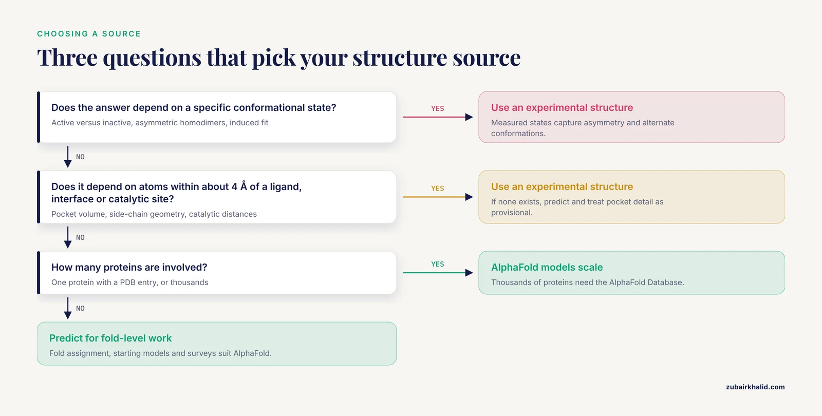
Use three questions to decide between alphafold or experimental structures for a specific task.
- Does the answer depend on a specific conformational state? If yes, experimental. If any reasonable conformation gives the same answer, prediction is fine.
- Does the answer depend on atoms within about 4 Å of a ligand, an interface, or a catalytic site? If yes, experimental, unless no structure exists, in which case predict, then treat every pocket detail as provisional.
- How many proteins are involved? One protein with an available PDB structure, use the structure. Thousands of proteins, AlphaFold models are the only scalable option.
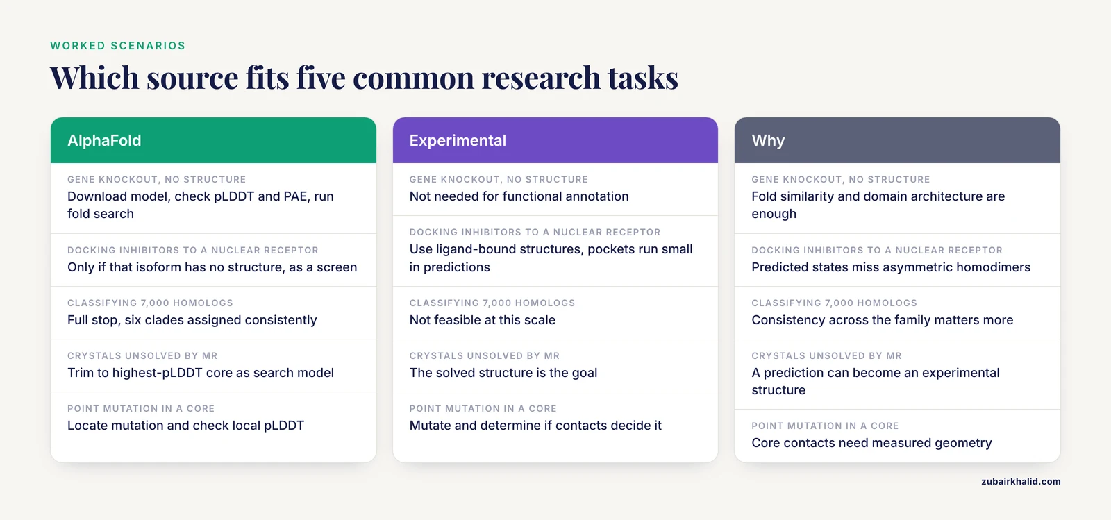
Scenario 1. You knock out a gene and want to know what its product does. No structure exists. Download the AlphaFold model from the AlphaFold Database, check pLDDT and PAE, note the confidently folded domains and the disordered tails, and run a fold-recognition search with AlphaFind or 3D-AF-Surfer to find structural relatives the sequence search missed [5] [1]. This is a textbook case of when to use alphafold. Functional annotation from fold similarity, domain architecture, and localization prediction is exactly what the models are good for.
Scenario 2. You are docking inhibitors against a nuclear receptor. Experimental structures of the ligand-binding domain exist for most receptor family members. Use them. The systematic analysis showed predicted pocket volumes run small and predicted states miss the asymmetric conformations experimental structures display in homodimers [2]. If your particular receptor isoform has no structure, you can dock into the AlphaFold model as a screen, but confirm hits against a measured structure or an experimental binding assay before drawing conclusions.
Scenario 3. You want to classify 7,000 homologs into structural subfamilies. AlphaFold, full stop. This mirrors the hydrophobin work, where AlphaFold models were benchmarked against the solved structures first and then used to assign every known member to one of six clades [4]. Consistency of method across the family matters more than absolute accuracy for each member, and AlphaFold provides exactly that consistency.
Scenario 4. Your crystals refuse to solve by MR. Pull the AlphaFold model, trim to the highest-pLDDT core, and try it as a search model alongside homologs found by MrParse, which ranks both experimental and predicted candidates by expected MR success [3]. If the prediction solves it, you have converted a prediction into an experimental structure, which is the ideal outcome.
Scenario 5. You need to know whether a point mutation breaks a protein. Start with the AlphaFold model to locate the mutation, check local pLDDT, and check whether the residue sits in a confidently folded core or a flexible loop. If it sits in a core, the model gives a reasonable first structural explanation. If your conclusion will rest on specific disrupted contacts, mutate and determine, or at minimum support the prediction with biophysical data such as thermostability or SAXS.
Getting the Structures: PDB, AlphaFold Database, and the AlphaFold Server
Both sources are free. Experimental structures live in the Protein Data Bank, searchable through RCSB and the EBI PDBe services, downloadable as PDB or mmCIF files, and accompanied by validation reports you should read. AlphaFold models live in the AlphaFold Database at EMBL-EBI, searchable by UniProt accession, with per-residue confidence colored into the viewer and PAE matrices downloadable as JSON.
For a single protein, the direct download pattern is stable and worth knowing. Every entry has files at predictable URLs under alphafold.ebi.ac.uk. For human p53 (UniProt P04637), a version-4 model fetches like this:
curl -O https://alphafold.ebi.ac.uk/files/AF-P04637-F1-model_v4.pdb
curl -O https://alphafold.ebi.ac.uk/files/AF-P04637-F1-predicted_aligned_error_v4.json
The B-factor column of the downloaded PDB file contains the pLDDT score per residue, so any structure viewer that colors by B-factor shows confidence directly.
For bulk retrieval, use a dedicated tool rather than scraping. The AlphaFoldDB Structure Extractor accepts taxonomy IDs, RefSeq accessions, locus tags, UniProt accessions, or AlphaFold accessions, returns structures in PDB, mmCIF, bCIF, or PAE JSON, and writes a mapping file plus a list of unmapped inputs, with an API for pipeline integration [11].
For complexes, pairs with nucleic acids, or proteins with ligands and modifications, the AlphaFold server (the hosted AlphaFold 3 service from Google DeepMind and ISMB) accepts composed inputs of proteins, nucleic acids, and small molecules and returns predicted complexes with confidence metrics. For high-throughput or customized use, the open-source AlphaFold code and community reimplementations such as ColabFold let you run predictions on your own hardware or in Colab notebooks. A minimal alphafold tutorial path for a newcomer looks like this:
- Look up your protein in UniProt and note the accession.
- Check whether a structure already exists in the PDB with a sequence coverage and identity search.
- Open the AlphaFold Database entry for the UniProt accession and inspect the pLDDT coloring and PAE matrix.
- If no model exists or the protein is a complex, submit to the AlphaFold server or run ColabFold locally with a multiple sequence alignment from your choice of sequence databases.
- Validate anything consequential against experimental data, whether that is a crystal structure of a homolog, SAXS profiles, crosslink distances, or biochemical assays.
A growing class of databases also ships AlphaFold models as part of their annotations. DRAVP, an antiviral peptide and protein database, used AlphaFold to predict structures for entries lacking experimental determination [15]. BoMiProt 2.0 includes AlphaFold predicted models alongside crystal structures for each bovine milk protein entry [16]. These are useful shortcuts, but the same rule applies. Confidence metrics travel with the model, so read them.
Common Mistakes and Limitations
Treating pLDDT like a resolution number. pLDDT is a prediction of agreement with a hypothetical experimental structure, not a measurement. A 95 pLDDT region is very likely to be modeled accurately, but the number does not certify side-chain rotamers, ligand compatibility, or the biologically relevant state. It certifies consistency with the training distribution.
Ignoring PAE between domains. A two-domain protein with beautiful pLDDT inside each domain can still have an arbitrarily wrong domain arrangement. Check the interdomain block of the PAE matrix before trusting any interdomain distance, pocket formed across domains, or quaternary geometry.
Docking into predicted pockets without experimental follow-up. Because AlphaFold models lack ligands and predict pockets in a ligand-free state, pocket volumes and side-chain positions can be off, as demonstrated quantitatively for nuclear receptors [2]. Use predicted pockets for triage. Use measured structures for claims.
Assuming the predicted state is the functional state. AlphaFold returns the dominant conformation in its training data. Alternate states, autoinhibited forms, asymmetric assemblies, and induced-fit conformations require experimental structures of those states [2].
Modeling complexes and expecting crystal-accuracy interfaces. Performance on antibody-peptide complexes was good in only about half of tested cases and degraded further in more complex contexts [14]. Treat every predicted interface as a hypothesis to test, not a result to report.
Interpreting structured predictions of disordered regions. Regions with pLDDT below about 50 are predictions of disorder, not low-quality structures. Trimming them before docking, MR, or refinement is standard practice, and their absence of stable structure is itself informative.
Forgetting that experimental structures have their own biases. Crystal packing, construct boundaries, purification tags, and Cryo-EM classification all shape what gets measured. The correct mental model is two fallible sources with different error structures, not one perfect truth and one approximate copy. Cross-validation against independent data, such as SAXS ensembles [13] or crosslink restraints [9], catches problems in both.
Discarding surprising predictions as noise. AlphaFold occasionally predicts alternative topologies that turn out to be structurally meaningful. In the whirlin PDZ tandem, AlphaFold predicted a domain-swapped arrangement differing from the experimental structure in exactly the regions that mutational analysis identified as energetic determinants of a transient misfolded state, suggesting the alternative topology may describe a real, sparsely populated state [17]. A mismatch between prediction and experiment is a lead, not automatically an error.
Skipping structure preparation. Whether you start from a PDB entry or an AlphaFold model, protonation states matter for simulation and docking. Tools such as PypKa handle pKa-based protonation for structures from either source, with precomputed results for a large fraction of both databases [10].
Frequently Asked Questions
Can an AlphaFold model replace a crystal structure?
No, not for questions that depend on a specific conformational state, ligand geometry, or an experimentally validated interface. AlphaFold models are excellent starting points, search models, and survey tools, but a measured structure remains the standard for structural claims about binding and mechanism.
What pLDDT cutoff should I use when trimming an AlphaFold model?
A common practice is to keep residues above about 70 pLDDT for backbone-level work and above about 90 when side-chain detail matters, and to treat residues below 50 as predicted disorder. Adjust the threshold to the task, and always inspect the PAE matrix for multidomain proteins.
Is the AlphaFold server free to use?
Yes. The AlphaFold Database and the hosted AlphaFold server are free to use through their web interfaces, the open-source AlphaFold code is publicly available for local prediction, and community tools such as ColabFold run in free notebook environments, subject to each platform's own usage limits.
Should I dock ligands into an AlphaFold model or a PDB structure?
Dock into the experimental structure whenever one exists for your target, especially a ligand-bound structure, because predicted pocket volumes and side-chain states can deviate from measured ones. Dock into an AlphaFold model only when no experimental structure covers the binding region, and treat the output as a hypothesis for experimental testing.
How do I download many AlphaFold models at once?
Use a bulk retrieval tool such as the AlphaFoldDB Structure Extractor, which accepts taxonomy IDs, RefSeq accessions, locus tags, or UniProt accessions and returns PDB, mmCIF, bCIF, or PAE JSON files with an ID mapping file, or use the AlphaFold Database API for programmatic access with your own accessions.
Can AlphaFold predict protein-protein and protein-ligand complexes?
AlphaFold-Multimer and AlphaFold 3 can predict complexes, and AlphaFold 3 extends prediction to nucleic acids and small molecules, but complex accuracy is consistently lower than single-chain accuracy, particularly for antibody-antigen and peptide interfaces. Validate predicted interfaces against experimental data before relying on them.
Sources
- Real-time structure search and structure classification for AlphaFold protein models.
- Bridging prediction and reality: Comprehensive analysis of experimental and AlphaFold 2 full-length nuclear receptor structures.
- MrParse: finding homologues in the PDB and the EBI AlphaFold database for molecular replacement and more.
- AlphaFold modeling uncovers global structural features of class I and class II fungal hydrophobins.
- AlphaFind: discover structure similarity across the proteome in AlphaFold DB.
- AlphaFold-driven structure-based drug discovery: Capabilities, benchmarks and limitations for medicinal chemistry.
- Human and zebrafish cross-species comparison on ligand affinity towards the aryl hydrocarbon receptor and Keap1 oxidative stress sensor: a molecular docking dataset.
- Novel PFAS Alternatives 6:2 Cl-PFESA and HFPO-TA as Plasma FXII Activators: Experimental Evidence and Computational Mechanistic Insights.
- AlphaCross-XL: A Seamless Tool for Automated and Proteome-Scale Mapping of Cross-Linked Peptides Onto 3D Protein Structures.
- PypKa server: online pKa predictions and biomolecular structure preparation with precomputed data from PDB and AlphaFold DB.
- AlphaFold Database Structure Extractor: a web server and API to download AlphaFold structures using common protein accessions.
- OpenAntigens: a structure-aware database for antigen construct design across the human cell-surface and secreted proteome.
- AlphaFold-predicted protein structures and small-angle X-ray scattering: insights from an extended examination of selected data in the Small-Angle Scattering Biological Data Bank.
- Evaluation of Alphafold modeling for elucidation of nanobody-peptide epitope interactions.
- DRAVP: A Comprehensive Database of Antiviral Peptides and Proteins.
- BoMiProt 2.0: An update of the bovine milk protein database.
- Residue-Level Energetic Analysis of Transient Misfolding in the Whirlin PDZ Tandem Reveals a Possible Connection to an AlphaFold-Predicted Alternative Topology.