Using AlphaFold Structures for Virtual Screening
By Dr. Zubair Khalid, DVM, MS, PhD ·

AlphaFold structures replace the missing crystal structure in a virtual screening campaign, but they are predictions, and docking into a prediction is not the same as docking into an experiment. The one-sentence difference is this: an experimental structure shows you where the protein actually was during crystallization, while an AlphaFold model shows you where the network thinks the protein most likely sits, with a per-residue confidence score telling you which parts to trust. The rule of thumb for choosing an approach: if your target has a high-confidence binding pocket and a rigid fold, dock against the AlphaFold model directly and validate the top hits. If the pocket is flexible, low in confidence, or depends on a cofactor or conformational change, refine the model first or use a method built to tolerate structural error.
This guide covers the alphafold virtual screening options available today, the tradeoffs of each, a step-by-step tutorial for a standard workflow, and the mistakes that most often sink predicted-structure campaigns.
What AlphaFold Gives You, and What It Does Not
AlphaFold2 and its successors predict protein structures from sequence with near-experimental accuracy for well-ordered domains. AlphaFold3 extended prediction to biomolecular complexes, including proteins with ligands, nucleic acids, and chemical modifications [1]. For drug discovery, the practical consequence is large: targets that lacked any structural model, including most bacterial virulence factors, orphan GPCRs, and pathogen enzymes, now have computable three-dimensional starting points. A recent review of AlphaFold-driven structure-based drug discovery frames the situation plainly. Predicted structures have expanded structural coverage across much of the known proteome, but prediction confidence and accuracy vary by target and by region within a target, and those differences matter enormously for docking [2].
Every residue in an AlphaFold2 model carries a pLDDT score, a per-residue confidence metric on a 0 to 100 scale. The AlphaFold Database at EMBL-EBI colors models by this score, with very high confidence above 90, confident backbone in the 70 to 90 band, and low confidence below 70, where the chain is usually disordered in isolation. The predicted aligned error (PAE) file complements pLDDT by describing expected positional error between residues, which matters when you judge whether two regions of the model are positioned relative to each other correctly. For binding pockets, the numbers that decide usability are the pLDDT values of pocket residues and the PAE between them, not the global average.
What AlphaFold does not give you is the holo state. Virtual screening performance depends on the protein conformation, and docking scores degrade substantially when the receptor conformation used for screening differs from the ligand-bound form. This is a general weakness of structure-based screening, not an AlphaFold-specific one, but predicted apo structures inherit it fully [3]. AlphaFold models are also static single conformations. Many proteins bind ligands through induced fit, and flexible peptides often adopt their bioactive conformations only upon binding, in states that a static prediction may not capture [4].
Getting Structures: The AlphaFold Server and the AlphaFold Database
Two entry points exist, and they serve different purposes.
The AlphaFold Server, operated by Google DeepMind, lets researchers submit sequences to AlphaFold 3 through a web interface and receive predicted structures, including complexes, without installing anything. It is free for noncommercial use and subject to usage limits and a terms-of-use framework. For a one-off target, a protein-ligand complex, or a quick look at whether a protein-peptide pair co-folds plausibly, the server is the fastest route. For anything reproducible at scale, you want the database and local tools instead.
The AlphaFold Database (AlphaFold DB), hosted by EMBL-EBI, holds precomputed structures for over 200 million proteins. Individual structures can be fetched directly by UniProt accession using the documented URL pattern, for example https://alphafold.ebi.ac.uk/files/AF-P12345-F1-model_v4.pdb for a PDB-format model. File formats include PDB, mmCIF, and binary mmCIF, alongside PAE confidence data.
For bulk work, downloading thousands of structures by hand is not practical. The AlphaFoldDB Structure Extractor is a web server and API built for this job. It accepts NCBI Taxonomy IDs, RefSeq accessions, locus tags, and UniProt or AlphaFold accessions, and returns structure files in PDB, mmCIF, bCIF, or PAE JSON formats, with an ID mapping file that traces inputs back to standard accessions and reports unmapped IDs separately. The authors tested it with individual inputs and batches of up to 5,000 accessions, and the API supports integration into automated pipelines [5].
MrParse offers a complementary check before you commit to a predicted model. Built for crystallographers choosing molecular replacement search models, it searches for homologues in both the Protein Data Bank and the EBI AlphaFold database and displays predicted secondary structure, coiled-coils, and transmembrane regions. Even outside crystallography, its homologue ranking is useful, because if a close experimental homologue exists in the PDB, that structure is usually a better docking receptor than a de novo prediction [6].
Alphafold Virtual Screening Options
Five workflow families now dominate published practice. They differ in what they assume about your target, how much compute they need, and how they handle the uncertainties of predicted structures.
Option 1: Dock Small Molecules Directly Against the AlphaFold Model
This is the default option and the most common in the literature. You retrieve the AlphaFold model, identify or predict a binding pocket, prepare the receptor for your docking engine, and screen a compound library. Docking engines used in recent AlphaFold-model campaigns include AutoDock Vina, an open-source empirical docking program, and Schrödinger's Glide in Extra Precision mode with Induced-Fit Docking refinement [7], [8].
Published examples span antimicrobial, antiviral, and anticancer targets. In work on Clostridioides difficile, researchers selected the essential cell-division protein FtsZ after a two-phase target identification process, docked StreptomeDB compounds against the AlphaFold-generated model with AutoDock Vina, and shortlisted 27 high-affinity ligands, with fasamycin C and formicamycin D showing favorable docking, drug-likeness, and ADMET profiles [7]. A study of the Listeria monocytogenes virulence protein ActA screened 106 licorice phytocompounds against the AlphaFold model, with top compounds docking at binding affinity values of -10.4 and -8.7 kcal/mol before molecular dynamics confirmation [9]. Against the Kyasanur Forest Disease Virus NS5 RNA-dependent RNA polymerase, a workflow in Schrödinger Maestro screened 1,388 compounds from DrugBank and MedChem libraries using Glide XP docking followed by Induced-Fit Docking, then validated complex stability with 200 ns molecular dynamics and MM/GBSA binding free energy calculations [8]. A dengue virus NS5 study built full-length models with homology modeling, ab initio modeling, and AlphaFold, selected the best model on RMSD and stereochemical quality, docked a library of 587 antiviral compounds, and found a top RNA-binding-site hit at -11.8 kcal/mol [10].
The strength of this option is simplicity. The weakness is that AlphaFold2 models often show errors in local structural details, especially within binding pockets, which limits their utility for small molecule docking [11]. Side-chain rotamers in the pocket may be wrong, and no docking engine can fix a pocket whose shape is wrong.
Option 2: Refine the Model Before Screening
A second family of workflows treats the AlphaFold model as a starting point and improves it with physics before docking.
Molecular dynamics (MD) simulation, which propagates atomic motions with a force field, relaxes strained side chains and lets pockets settle into physically plausible conformations. The MurA study from Staphylococcus aureus work followed this logic in sequence. The researchers used AlphaFold 3 to predict the structure of S. aureus MurA, an enzyme in peptidoglycan synthesis with no published crystal structure, constructed a MurA-UNAG complex model through active site prediction and docking, then ran molecular dynamics to refine and optimize the complex before using it for virtual screening. The resulting hit, compound X14, showed an IC50 of 20.41 ± 1.10 µM in a MurA inhibition assay and represented a novel scaffold by Tanimoto coefficient comparison against known MurA inhibitors [12].
A Bayesian refinement method called bAIes goes further by combining physics-based force fields with the data-driven information in AlphaFold2 predictions. In benchmarks, bAIes discriminated between binders and nonbinders in virtual screening better than both raw AlphaFold2 models and molecular dynamics-refined models, and it achieved this without requiring extensive experimental or computational resources [11]. The key insight from that work is that raw prediction and long MD refinement sit at two ends of a spectrum, and an intermediate that constrains the simulation with the prediction's information can outperform both.
Model selection across predictors is another refinement strategy. For the Acinetobacter baumannii siderophore transporter subunit BauE, no experimental structure existed, so the authors generated models with AlphaFold, Robetta, and trRosetta, validated them, and ran 500 ns MD simulations. The AlphaFold-predicted model showed the highest structural stability and was selected for pocket prediction and screening of 601 antimicrobial peptides [13]. Comparing several predictors costs little compute and protects you from betting a campaign on a single unlucky prediction.
Option 3: Co-Fold the Ligand With the Target in AlphaFold 3
AlphaFold 3 can predict complexes, which opens a screening mode that did not exist with AlphaFold 2. Instead of docking a ligand into a preformed pocket, you ask the network to fold the protein and candidate ligand together and judge the result by interface confidence metrics.
A review of AlphaFold in natural product research describes co-folding strategies as showing considerable potential for improving early enrichment in natural product screening and for elucidating binding mechanisms of complex natural products with their targets, beyond conventional structure-based virtual screening [1]. For peptide targets, the distinction matters even more. A review of the AlphaFold series for peptide structure prediction notes that the progression from AlphaFold2 through AlphaFold-Multimer to AlphaFold3 improved geometric reasoning and introduced interface confidence metrics such as the ipTM score, while static predictions still struggle with peptides that fold only on binding [4].
Co-folding has already powered a complete discovery method. DeorphaNN, a graph neural network for orphan GPCR deorphanization, integrates active-state GPCR-peptide structural predictions, interatomic interactions, and deep learning embeddings. Its builders showed that AlphaFold-Multimer confidence metrics partially discriminate agonist from non-agonist complexes, with improved discrimination using AF-Multistate-derived active-state templates, and that pair representations outperform single representations. The method generalized across species in retrospective benchmarks on annelid and human data, and experimental validation confirmed predicted agonists for two orphan GPCRs [14]. The lesson generalizes: co-folding confidence alone is a weak classifier, but co-folding features fed into a learned model can be strong.
Co-folding is not free of cost at scale. Each candidate requires its own prediction run, so screening a 100,000-compound library this way is impractical for most groups. It works best as a reranking step on tens to hundreds of candidates already prioritized by docking or sequence similarity.
Option 4: Embedding-Based Screening Built for Predicted Structures
Contrastive learning methods treat virtual screening as a retrieval problem. They learn embeddings that place true protein-ligand pairs close together in a vector space, then screen by nearest-neighbor search rather than by docking every molecule.
DrugCLIP is the landmark implementation. It aligns protein pocket and small-molecule representations through a contrastive framework, paired with a pocket refinement module targeting predicted structures, and reimagines virtual screening as fast embedding-based dense retrieval. Benchmarks validate its performance relative to molecular docking and machine-learning baselines, and it addresses the computational cost barrier that has restricted genome-wide application of screening, especially as AlphaFold-predicted structures have proliferated [15]. A commentary placing DrugCLIP in context with tools including RosettaVS, Deep Docking, and AF2-RAVE also flags the unresolved challenge: translating binding predictions into functional activity remains the field's core difficulty [15].
This option suits proteome-scale prioritization, where you want to rank thousands of pockets against a ligand set and accept some false positives in exchange for speed and coverage.
Option 5: Conformation-Tolerant Screening Methods
When you suspect the predicted structure is in the wrong conformational state for binding, surface-based methods offer a hedge. PL-PatchSurfer3 represents binding sites as surface patches described by 3D Zernike descriptors, compact rotation-invariant mathematical encodings of shape and physicochemical properties such as electrostatic potential. Because it compares molecular surfaces rather than fitting ligands into rigid pockets, its screening performance is more stable when the receptor structure used for screening differs from the ligand-bound form, a weakness that causes docking performance to drop substantially for conventional methods [3]. For AlphaFold models, which may not capture the holo conformation, this robustness is directly relevant.
Side-by-Side Comparison
| Option | Input required | Best suited for | Main weakness | Typical tools | Evidence base |
|---|---|---|---|---|---|
| Direct docking on the AF model | Target sequence or AFDB model, compound library | Rigid, high-confidence pockets, enzymes with conserved catalytic sites | Pocket side chains may be wrong, apo vs holo mismatch [11], [3] | AutoDock Vina, Glide XP, Induced-Fit Docking | Multiple published hit campaigns [7], [9], [8], [10] |
| Model refinement before docking | AF model plus compute for MD or refinement | Targets with flexible or low-confidence pockets, cofactor-dependent enzymes | Compute cost, MD can drift from the true fold if unconstrained | MD force fields, bAIes Bayesian refinement | MD-refined MurA model yielded a 20 µM inhibitor [12], bAIes outperformed both raw AF2 and MD-refined models [11] |
| Co-folding target and ligand | Target and candidate ligand sequences or structures | Peptides, protein-protein and protein-natural-product interactions, reranking shortlists | Too slow for large libraries, confidence alone is a weak classifier | AlphaFold 3, AlphaFold-Multimer, ipTM scoring | Validated in GPCR deorphanization with experimental hits [14], reviewed for natural products [1] |
| Embedding-based retrieval | Pocket representations and ligand embeddings, precomputed models | Proteome-scale prioritization, genome-wide campaigns | Binding prediction does not equal functional activity [15] | DrugCLIP with GenPack pocket refinement | Benchmarked against docking and ML baselines with wet-lab successes [15] |
| Conformation-tolerant surface matching | AF model or any receptor structure | Targets where holo and apo states differ, uncertain conformational state | Coarser than atom-level docking, less precise poses | PL-PatchSurfer3 | Designed specifically to stay stable across conformational variation [3] |
Choosing Between Options: The Criteria That Decide
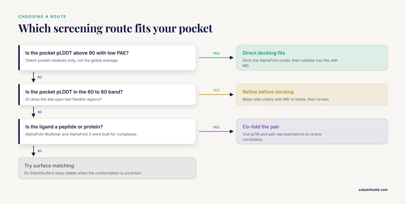
Four questions separate these options in practice.
How confident is the pocket? Check pLDDT over pocket residues and the PAE connecting them. A pocket at pLDDT above 90 with low PAE to the rest of the catalytic domain is a direct-docking candidate. A pocket threaded with 60 to 80 pLDDT residues, or a binding site spanning two flexible regions, needs refinement or a tolerant method. The KFDV NS5 study illustrates the good end of the spectrum, with a model showing pLDDT scores approaching 90 and a pTM score of 0.8, validated further with Verify3D, PROCHECK, and ERRAT before screening [8].
Does the target need a cofactor or bound ligand to be in a screenable state? Many enzymes only adopt their ligand-binding pocket geometry when the substrate or cofactor is present. The VbrK histidine kinase study grafted the essential ligand and metal cofactor onto AlphaFold predictions before screening 7,665 natural products, then confirmed phlorizin, diprophylline, and baicalin as micromolar binders in a competitive fluorescence polarization assay, with two of them enhancing carbenicillin susceptibility in resistant strains [16]. If your target is in this category, plain docking into an empty predicted pocket will mislead you.
Is the ligand a small molecule, a peptide, or a macromolecule? Small molecules suit conventional docking. Peptides and protein ligands suit co-folding, since AlphaFold-Multimer and AlphaFold 3 were built for complexes [4], [1]. Bacteriocin screening against the monkeypox virus DNA polymerase shows the pattern: ligand structures came from the PDB or AlphaFold prediction, quality was scored with LGscore, and protein-peptide docking ran on the HADDOCK platform [17].
How big is the library and the target set? One target and 1,000 compounds fit on a workstation with direct docking. One thousand AlphaFold models and a million compounds push you toward embedding methods [15] and bulk retrieval infrastructure [5].
Worked Scenarios
Scenario 1: A bacterial enzyme with no crystal structure. You want inhibitors of an essential S. aureus enzyme. Follow the MurA playbook. Predict the structure with AlphaFold 3, predict or literature-annotate the active site, dock the native substrate to build a reference complex, run MD to relax the complex, then screen your library against the refined pocket. Confirm hits with an enzymatic assay, not docking scores alone. The published result was a 20.41 ± 1.10 µM inhibitor with a scaffold unlike any known MurA inhibitor [12].
Scenario 2: An orphan GPCR and its peptide ligand. You want to find the natural agonist of a receptor with no characterized homolog. Sequence similarity will not help. Build active-state models using AF-Multistate templates, co-fold candidate peptides from the proteome, and use the AF-Multimer pair representations as features for a learned ranker, as DeorphaNN does [14]. Validate top predictions with a signaling assay. This method is specifically designed for the case where gene-family relationships give limited predictive insight [14].
Scenario 3: Prioritizing drug targets across a pathogen genome. You have a pathogen proteome and want to know which proteins are both essential and druggable. Run target identification to shortlist candidates, as in the C. difficile work that used EDGAR for prevalence, PSORTb for subcellular localization, and human and gut microbiome homology filters before selecting six targets including FtsZ [7]. Download all shortlisted AlphaFold models in bulk through the extractor's API [5]. Screen a curated library against each model with AutoDock Vina, then take the top hits to MD and ADMET filtering [7].
A Practical Alphafold Tutorial: Direct Docking Workflow
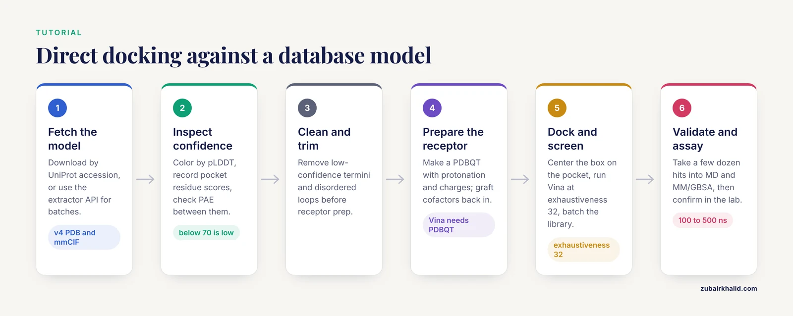
This walkthrough covers the most common option, direct docking against an AlphaFold Database model, using free and open tools. Commands assume a Linux environment.
Step 1. Retrieve the model.
# Replace P12345 with your target's UniProt accession
wget https://alphafold.ebi.ac.uk/files/AF-P12345-F1-model_v4.pdb
wget https://alphafold.ebi.ac.uk/files/AF-P12345-F1-model_v4.cif
The mmCIF file carries confidence annotations that plain PDB files may drop. For batches, use the AlphaFoldDB Structure Extractor web server or its API, which maps RefSeq accessions, locus tags, and Taxonomy IDs to structures and returns a mapping file [5].
Step 2. Inspect confidence before anything else.
Open the structure in a molecular viewer and color by pLDDT. Identify your intended binding pocket from literature, UniProt annotations, or a pocket-detection tool such as fpocket or P2Rank. Record the pLDDT of every residue lining the pocket. If several pocket residues sit below the 70 to 90 confidence band, plan on refinement rather than direct docking. Confirm that the pocket residues are mutually well aligned by checking the PAE file.
Step 3. Clean and trim the model.
Remove low-confidence termini and disordered loops that would otherwise complicate receptor preparation. Keep the confidence record from step 2 so you can interpret docking results in context.
Step 4. Prepare the receptor.
Convert the model to the format your docking engine needs. For AutoDock Vina, that means a PDBQT file with assigned protonation states and partial charges. Set protonation consistent with the pH of your assay and any known catalytic residues. If your target needs a cofactor or metal, graft it in before docking, as the VbrK study did with its ligand and metal cofactor on AlphaFold predictions [16].
Step 5. Define the docking box.
Center the box on the pocket. If a substrate site is known from a homologue, align your model to the homologue's ligand-bound structure and copy the box coordinates. Homologue searching in the PDB and AlphaFold DB, which MrParse automates, helps here [6]. Otherwise, use pocket detection to place the box, and make it generous enough for induced fit on the ligand side.
Step 6. Dock and screen.
For a single ligand with Vina:
vina --receptor receptor.pdbqt \
     --ligand ligand.pdbqt \
     --center_x X --center_y Y --center_z Z \
     --size_x SX --size_y SY --size_sz SZ \
     --exhaustiveness 32 --out out.pdbqt
For library screening, run Vina in batch mode across a prepared ligand set, or use a screening manager to parallelize. Reserve more expensive protocols, such as Glide XP with Induced-Fit Docking as used in the KFDV NS5 campaign [8], for a filtered shortlist.
Step 7. Validate computationally.
Take the top fraction of ranked compounds, typically a few dozen, into molecular dynamics to test complex stability, as done over 200 ns for KFDV NS5 hits [8] and over 100 ns for the ActA study [9]. Follow with MM/GBSA binding free energy estimates to rerank, a step used in the MurA [12], VbrK [16], and KFDV [8] workflows. Then, and only then, test experimentally. The MurA and VbrK campaigns both closed the loop with assay confirmation, which is what separates a screening result from a screening claim [12], [16].
Alphafold Virtual Screening Pros and Cons
Pros.
- Structural coverage. Targets without experimental structures, the historical bottleneck of structure-based drug discovery, now have usable models, which is the central advance documented across the drug discovery literature [2].
- Demonstrated hit discovery. Peer-reviewed campaigns have converted AlphaFold models into experimentally confirmed inhibitors, including a novel MurA scaffold at 20.41 µM [12] and micromolar VbrK binders with phenotypic resistance reversal [16].
- Complex prediction. AlphaFold 3 and AlphaFold-Multimer extend screening beyond small molecules into peptides, proteins, and natural products [1], [4].
- Scale. Bulk retrieval tools [5] and embedding-based methods [15] make proteome-scale campaigns feasible.
- Free access. The database, the noncommercial server, and open-source docking engines let academic groups run complete campaigns at zero licensing cost.
Cons.
- Pocket-level accuracy is not guaranteed. Local errors in binding pockets limit small molecule docking utility, and this limitation motivated the development of refinement methods that outperform raw models [11].
- Apo versus holo mismatch. Predicted structures are unliganded, and screening performance drops when the receptor conformation differs from the binding-competent state [3].
- Static snapshots. Induced fit, transient interactions, and binding-induced peptide folding are invisible to a single static prediction [4].
- Docking scores overpromise. Reported binding energies, such as -11.8 kcal/mol in a dengue NS5 study [10] or -10.4 kcal/mol in the ActA study [9], are ranking heuristics, not free energies, and require experimental confirmation.
- Co-folding does not scale. Asking AlphaFold 3 to fold every library compound with the target is computationally prohibitive beyond small candidate sets [1].
Common Mistakes and Limitations
Reading the global pLDDT as pocket quality. A model can average 90 while the pocket sits at 65. Always inspect the pocket residues specifically, and the PAE between them.
Docking side chains that are confidently wrong. A side chain can be placed at high pLDDT and still be wrong for binding. If the pocket contains residues whose rotamers matter, relax them with MD or constrained refinement before screening. The bAIes result, where Bayesian refinement outperformed both raw AlphaFold2 and MD-refined models, shows that refinement strategy itself is a choice worth making deliberately [11].
Ignoring cofactors, metals, and waters. A predicted structure contains none of them. If the functional site needs them, add them back deliberately, as the VbrK study did before its successful screen [16].
Screening without any experimental structure check. If a close homologue exists in the PDB, compare your model against it and, where they disagree in the pocket, prefer the homologue or a hybrid. MrParse automates exactly this comparison. Validation tools such as Verify3D, PROCHECK, and ERRAT add an independent quality read on the model before you trust it [8].
Trusting docking score magnitudes. Compounds scoring several kcal/mol apart can be indistinguishable experimentally. Use scores to rank within a campaign, not to compare across campaigns or to predict potency.
Screening flexible peptides against a static model. Short and disordered peptides often adopt functional conformations only on binding, in low-probability states that static predictions miss, which leads to failures in virtual screening [4]. Ensemble approaches combining AlphaFold predictions with MD and free energy calculations are the emerging answer [4].
Treating computational hits as drugs. Every published success in this list involved an assay. Docking into a predicted structure generates hypotheses. The 20 µM MurA inhibitor [12], the VbrK binders [16], and the confirmed GPCR agonists [14] all became results through benchwork.
Forgetting the pipeline around the structure. The strongest published campaigns combined target prioritization [7], library curation with ADMET filtering [7], MD validation [12], [9], [13], [18], and free energy calculations [19] around the AlphaFold model. The model is one component, not the campaign.
Frequently Asked Questions
Can I use an AlphaFold model instead of a crystal structure for docking?
Yes, and published campaigns have done so successfully, but treat the model as a hypothesis with quantified uncertainty. Check pLDDT over the binding pocket, refine side chains if needed, and confirm hits experimentally, because pocket-level errors in AlphaFold2 models are a documented limitation for small molecule docking [11].
What is the best alphafold virtual screening workflow?
The best workflow depends on the pocket. For rigid, high-confidence pockets, direct docking followed by MD and MM/GBSA reranking is the most validated path, with confirmed hits across multiple studies [12], [8]. For flexible or low-confidence pockets, refinement before docking performs better, and for peptides or complexes, co-folding with interface confidence metrics is the appropriate choice [14], [1].
Which AlphaFold confidence scores matter for a binding pocket?
Focus on pLDDT for each pocket residue and the PAE between pocket residues. In the AlphaFold Database color scheme, pLDDT above 90 indicates very high confidence and the 70 to 90 band indicates confident backbone. One viral polymerase study reported a model with pLDDT approaching 90 and pTM of 0.8 as high confidence before screening [8].
Does AlphaFold 3 replace molecular docking?
No. AlphaFold 3 predicts complexes and enables co-folding screens, but running a prediction per library compound is impractical at screening scale, and docking remains the workhorse for ranking large libraries. The current role of co-folding is enriching and reranking candidate interactions, particularly for peptides and natural products [1].
How do I download AlphaFold structures in bulk?
Use the AlphaFoldDB Structure Extractor, a web server and API that accepts NCBI Taxonomy IDs, RefSeq accessions, locus tags, and UniProt or AlphaFold accessions, and returns PDB, mmCIF, bCIF, or PAE JSON files with an ID mapping file. It has been tested with batches up to 5,000 accessions [5]. For individual structures, download directly from the AlphaFold Database by UniProt accession.
Do I need molecular dynamics after docking?
For anything you intend to publish or pursue, yes. MD tests whether the docked complex is stable, relaxes artifacts of rigid docking, and enables MM/GBSA free energy reranking. Recent campaigns used 100 to 500 ns simulations to confirm complex stability before experimental work [12], [9], [13].
Sources
- From structure prediction to ligand-target recognition: expanding the reach of AlphaFold in natural product research.
- AlphaFold-driven structure-based drug discovery: Capabilities, benchmarks and limitations for medicinal chemistry.
- PL-PatchSurfer3: improved structure-based virtual screening for structure variation using 3D Zernike descriptors.
- AlphaFold-based peptide structure prediction: Opportunities, limitations, and future directions.
- AlphaFold Database Structure Extractor: a web server and API to download AlphaFold structures using common protein accessions.
- MrParse: finding homologues in the PDB and the EBI AlphaFold database for molecular replacement and more.
- Discovering Novel Drug Targets in Clostridioides difficile and Proposing FtsZ Inhibitors via Structure-Based Virtual Screening.
- AlphaFold-driven structure-guided identification of NS5 RdRp-targeting antiviral leads against Kyasanur Forest Disease Virus.
- In silico identification of phytocompounds derived from Glycyrrhiza glabra as potential inhibitors of actin assembly-inducing protein in Listeria monocytogenes: a virtual screening and molecular dynamics study.
- Identification of potential inhibitors of dengue virus ns5 methyltransferase and polymerase domains through virtual screening and molecular dynamics studies.
- Advancing In Silico Drug Design with Bayesian Refinement of AlphaFold Models.
- Discovery of a novel MurA inhibitor via AlphaFold 3 model-based virtual screening.
- Structural modeling and in silico screening of antimicrobial peptides targeting BauE in Acinetobacter baumannii.
- DeorphaNN: Virtual screening of GPCR peptide agonists using AlphaFold-predicted active-state complexes and deep learning embeddings.
- Contrastive Learning-Powered Ultrafast Genome-Wide Virtual Screening: The Rise of DrugCLIP.
- Structure-based virtual screening identifies naturally derived histidine kinase VbrK inhibitors as potential countermeasures against β-lactam antimicrobial resistance.
- Virtual Screening of Bacteriocins From Lactic Acid Bacteria Against Monkeypox DNA Polymerase: Sakacin P and Mundticin KS Emerge as Promising Candidates.
- Deep learning-based structural prediction (AlphaFold 3) of shrimp IMNV RdRp and identification of seaweed-derived metabolites for antiviral intervention in aquaculture.
- Integrative transcriptomics and structure-based screening identifies Phyllanthus amarus phytocompounds as potential WNT5A modulators in ovarian cancer.