Common AlphaFold Sequence Errors and How to Fix Them
By Dr. Zubair Khalid, DVM, MS, PhD ·

Most AlphaFold sequence errors trace back to one root cause: the sequence you submitted is not the sequence you think it is. The single most common version of this problem is a mismatch between your input FASTA and the current reference sequence, because reference databases revise protein records over time while cached structures and old local files stay frozen. Nearly 3 percent of the sequences tied to AlphaFold Database models were revised in UniProt while the database itself went four years without an update, until September 2025 [1]. If your model looks wrong, your first diagnostic step should therefore be a direct sequence comparison between your input file and the current UniProt record, not a rerun with different parameters.
This article covers the full range of AlphaFold sequence errors, ordered from the most common cause to the rarest. Each section explains the mechanism behind the failure, how to confirm it, and how to fix it. A troubleshooting table, a short diagnostic sequence, and a set of prevention practices give you a workflow you can apply to any prediction, whether you run it locally or through an AlphaFold server such as ColabFold.
How AlphaFold Reads Your Sequence
AlphaFold never validates your input against biology. The model consumes whatever residues you provide, builds a multiple sequence alignment (MSA, a set of evolutionarily related sequences aligned to your query) and, if available, structural templates, and then produces coordinates plus two confidence outputs. The predicted local distance difference test (pLDDT) score, reported per residue on a 0 to 100 scale, estimates local coordinate accuracy. The predicted aligned error (PAE) matrix estimates the expected positional error between pairs of residues, and for complexes the interchain PAE blocks and the ipTM score estimate interface quality [2][3].
This design has two consequences for sequence errors. First, a wrong, truncated, or contaminated sequence will still yield a confident-looking model if the MSA supports the fold that the wrong sequence implies. Second, confidence metrics measure agreement with the sequence you gave the model, not with the protein you intended to model. High pLDDT on the wrong sequence is still high pLDDT. Every fix in this article therefore starts upstream of the neural network, at the sequence itself.
Quick Diagnostic Sequence
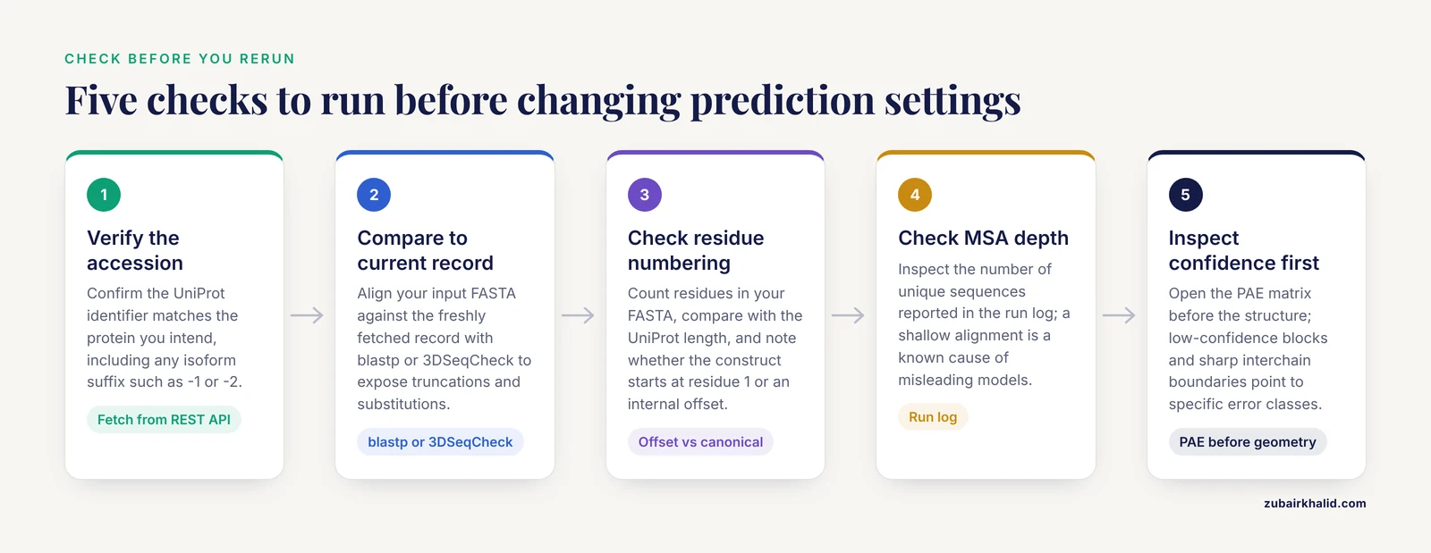
Run these checks in order before touching prediction parameters.
- Verify the accession. Confirm that the UniProt identifier in your input matches the protein you intend to model, including any isoform suffix such as -1 or -2. Fetch the current record directly from the UniProt REST API rather than reusing a local file:
curl https://rest.uniprot.org/uniprotkb/Q9Y6K9.fasta -o Q9Y6K9_current.fasta- Compare your input to the current record. A pairwise alignment or a simple identity calculation exposes truncations, insertions, and silent substitutions:
seqkit seq -n input.fasta
blastp -query input.fasta -subject Q9Y6K9_current.fasta -outfmt 6- Check residue numbering. Count the residues in your FASTA and compare the total against the UniProt length. Then check whether your construct starts at residue 1 of the canonical sequence or at an internal offset, because most downstream mapping errors come from this offset.
- Check the MSA depth. If you run ColabFold locally, inspect the reported number of unique sequences in the alignment. A shallow MSA (fewer than a few hundred effective sequences for a typical globular domain) is a known cause of poor or misleading models [4].
- Inspect confidence before geometry. Open the PAE matrix first, then the structure. Low-confidence blocks, sharp interchain PAE boundaries, and long high-pLDDT tails of disorder all point to specific error classes described below [3].
Troubleshooting Table
| Symptom | Likely cause | How to confirm | Fix |
|---|---|---|---|
| Model disagrees with experimental structure or known domain boundaries | Outdated or wrong input sequence (isoform mismatch, database revision) | Align input FASTA against the current UniProt record with blastp or 3DSeqCheck | Redownload the current sequence, rerun the prediction, remap any annotations [1] |
| Model lacks the N-terminal region or fails at the catalytic site | Gene annotation error in the source record (wrong start codon) | Compare your sequence to homologs and to any experimental structure of the family | Extend the sequence to the experimentally supported start, rerun, and report the corrected annotation [5] |
| High pLDDT on a very short hypothetical ORF that shows no homology | Spurious sequence from gene prediction, not a real protein | Search against AntiFam, check length (discrimination improves beyond 100 amino acids), check genomic context | Treat the model as unvalidated, flag the ORF for curation, combine structure scores with Foldseek and InterProScan evidence [6][7] |
| Globular domain distorted, secondary structure incoherent | MSA too shallow or dominated by narrow taxonomy | Check MSA depth in the run log, reformat MSA with a broader database | Expand the MSA search, add sequences from a wider database, or supply templates [4] |
| ipTM jumps upward after you trim the construct, with no change in the modeled interface | ipTM formulation penalizes unstructured or accessory regions on the same chains | Compare ipTM before and after trimming while inspecting interchain PAE | Judge interfaces by interchain PAE, not raw ipTM, and report both trimmed and full-length values [8] |
| Complex prediction misses a known interaction partner or adds a phantom chain | Incorrect oligomeric state or modified residues in the input | Check stoichiometry against literature and the stoichiometry in similar PDB entries | Submit each chain in the correct copy number and revert engineered mutations to wild type where possible [9] |
| Residue numbers in the model do not match UniProt annotations | Numbering offset between the construct and the canonical sequence | Map model numbering to UniProt with a sequence alignment tool such as PDBminer | Renumbers the model to the UniProt frame before mapping variants or ligand contacts [10] |
| Flexible regions look chaotic, termini are extended and floppy | Real disorder, not an error | pLDDT below about 50 in those regions, high PAE to the rest of the protein, PAE pattern matches dynamic behavior | Interpret those regions as conformational ensembles, not coordinate errors [3][11] |
| Side chains in the core look plausible but distant side chains are wrong | Known side chain prediction limits | Check chi1 and chi3 rotamers against experimental structures of homologs | Energy-minimize or experimentally resolve side chains that matter for your analysis [12] |
Error 1: Outdated or Mismatched Reference Sequences
This is the most frequent AlphaFold sequence error and the easiest to fix. Reference databases are living objects. UniProt entries are updated several times a year as new sequencing evidence accumulates, while structure resources evolve on their own schedule. The AlphaFold Database went four years without a content update, until September 2025, during which nearly 3 percent of the sequences associated with its models were revised in UniProt [1]. Any pipeline that pairs an AlphaFold model with UniProt annotations during that window could map annotations onto residues that no longer exist at those positions.
The same drift hits locally cached FASTA files. A sequence downloaded years ago for one project tends to propagate through personal folders, lab wikis, and supplementary materials, accumulating isoform swaps and stale versions along the way. Isoform mismatch is the classic variant of this error: many UniProt entries carry multiple isoforms that differ by whole exons, and predicting isoform 1 while annotating isoform 2 produces a model with confidently built sequence that simply is not in the protein you meant.
How to confirm. Fetch the current UniProt record and align it against your input. A web tool such as 3DSeqCheck compares the sequence embedded in a structure file against the latest UniProt entry and displays the alignment and residue numbering differences interactively [1]. A command line check with blastp takes seconds and gives you percent identity plus gap positions.
How to fix. Redownload the current canonical or isoform-specific sequence, rerun the prediction, and regenerate any annotation mapping. For database-scale work, note that the AlphaFold Database itself now integrates with UniProt, Ensembl, InterPro, and MobiDB, and offers programmatic access alongside bulk FTP downloads, so a scripted pull of the current record is safer than a hand-copied file [13]. The database has grown from roughly 360,000 structures across 21 model organism proteomes at launch [2] to more than 214 million sequences [13], which means most proteins you will ever need have a current model available under the current sequence.
Error 2: Gene Annotation Errors in the Source Sequence
Sometimes the reference record itself is wrong. Genome annotation pipelines mispredict start codons, splice junctions, and exon boundaries, and those errors flow directly into protein sequences and then into AlphaFold predictions. The model will faithfully fold an incorrectly annotated sequence, and the error is invisible unless you look outside the record.
A documented example comes from Borrelia burgdorferi. The plasmid-encoded gene bbe22 encodes a nicotinamidase, and translation initiates from a rare non-canonical AUU start codon, producing a protein 24 residues longer than the sequence annotated in major databases. UniProt and KEGG still listed a truncated protein beginning at residue 36, a form that lacks part of the N-terminal region required for enzymatic activity. The crystal structure of the full-length enzyme, determined at 3.2 angstrom resolution, confirmed that the longer N-terminal sequence is required for the correct fold [5]. Any AlphaFold model built from the truncated database sequence would have inherited that defect.
How to confirm. Align your sequence against homologs across species. A conserved catalytic N-terminus in every homolog while your query starts short is a strong signal of a start codon misannotation. Where an experimental structure of a homolog exists, compare the secondary structure at the N-terminus.
How to fix. Extend the sequence to the experimentally or evolutionarily supported start, rerun the prediction, and treat the corrected annotation as a finding worth reporting. Structure prediction is increasingly used for exactly this purpose. In a benchmark across Fusarium graminearum, Toxoplasma gondii, and Aspergillus fumigatus, AlphaFold 3 scores favored manually improved gene models over pre-improvement models and supported 65 to 84 percent of manually curated changes, with the best discrimination coming from combining AlphaFold 3 scores with Foldseek structural searches and InterProScan domain identification [6]. The same logic works in reverse: a low-quality score on an annotated gene model is evidence that the translation, not the protein, is at fault.
Error 3: Spurious Sequences That Fold Confidently
A more insidious failure mode is the sequence that folds well but never existed. Spurious protein sequences arise from gene prediction errors, and theory says they should not produce folded structures. In practice, AlphaFold2, ESMFold, and AlphaFold3 all assign unexpectedly high pLDDT scores to short spurious sequences drawn from AntiFam, a database of decoy protein families. Discrimination between spurious and real proteins improves only beyond 100 amino acids, which is exactly the length range where many gene prediction artifacts live [7]. The practical consequence is that a high pLDDT score is not evidence of biological reality for a short ORF.
How to confirm. Check three things. First, search the sequence against AntiFam and standard homology databases. Second, check genomic context for coding evidence such as conservation across related species. Third, look at the score profile, since analyses of proteins with disparate pTM and pLDDT values have identified suspected spurious shadow ORFs even inside curated Swiss-Prot entries [7].
How to fix. Treat the model as unvalidated until independent evidence supports the ORF. Combine structure prediction scores with Foldseek and InterProScan results, an approach whose discriminative power for gene model assessment has been demonstrated at scale [6]. If the ORF fails all checks, remove it from downstream use rather than building biology on top of a confidently folded artifact.
Error 4: MSA Depth and Composition Problems
AlphaFold's accuracy depends heavily on the multiple sequence alignment, and MSA failures are among the most common causes of distorted or implausible models. A key insight from recent evaluations is that the limitation stems from reliance on MSA depth rather than mere taxonomic composition of the Protein Data Bank, meaning that the number of effective homologous sequences matters more than which organisms they come from [4]. A shallow MSA starves the model of coevolutionary signal, and the output can range from subtly wrong loop conformations to completely misarranged domains.
How to confirm. Check the MSA statistics in your run log. ColabFold reports the number of unique sequences and the MSA depth when it runs. If you use an AlphaFold server through a browser interface, look for the same statistics in the output bundle. Depth in the low hundreds of sequences for a single domain, or the absence of detectable homologs for a novel fold, predicts trouble.
How to fix. Expand the search. Run the MSA search against a broader database, lower the sequence identity filtering threshold to retain more distant homologs, or supply structural templates so the model has geometric information even when coevolutionary signal is thin. Templates measurably improve side chain accuracy as well [12]. For proteins from poorly sampled phylogenetic space, temper expectations and validate the model against experimental data where any exists.
Error 5: Construct Trimming and ipTM Artifacts
For protein-protein interaction studies, a specific sequence-level decision produces a well-known misleading score. Users routinely trim full-length sequences down to the interacting domains before predicting a complex, and many notice that ipTM (interchain predicted TM score, a confidence metric for interfaces) rises after trimming even though the predicted interface itself is unchanged. The cause is mathematical. ipTM in AlphaFold2 and AlphaFold3 scores interactions across whole chains, so large disordered regions or accessory domains that do not participate in the primary interface drag the score down. The score then fails to represent either the accuracy of the interface prediction or the likelihood that the two proteins actually interact [8].
How to confirm. Run the same complex twice, once with full-length sequences and once trimmed, and compare the interchain PAE matrix in both runs. If the interface geometry and PAE values are essentially identical while ipTM shifts substantially, you are looking at the scoring artifact.
How to fix. Judge interface quality by the interchain PAE values over the contacting residue pairs rather than by raw ipTM. Where a single number is needed, the corrective approach in the literature restricts the ipTM calculation to residue pairs with good interchain PAE values, adjusts the d0 length parameter accordingly, and uses PAE values directly rather than full probability distributions [8]. Practically, report both the full-length and trimmed predictions, and cite the PAE pattern at the interface as your primary evidence.
Error 6: Wrong Oligomeric State and Modified Input Sequences
AlphaFold models whatever stoichiometry you specify. Submit a monomer when the protein works as a dimer, or leave an engineered mutation in the construct, and the model can be globally high quality while locally wrong at exactly the sites you care about. Studies of AlphaFold 3 on natural variants of Helicobacter pylori catalase KatA found that user inputs such as incorrect oligomeric states or sequence modifications influenced prediction quality, with deviations concentrated at flexible or interface-exposed sites even when the overall model remained strong [9]. The inverse also holds: when the oligomeric state is correctly specified, AlphaFold can resolve real biological assemblies, as shown for the archaeal PolB2 complex, where a heteropentameric composition emerged from careful combined modeling with high confidence scores [14].
How to confirm. Cross-check the stoichiometry against the literature and against PDB entries of homologs. Compare interchain PAE between your assumed state and plausible alternatives.
How to fix. Submit each chain in the biologically supported copy number, revert construct artifacts (purification tags, point mutations, truncations) to the wild-type sequence where the goal is the native protein, and reserve engineered constructs for questions about the engineered state specifically [9]. For polyprotein systems, an alternative is to model the full polyprotein as one chain, since PAE plots between domains of a single polyprotein can reproduce protein-protein interactions that persist after proteolytic cleavage [15].
Error 7: Residue Numbering and Mapping Errors Downstream
The model itself can be correct and the analysis still wrong if residue numbering is off. Experimental PDB structures are numbered against the construct the crystallographer crystallized, which often differs from the UniProt canonical sequence by expression tags, missing disordered termini, or internal gaps. AlphaFold models carry their own numbering, conventionally the input sequence positions. Mapping variants, domains, or ligand contacts across these frames without an alignment produces silent errors.
How to confirm. Take one annotated residue, such as an active-site cysteine, and verify that its position in the model file corresponds to the same residue in the UniProt entry. Tools built for this purpose, such as PDBminer, search the AlphaFold Database and the PDB for structures of a target protein and report the coverage of each structure aligned to the UniProt sequence, explicitly to overcome numbering differences [10].
How to fix. Reindex the model into the UniProt frame before any annotation mapping. Domain parsing tools help here too, since correct domain boundaries in the UniProt frame depend on correct segmentation of the model, and parsers such as DPAM-AI combine inter-residue distances, PAE values, and sequence and structural alignment to assign domains reliably [16].
Error 8: Conformational Diversity Misread as Error
Not every disagreement between an AlphaFold model and an experimental structure is a sequence error, and treating dynamics as an error leads to wasted effort. Proteins are ensembles, and AlphaFold2 predicts the holo (ligand-bound) conformation in roughly 70 percent of apo-holo pairs, meaning the model frequently snapshots one state of a two-state protein. Prediction performance worsens as the conformational diversity of the target increases, and that impairment tracks the heterogeneity of conformational diversity within the protein's homologous family [11]. Meanwhile, pLDDT values in a single model correlate with main-chain flexibility, so low pLDDT in a loop is often a truthful report of motion rather than a defect [11].
The dynamical reading of confidence scores is supported by molecular dynamics comparisons. PAE maps from AlphaFold 2 correlate with distance variation matrices from MD simulations, and AF2-scores derived from pLDDT correlate with RMSF for most globular proteins and complexes, though not for intrinsically disordered proteins [3].
How to confirm and fix. Before editing the sequence, check whether the suspicious region sits in a low-pLDDT, high-PAE zone that would be expected for a flexible element. If it does, the correct fix is interpretation: treat that region as an ensemble, complement the static model with MD sampling where needed, and resist the temptation to delete flexible regions from the input, which merely converts honest uncertainty into a misleadingly tidy model. Note that deleting disorder has a separate scoring cost in complexes, as described in the ipTM section [8].
Error 9: Side Chain Errors
Backbone accuracy and side chain accuracy are different quantities, and sequence-level surprises concentrate in side chains. Benchmarking with ColabFold across ten proteins spanning populated fold families found a side chain conformation prediction error of about 14 percent for chi1 dihedral angles, rising to about 48 percent for chi3 dihedral angles. Nonpolar side chains predict better than polar ones, structural templates modestly improve accuracy, and ColabFold shows a bias toward the most prevalent PDB rotamer states, limiting its ability to capture rare conformations [12].
How to confirm. Compare predicted rotamers for residues that matter to your hypothesis (catalytic residues, contact residues) against experimental structures of close homologs. Trust backbone placement more than side chain placement.
How to fix. Where a specific side chain conformation drives your conclusion, relax the model with a force field, use experimental structures as templates, or obtain experimental data. Do not quote chi3-level detail from an AlphaFold model as if it were resolved.
Common Mistakes and Limitations
Several recurring mistakes turn manageable sequence errors into incorrect conclusions.
- Trusting pLDDT as a validity certificate. High pLDDT means the model agrees with its own sequence and MSA, not that the sequence is real or biologically active. Spurious short ORFs fold confidently [7], and an IDR attached to a folded domain gets a low pLDDT that reflects real disorder, not a broken prediction [3].
- Optimizing scores instead of questions. Trimming constructs to raise ipTM changes the metric, not the structure [8]. Choose the input that represents the biological question, then report the score honestly.
- Ignoring the database version. A model downloaded once and reused for years can silently diverge from the current UniProt record [1]. Record the retrieval date and accession version for every sequence.
- Assuming the MSA is fine because the model finished. Completion is not quality. Shallow MSAs produce plausible-looking but wrong models, and MSA depth, not database identity, is the limiting variable [4].
- Overreading single-residue predictions. AlphaFold 3 models natural variants with high global fidelity but variable accuracy at flexible or interface-exposed sites [9], and distant side chains carry substantial error [12]. Treat residue-level claims for such positions as hypotheses.
- Docking directly into apo-shaped models. Predicted structures often represent holo-like states [11], and direct molecular docking onto them is unreliable because of apo conformation issues and protein dynamics [4].
- Forgetting that confidence metrics themselves can be miscalibrated for complexes, as the ipTM analysis shows [8]. Metrics are estimates with known failure modes, not grades.
Prevention Practices
A short routine prevents almost all of the errors above.
- Fetch sequences programmatically from UniProt at prediction time, and store the accession, isoform, and retrieval date alongside every model [13].
- Run an automatic input-versus-reference identity check in your pipeline, using a tool such as 3DSeqCheck for structure-to-UniProt comparisons [1].
- For newly annotated genomes, screen predicted proteins with structure scores plus Foldseek and InterProScan before biological interpretation, since combined scores identify poor gene models effectively [6].
- Record MSA depth in your lab notebook or pipeline log for every prediction, and treat low depth as a flag requiring either broader search or template use [4].
- Specify stoichiometry deliberately, with a literature citation for the state you choose, and model alternatives when the state is uncertain [9].
- Archive the exact input FASTA, MSA, run parameters, and version of the prediction software with every model so results stay reproducible.
- Interpret pLDDT and PAE as measurements of flexibility and uncertainty, not as pass-fail grades, and pair them with dynamics data or experimental validation for conclusions that depend on conformation [3][11].
Frequently Asked Questions
Why does my AlphaFold model look wrong even though pLDDT is high?
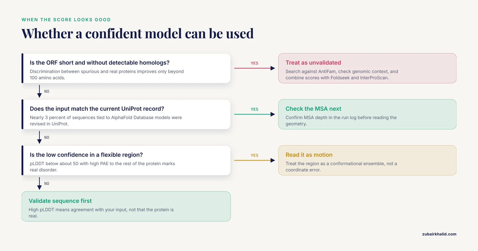
Because pLDDT measures agreement with your input sequence and MSA, not with the protein you intended to model. A wrong, outdated, or spurious sequence can fold with high confidence, so validate the sequence itself against the current UniProt record before trusting the score [1][7].
How do I check whether my AlphaFold input sequence matches the current UniProt entry?
Fetch the current record from the UniProt REST API and align it against your input FASTA with blastp, or submit the structure file to 3DSeqCheck, which compares the embedded sequence to the latest UniProt entry and displays numbering differences [1].
Why did trimming my construct raise my ipTM score?
The ipTM formulation scores whole chains, so unstructured regions and accessory domains that do not form the interface lower the score. Trimming removes them and raises the number without changing the interface. Judge interfaces by interchain PAE values instead [8].
Does a low pLDDT mean my sequence is wrong?
Usually no. Low pLDDT most often marks genuine flexibility, disorder, or isolated domains rather than a sequence error. Intrinsically disordered regions receive low scores by design, and pLDDT correlates with backbone flexibility [3][11].
Should I trust AlphaFold models of short ORFs from gene prediction?
Be cautious. AlphaFold2, ESMFold, and AlphaFold3 all assign high pLDDT to short spurious sequences, and reliable discrimination improves only beyond 100 amino acids. Require homology, genomic context, or expression evidence before treating such a model as a real protein [7].
Why is my AlphaFold model missing the N-terminal region I expected?
Check the source record for a start codon misannotation. Documented cases exist where databases list a truncated protein beginning tens of residues downstream of the true translation start, and the missing region can be required for the correct fold [5]. Compare your sequence to homologs and extend to the supported start.
Does AlphaFold make side chain errors?
Yes, more often than backbone errors. Reported prediction error is roughly 14 percent for chi1 dihedral angles and about 48 percent for chi3 angles, with a bias toward common rotamer states. Verify side chains that matter for your analysis against homolog structures or experimental data [12].
Can AlphaFold predict alternative conformations?
A single run generally returns one state, biased toward the holo form in about 70 percent of apo-holo pairs. Conformational diversity within a protein family worsens prediction, so treat the output as one member of an ensemble and use dynamics methods to explore alternatives [11].
Sources
- 3DSeqCheck: A Web-based Tool for Verifying Sequence Consistency Between a 3D Structure File and the Corresponding UniProt Entry.
- AlphaFold Protein Structure Database: massively expanding the structural coverage of protein-sequence space with high-accuracy models.
- AlphaFold2 models indicate that protein sequence determines both structure and dynamics.
- From Sequence to Structure: AlphaFold and the AI Revolution in Aquaculture Drug Discovery and Vaccine Design.
- The crystal structure of the Borrelia burgdorferi nicotinamidase BBE22 resolves a long-standing annotation error.
- The promise of AlphaFold for gene structure annotation.
- Folding the unfoldable 2: using AlphaFold and ESMFold to explore spurious proteins.
- Rēs ipSAE loquunt: What's wrong with AlphaFold's ipTM score and how to fix it.
- AlphaFold 3 accurately models natural variants of Helicobacter pylori catalase KatA.
- PDBminer to Find and Annotate Protein Structures for Computational Analysis.
- Impact of protein conformational diversity on AlphaFold predictions.
- Predicting side chain conformations in folded proteins by AlphaFold: Perspective and challenges.
- AlphaFold Protein Structure Database in 2024: providing structure coverage for over 214 million protein sequences.
- Common themes in architecture and interactions of prokaryotic PolB2 and Pol V mutasomes inferred from in silico studies.
- AlphaFold Prediction of Protein-Protein Interactions in the Flaviviridae Proteomes.
- DPAM-AI: a domain parser for AlphaFold models powered by artificial intelligence.