AlphaFold for Protein Engineering
By Dr. Zubair Khalid, DVM, MS, PhD ·

By the end of this guide you will be able to pull a predicted structure for any protein of interest, run your own prediction when the database model is not enough, read the confidence metrics the way a structural biologist does, plan mutations against a predicted model, and screen a designed variant with a second predictor before you order a single oligo. You will need a target sequence (a UniProt accession or a FASTA file), access to a Linux or macOS terminal, Python 3 with a few common packages, and either a web browser for the hosted prediction services or a GPU if you want to run predictions locally.
AlphaFold changed the economics of protein engineering. A workflow that once required a crystallography collaboration, months of cloning and expression trials, and a series of structure-based redesign cycles can now start with a prediction completed in minutes. Reviews of engineered enzymes and biocatalytic systems now treat AlphaFold-assisted structure prediction, molecular docking, molecular dynamics, and machine learning guided mutagenesis as standard components of the engineering toolkit rather than specialist extras [1], and enzyme engineering programs built around AlphaFold-assisted rational design have reported large activity gains with reduced experimental screening [2], [3]. The catch is that a prediction is a hypothesis, not a structure, and most failed engineering projects that use AlphaFold fail because the confidence metrics were misread. This guide covers both the mechanics and the judgment.
What AlphaFold Actually Gives You
AlphaFold predicts the three-dimensional coordinates of a protein from its amino acid sequence. For engineering purposes, the coordinates matter less than the two numbers attached to them.
pLDDT (predicted local distance difference test) is a per-residue score from 0 to 100 that estimates how accurately each residue is positioned. In AlphaFold Database files, pLDDT is stored in the B-factor column of the PDB file, which means you can read it with any structure viewer without any extra parsing.
PAE (predicted aligned error) is a matrix that estimates, for every pair of residues, the expected positional error of one residue when the structure is aligned on the other. Low PAE within a domain means the domain is confidently folded. Low PAE between two domains or two chains means their relative orientation is confidently predicted. High PAE between domains means the model is confident about each piece but guessing about how the pieces connect, which is exactly the region where engineering decisions go wrong.
ipTM (interface predicted TM-score) is a multimer metric from 0 to 1 that estimates whether the predicted interface between chains is correct. It is the primary screen for designed binders and oligomers.
A single number worth internalizing: high pLDDT on a residue tells you the residue sits where AlphaFold thinks it sits. It does not tell you the residue is in that position in the protein you are actually studying, in the state you care about, with your ligand bound. The prediction is a conditional statement, and the conditions are the ones the next sections help you check.
Choose Your Route: AlphaFold Server, ColabFold, or a Local Install
There are three practical ways to get a prediction, and the right one depends on scale.
The AlphaFold Server is the hosted service from Google DeepMind for AlphaFold 3. You paste sequences for proteins, nucleic acids, ions, and small molecule ligands, and it returns coordinates plus confidence files. It is the fastest route for occasional predictions of complexes with ligands, and it requires no installation. For engineering work where you will iterate on many variants, check the service's current usage limits on the official site, because batch screening can exhaust them quickly.
ColabFold is the community implementation of AlphaFold 2 and AlphaFold-Multimer that runs in a free Google Colab notebook or on your own machine through pip. It exposes MSA depth, template usage, and model presets as controllable options, which you need when you are deliberately testing whether a result depends on the MSA.
A local AlphaFold or AlphaFold 3 installation makes sense for high-throughput screening, for air-gapped environments, or when you need custom MSA inputs. Expect to provision a multi-terabyte reference database and an A100-class or equivalent GPU.
For most engineering projects the pattern is: use the AlphaFold Database for the parent protein, use the AlphaFold Server or ColabFold for variants and complexes, and reserve local installs for screening campaigns of hundreds of designs.
AlphaFold Protein Engineering Step by Step
This walkthrough follows a single concrete target, human epidermal growth factor receptor EGFR (UniProt P00533), but every command works with your own accession. The same pattern applies to a novel sequence that has no database entry, in which case you start at step 4.
Step 1: Confirm the Sequence and Annotations
Start from UniProt rather than a sequence you copied from a figure. UniProt records the canonical isoform, signal peptides, domains, and variants, all of which affect what you should model. Pull the FASTA directly:
curl -s https://rest.uniprot.org/uniprotkb/P00533.fasta > egfr.fasta
head -c 300 egfr.fasta
Expected output starts with:
>sp|P00533|EGFR_HUMAN Epidermal growth factor receptor OS=Homo sapiens OX=9606
PEPPTCVSVNIGLY...
Decide now which residues you are engineering. If your target has a signal peptide, propeptide, or transmembrane anchor that will not be present in your construct, truncate the FASTA before predicting. Predicting the full membrane-spanning receptor when you plan to express a soluble ectodomain wastes compute and produces a model whose confidence reflects the full-length context rather than your construct.
Step 2: Check Whether You Need to Predict at All
The AlphaFold Database holds predicted structures for catalogued proteins across the tree of life, indexed by UniProt accession. Before spending compute, download the existing model:
curl -s -o AF-P00533-F1-model.pdb \
  https://alphafold.ebi.ac.uk/files/AF-P00533-F1-model_v4.pdb
The version suffix (v4 here) reflects the current database build and can differ between entries, so if the URL returns an error, pull the file links from the entry page at alphafold.ebi.ac.uk for your accession. The same directory pattern serves the predicted aligned error file as JSON.
If you need structures for many proteins at once, for example all enzymes in an operon you are rewiring, the AlphaFoldDB Structure Extractor web server and API accepts NCBI Taxonomy IDs, RefSeq accessions, locus tags, and UniProt or AlphaFold accessions, and returns coordinates in PDB, mmCIF, or binary CIF format together with PAE JSON, plus an ID mapping file that traces your input identifiers back to standard accessions. It has been tested with individual inputs and batches of up to 5,000 accessions, which makes it useful for pipeline integration [4].
Step 3: Search for Structural Homologues
A predicted structure is most useful when you know its relatives. Homologues with experimental structures anchor your interpretation and provide templates. MrParse searches the PDB and the EBI AlphaFold database for structural homologues, ranks candidates for molecular replacement suitability using eLLG when reflection data are supplied, and displays predicted secondary structure, coiled-coil, and transmembrane regions in one report [5]. Even without crystallographic intent, its homologue ranking tells you whether your protein is in a well-characterized fold family or floating alone.
For a fold-level question, for example whether your new enzyme adopts a fold with a known engineered scaffold, 3D-AF-Surfer provides real-time structure-based search over AlphaFold models using 3D Zernike descriptors, a rotation-invariant mathematical representation of molecular shape, and a neural network that retrieves proteins of the same fold more accurately than direct descriptor comparison [6]. That same study documents a pattern you should expect to see yourself: low-confidence AlphaFold models correlate strongly with intrinsically disordered regions, which is a diagnostic as much as a warning.
Step 4: Run a Prediction
For a variant sequence, a chimera, or a protein with no database model, run a fresh prediction. The fastest route is the AlphaFold Server (paste sequences, optionally add ligands or cofactors as inputs, download the result). For controllable iteration, use ColabFold. Install it locally with:
python -m pip install "colabfold[alphafold-minus-jax]"or open the ColabFold notebook in Google Colab and use the free GPU runtime. Prepare your variant as a FASTA:
printf ">egfr_variant\n%s\n" "$(tail -n +2 egfr.fasta | tr -d '\n')" > input.fastaThen run the batch predictor with the output directory as the second argument:
mkdir results && colabfold_batch input.fasta resultsColabFold accepts optional flags for template usage, MSA mode, number of recycles, and model presets, all documented in its repository. Its defaults are sensible for monomers. A completed run produces ranked models and score files that look like this when you list the directory:
results/
  rank_001_alphafold2_multimer_v3_model_1_seed_000.pdb
  rank_001_alphafold2_multimer_v3_model_1_seed_000_scores.json
  rank_002_alphafold2_multimer_v3_model_2_seed_000.pdb
  ...
  rankings_env.log
The rank ordering is by model confidence. Exact file names vary with the model preset and seed, so treat the listing above as representative rather than literal.
Step 5: Read the Confidence Metrics Before the Structure
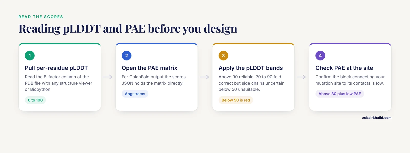
This is the step that separates useful engineering from expensive guesswork. Extract per-residue pLDDT from the PDB B-factor column:
from Bio.PDB import PDBParser

parser = PDBParser(QUIET=True)
structure = parser.get_structure("af", "AF-P00533-F1-model_v4.pdb")

for residue in structure.get_residues():
    ca = residue["CA"]
    print(residue.id[4], round(ca.bfactor, 1))
Expected output is two columns, residue number and pLDDT, for example:
1 41.7
2 55.2
3 68.9
...
310 94.1
Then read the PAE matrix. For ColabFold output, the scores JSON holds the matrix directly:
import json

with open("results/rank_001_alphafold2_multimer_v3_model_1_seed_000_scores.json") as fh:
    scores = json.load(fh)

pae = scores["pae"]
print(len(pae), min(min(r) for r in pae), max(max(r) for r in pae))
Expected output is the matrix dimension and the PAE range, for example 1130 0.31 28.4.
Apply the working rules. Residues with pLDDT above 90 are modeled at backbone accuracy comparable to low-resolution experimental structures and are reliable anchors for design. Residues between 70 and 90 are usually correct in fold but uncertain in side-chain detail, so redesign there needs caution. Residues below 50, conventionally drawn in red in AlphaFold Database visualizations, are expected to be disordered or wrongly placed, and mutations there cannot be reasoned about from the model. If your mutation site and its packing partners all sit above 80 and the PAE block connecting them to the rest of the fold is dark (low), you have a model worth designing against.
Step 6: Plan Mutations Against the Model
Open the model in a molecular viewer (ChimeraX, PyMOL, or the web viewer at the AlphaFold Database). For a stability or specificity mutation, check three things at the position: packing density, whether the position sits at an interface or active site, and whether the local pLDDT supports detailed reasoning. A worked judgment example: if residue 790 of your construct shows pLDDT 94 and sits in a packed hydrophobic core with three contacts, a conservative substitution reasoned from the model has a solid structural basis. If the candidate position shows pLDDT 52 in a loop, the model is describing a plausible guess, and structure-informed mutagenesis at that site is speculation wearing a structure's clothes.
For systematic redesign rather than single mutations, pair AlphaFold with a sequence design model. ProteinMPNN generates sequences compatible with a fixed backbone, and the ProteinMPNN-AlphaFold pipeline has produced striking enzyme improvements in published workflows. In one recent example, a genome-mined beta-thioglucosidase scaffold was redesigned at flexible loops near the active site using this pipeline, and the improved variant raised kcat from 98.5 plus or minus 3.2 to 421.3 plus or minus 9.1 per minute while lowering Km from 4.6 plus or minus 0.2 to 0.9 plus or minus 0.1 mM, with molecular dynamics and QM/MM analysis attributing the gain to loop rigidification, improved hydrophobic packing, and a reshaped electrostatic environment [3]. Reviews of transaminase engineering describe the same pattern at the field level, with AlphaFold-assisted workflows reducing experimental effort in semi-rational campaigns [2].
For scaffold-constrained binder design, recent work offers a cautionary benchmark. A DARPin design framework restricted sequence generation to grammar-permitted residue pools, screened 15,000 generated variants against mesothelin, VEGF-A, and HER2 using AlphaFold2-Multimer, then re-evaluated 149 library designs with AlphaFold 3. Only eight designs passed the Ranking Score threshold of 0.70, all against mesothelin, a pass rate of roughly 0.05 percent of the generated library, and the median corrected scores reached 0.794 for the best library design against a positive control at 0.866 [7]. The throughput math matters: screening compute consumed four orders of magnitude more designs than produced passing candidates.
Step 7: Re-Screen with a Second Predictor and Validate
The DARPin study produced one of the most practically useful findings in current design literature. When the same 149 designs were scored by AlphaFold 2 and AlphaFold 3, the AF3 median chain-pair ipTM was lower than the AF2 ipTM for 148 of the 149 designs [7]. In other words, the two predictors disagree systematically on designed complexes, and passing one predictor's filter does not mean the interface exists. The practical rule is to screen with one predictor and confirm with another, and to treat designs that pass both as a much smaller, much better set. Those candidates still require biophysical and cellular validation before any claim of binding [7].
Experimental validation should be planned at design time, not after. Structure-guided engineering does not remove the expression, purification, and assay work. It concentrates it. A well-run cycle is predict, design a small library, assay, and feed failures back into the next prediction, and published enzyme and antimicrobial engineering studies in this cluster all followed that loop [8], [2], [3].
The table below summarizes the outputs you will handle and what each one licenses you to conclude.
| Output | Range or format | What it tells you | Use it for |
|---|---|---|---|
| , - | , - | , - | , - |
| pLDDT (B-factor column) | 0 to 100 per residue | Local positional accuracy of each residue | Deciding whether a mutation site is trustworthy |
| PAE matrix | Angstroms, residue pair matrix | Confidence in relative positions of residue pairs | Judging domain orientations and interfaces |
| ipTM (multimer) | 0 to 1 per model | Whether a predicted chain interface is correct | Screening designed binders and oligomers |
| ptm (multimer) | 0 to 1 per model | Global fold correctness of the assembly | Comparisons across models of one complex |
| Ranked PDB files | 5 models by default | Alternative fold hypotheses | Checking whether designs are consistent across models |
| PAE JSON | Structured scores file | Programmatic access to pair confidence | Pipelines and automated filtering |
Working with Complexes and Oligomers
Many engineering targets function as multimers, and the monomer database model tells you nothing about the quaternary state. AlphaFold-Multimer predicts complexes from chained input sequences. A large-scale example of what this enables is PHLEGM, a framework that infers homooligomeric states directly from phage protein sequences by combining AlphaFold-Multimer modeling with inter-subunit interface quality assessment, then validated predictions experimentally for a predicted dimer and trimer using size exclusion chromatography and hydrodynamic methods. Applied to more than 22,000 phage protein sequences in the PHROGs database, it revealed extensive homooligomeric diversity and outperformed sequence-based oligomer state predictors [9]. For your own target, the analogous practice is to run Multimer over the plausible oligomer states (dimer through pentamer for a small protein), compare inter-chain ipTM, and confirm the winning state experimentally if the stoichiometry matters for your design.
Interfaces deserve the same confidence discipline as folds. High inter-chain ipTM with low inter-chain PAE across a broad interface is the signature of a trustworthy complex prediction. A single-chain ipTM value with a thin strip of low PAE at one contact point is not.
Ligand Binding: Docking and Co-Folding
If your engineering goal touches small molecule binding, either to preserve an active site or to alter one, the evidence on AlphaFold models as docking receptors is encouraging with conditions. A systematic assessment using Glide docking with unbiased binding site identification by FTMap found that fragment docking to AlphaFold models outperformed docking to the corresponding unbound protein crystal structures and performed comparably to docking to ligand-bound structures, and that using ensembles of AlphaFold models improved larger-ligand docking over any single model while also outperforming unbound structures [10]. Two practices follow directly. First, use AF model ensembles rather than a single conformer, because the ranked models differ mainly in loop and side-chain geometry, which is what docking is sensitive to. Second, identify the binding site computationally rather than assuming the crystallographic site, since the AF model may place flexible side chains differently than any single experimental structure.
AlphaFold 3 extends prediction to complexes with ligands and modified chemistry, but benchmarking shows real limits. On the MODNAP benchmark of 1,039 nucleic acid and nucleic acid-protein complex structures, AlphaFold 3 exceeded 80 percent success at a 2.5 Å RMSD threshold overall, yet its accuracy in positioning modified nucleotides was variable and dropped for chemistries not represented in training, and PoseBusters analysis found frequent intramolecular validity failures arising from non-ideal ribose ring geometries [11]. The lesson generalizes beyond nucleotides. For any modified residue, noncanonical amino acid, or unusual ligand in your engineered system, AF3 coordinates on that chemistry are hypotheses that need inspection of bond geometries and, ideally, quantum or empirical refinement before you reason about them.
Conformational States and Dynamics
AlphaFold predicts a single dominant state, and many engineering problems are about states: an active versus inactive kinase, an open versus closed carrier, a ligand-bound versus apo loop. Two lines of work address this. PromptGPCR provides AlphaFold-Multimer and AlphaFold 3 with biological sequences as prompts informed by structural knowledge, such as pairing the receptor with its G protein for the active state, and predicted both active and inactive GPCR conformations more accurately than baselines, with higher docking success rates for the predicted states [12]. The generalizable idea is that you can bias a prediction toward a state by adding the interaction partners or constraints that define that state.
For dynamics beyond two states, diffusion-based ensemble methods use AlphaFold Database structural profiles as conditional guidance to generate conformational ensembles. DiffEnsemble, benchmarked on 72 proteins from the ATLAS molecular dynamics dataset, improved Pearson correlation of ensemble pairwise RMSD by 28.9 percent and RMSF by 7.5 percent relative to AlphaFLOW, indicating that static structural archives carry usable dynamic information [13]. For an engineering project, a generated ensemble is most useful for identifying which loop or domain motions correlate with the property you are changing, before you decide which conformer to design against.
Solubility and Developability Predictions
Expression and solubility failures kill more engineering projects than misfolding does, and AlphaFold-derived features are now part of the prediction toolkit. SeqStruct-XAttn integrates frozen ESM-2 protein language model embeddings with AlphaFold-derived structural cues through bidirectional cross-attention to predict Escherichia coli protein solubility, reaching an R squared of 0.560 and RMSE of 0.198 on the eSOL independent test set, with a five-model ensemble improving this to 0.575 and 0.190 [14]. The numbers are moderate rather than spectacular, which is itself informative: solubility depends on construct boundaries, expression conditions, and intermolecular contact chemistry that no sequence-level predictor fully captures. Treat solubility predictions as one more filter in a screening cascade, not a verdict. When engineering antimicrobial lysins for Gram-negative activity, structure prediction and charge analysis used in combination to choose peptide fusions showed a concrete structural correlate: variants fused to peptides predicted as alpha-helical and positively charged retained activity in physiological salt and serum, while fusions with non-helical, charge-poor peptides did not [8]. That study is a good model of using prediction for a developability property, not just a fold.
Protein Engineering Tips That Pay Off
These practices come up repeatedly in working pipelines.
Anchor every design decision to a confidence number. Before justifying a mutation from a model, write down the pLDDT at the site and the PAE between the site and its contacts. If either is poor, change your design strategy for that site, for example by targeting a nearby well-resolved position with the same functional effect.
Predict your construct, not the database entry. Truncations, tags, and linkers change local context. A fusion design, such as the BRIL fusion strategy that enabled cryo-EM of small GTPases by extending the RAS alpha-5 helix into the fusion partner while leaving most RAS surfaces unperturbed [15], succeeds or fails in the junction region, so model the junction explicitly rather than trusting two separately predicted parts.
Use multiple models and multiple seeds. The five ranked models from a ColabFold run sample the prediction uncertainty. If your mutation site has the same environment in all five, trust it. If the environment changes across models, you are designing against noise.
Keep the MSA in mind. A shallow MSA produces low-confidence models with misleading local geometry for distant homologues. If your protein comes from a poorly sampled family, expect low pLDDT even when the eventual structure is fine, and weight your interpretation accordingly.
Check chemistry before computing on modified residues. Whether you are designing around a cofactor, a noncanonical amino acid, or a modified nucleotide, inspect bond lengths and ring geometries in the output, because co-folding models can place unfamiliar chemistry in physically invalid poses [11].
Document the predictor and version. Design decisions should be traceable to a specific model, version, and score threshold, because the cross-predictor discordance documented for designed complexes means results are not portable between predictors without re-evaluation [7].
Common Mistakes and Limitations
These are the failure patterns that account for most wasted engineering effort on AlphaFold-based projects.
Treating pLDDT as experimental resolution. A model with mean pLDDT 92 is not a 1.5 Å crystal structure. It is a prediction whose local accuracy is estimated to be high. Side-chain rotamers, protonation states, and bound water are not resolved, and hydrogen bonding networks inferred from a model are inferences. Enzyme mechanism arguments that hinge on a 0.5 Å difference in a side-chain position are outside what the prediction supports.
Ignoring PAE for domain and chain relationships. The single most common serious error is designing a linker, domain fusion, or interface mutation against a model whose inter-domain PAE is high. The individual domains may be beautifully predicted while the relative orientation is a guess. Always check the off-diagonal PAE blocks before committing to any design that spans domains or chains.
Assuming one predictor's score transfers to another. The AF2 versus AF3 comparison on 149 designed binders found disagreement in 148 of them, with AF3 scoring lower [7]. If your screen used AF2, an AF3 evaluation is not a formality, it is a different and usually stricter filter.
Designing against disordered or low-confidence regions. Low pLDDT tracks intrinsic disorder [6]. If your target has long low-confidence segments, either those regions are genuinely disordered (and structure-based design there is meaningless) or the MSA is too shallow (and the model is unreliable). Both possibilities forbid confident design. Resolve which one applies before spending wet lab effort.
Expecting accurate modified-chemistry poses. AlphaFold 3 handles modified nucleotides unevenly and produced non-ideal ribose ring geometries in benchmark analysis [11]. The same caution applies to noncanonical residues in engineered proteins. Do not reason about a modified residue's interactions from coordinates you have not geometrically validated.
Using a single conformer for docking. Docking against one AF model underperforms docking against AF model ensembles, which themselves outperform unbound experimental structures for larger ligands [10]. Single-model docking is a self-inflicted handicap.
Confusing the database model with your protein's state. The AlphaFold Database model is a predicted dominant state of the canonical sequence. It does not represent your ligand-bound state, your engineered mutant, or an alternative conformation. Where state matters, add the state-defining partners as inputs or generate ensembles, rather than assuming the single model answers the question.
Skipping validation. Every prediction-driven study in the engineering literature discussed here either validated experimentally or explicitly labeled its candidates as requiring validation [7], [9],. A prediction that survives a second predictor and a docking calculation is still not a measured result. Plan the assay when you plan the design.
A note on scope. AlphaFold predicts structure from sequence. It does not predict your expression yield, your protein's behavior in your host strain, or the kinetics of your reaction, although solubility classifiers built on its features now exist as partial aids [14]. Those require experiments.
Frequently Asked Questions
Can I use AlphaFold models directly for docking?
Yes, and fragment docking against AlphaFold models has performed comparably to docking against ligand-bound experimental structures when the binding site is identified unbiasedly. Use an ensemble of AF models for larger ligands rather than a single conformer.
What pLDDT cutoff should I trust for design decisions?
Treat residues above 90 as reliable for detailed reasoning, residues from 70 to 90 as correct in fold but uncertain in side-chain detail, and residues below 50 as unsuitable for any structure-based argument. Check the PAE context around the site as well, since a high pLDDT residue in a poorly connected domain can still be misoriented.
Does AlphaFold predict the effect of my mutation?
Not reliably. AlphaFold is trained to predict native structures from native-like sequences, and point mutations usually leave the fold unchanged, so a prediction cannot tell you whether your mutation improves activity, stability, or binding. Predict the variant structure to confirm the fold is not broken, then assess the property with dedicated tools and experiments.
What is the difference between AlphaFold 2 and AlphaFold 3 for engineering work?
AlphaFold 3 extends prediction to complexes with nucleic acids, ions, and small molecule ligands, which matters for co-folding designs. AlphaFold 2 and its Multimer implementation remain widely used for screening because community tooling exposes more controls. Expect the two to disagree on designed interfaces, so screen with one and confirm with the other.
Can I use AlphaFold Database structures instead of running predictions?
For any protein with a UniProt accession, the database model is the right starting point because it is free, immediate, and standardized. Run your own predictions when you are modeling variants, constructs, truncations, complexes, or proteins from under-sampled organisms whose family coverage may be poor.
How much compute do I need to run AlphaFold myself?
The hosted AlphaFold Server needs only a browser. ColabFold runs on a free Colab GPU for single predictions. A local installation wants a multi-terabyte database store and a data-center-class GPU, which is justified mainly for screening hundreds to thousands of designs.
Can AlphaFold design sequences for me?
No. AlphaFold evaluates candidate structures from sequences. Sequence generation comes from tools such as ProteinMPNN, and published design pipelines pair the two by generating sequences against a scaffold and screening them with AlphaFold predictions.
How do I cite an AlphaFold model in a paper?
Cite the AlphaFold database and the underlying prediction paper for the model version you used, list the UniProt accession and database version, and state clearly in the methods that the structure is a prediction with its associated confidence metrics rather than an experimental structure.
Sources
- Programmable synthetic engineering of adaptive microbial cell factories: from amide transforming enzymes to advanced biocatalytic systems.
- Engineering of ω-transaminases for advanced applications.
- Generative AI-driven enzyme engineering unlocks sustainable valorization of broccoli waste into sulforaphane.
- AlphaFold Database Structure Extractor: a web server and API to download AlphaFold structures using common protein accessions.
- MrParse: finding homologues in the PDB and the EBI AlphaFold database for molecular replacement and more.
- Real-time structure search and structure classification for AlphaFold protein models.
- Scaffold-Constrained Generative Design Overcomes Failure Modes in Computational Protein Engineering.
- Experimental screening and structure-informed engineering of peptide-fused bacteriophage lysins with enhanced activity against Gram-negative bacteria.
- Computational prediction resolves thousands of homooligomeric phage protein structures.
- Assessment of Alphafold Protein Models for Small-Molecule Ligand Docking versus Co-Folding.
- The MODNAP benchmark set for modeling ribose-based modified nucleic acid and nucleic acid-protein complexes: a case study with AlphaFold 3.
- Multi-state Structure Prediction of G Protein-Coupled Receptor Proteins via Prompting on AlphaFold.
- Exploring Protein Conformational Ensembles Using Evolutionary Conditional Diffusion.
- Sequence-structure cross-attention model integrating ESM-2 embeddings and AlphaFold cues for accurate prediction of Escherichia coli protein solubility.
- Protein Engineering-Enabled Cryo-EM Investigation of Small GTPases.