AlphaFold Low Homology Sequence Preparation: Common Problems and Fixes
By Dr. Zubair Khalid, DVM, MS, PhD ·
Most AlphaFold predictions that come back looking like unstructured spaghetti on a protein you know should fold trace back to one root cause: the multiple sequence alignment (MSA) was too shallow. Before you change a single setting, open the per-residue confidence plot and count how many homologous sequences AlphaFold found for your query. Those two reads take five minutes and resolve the majority of low-homology failures.
Low homology sequence preparation is the work of getting a query sequence ready for AlphaFold when that sequence has few or no close relatives in public sequence and structure databases. The preparation matters because AlphaFold 2 is an MSA-driven method. It reads evolutionary covariation, the pattern of positions that mutate together across homologs, to infer which residues contact each other in the folded state. Strip away the homologs and the network falls back on statistical priors learned during training, which produces either honest low confidence or, worse, a confident wrong model.
This guide walks through the causes of failure in the order they typically occur, gives a short diagnostic sequence you can follow, and provides concrete fixes, from deepening the MSA and cleaning the input to recognizing intrinsic disorder that no parameter change will remove. It closes with prevention practices and the trade-offs between running a notebook, a local install, and the AlphaFold server.
What Low Homology Sequence Preparation Means for AlphaFold
AlphaFold 2 builds its prediction from three inputs. First, it searches the query against large sequence databases with iterative profile methods, typically jackhmmer against UniRef90 and the BFD database, with metagenomic sequences from Mgnify added in the full database preset. Second, it searches for structural templates, known structures in the Protein Data Bank (PDB) that resemble the query, using HHsearch against PDB70. Third, it feeds the MSA, the templates, and the raw sequence through a deep learning model that outputs 3D coordinates plus confidence scores. ColabFold, the popular notebook and command-line reimplementation, replaces the database searches with MMseqs2, a faster search tool run against UniRef30 and large environmental sequence databases.
Three confidence scores come with every model, and you need all three to diagnose problems:
- pLDDT is a per-residue score from 0 to 100 that estimates local backbone accuracy around each residue.
- PAE (predicted aligned error) is a residue-pair error measured in angstroms. It tells you how confidently the model places any two residues, domains, or chains relative to each other.
- pTM and ipTM are fold-level scores from 0 to 1, used mainly for multimer predictions, where ipTM reflects interface confidence.
Low homology is not an edge case. It is the normal situation for orphan receptors, small cysteine-rich secreted proteins, fungal effectors, rapidly evolving surface proteins, and proteins from poorly sampled branches of life. A 2025 study of fungal hydrophobins, small secreted proteins used in biotechnology and medical device applications, noted that few fungal hydrophobin structures are available in the PDB and fewer still were solved by homology modeling, which is exactly why the authors turned to AlphaFold [1]. In this regime, preparation decisions dominate the outcome. The same sequence submitted with a deep, clean MSA and submitted with nothing else can produce completely different models.
One time-saver before anything else: check the AlphaFold Protein Structure Database hosted by EMBL-EBI. If your protein is in a UniProt proteome, a precomputed structure may already exist, and you can spend your compute budget elsewhere.
First Moves: A Diagnostic Sequence You Can Follow
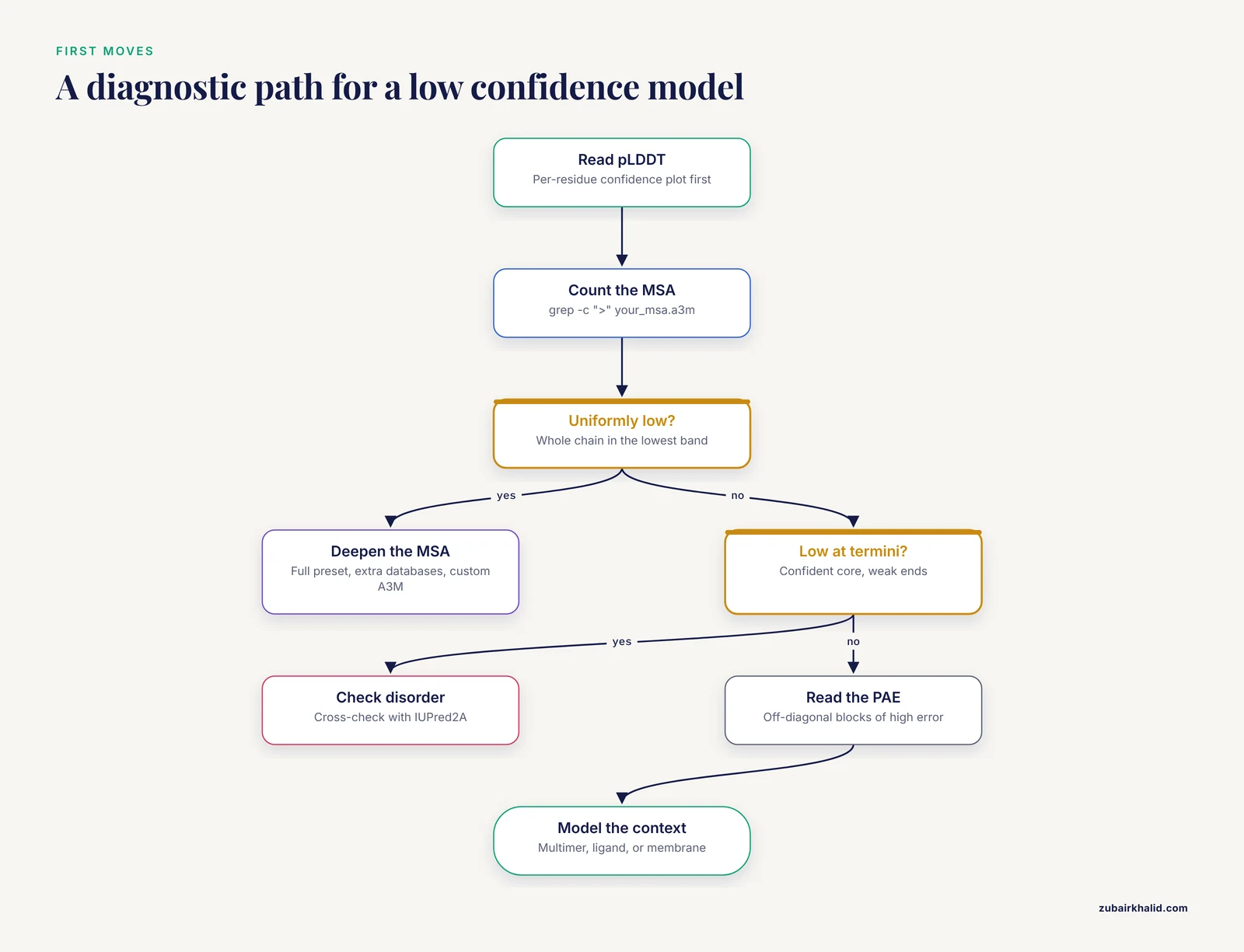
Work through these six steps in order. Each one eliminates a class of causes.
- Read the pLDDT curve. The AlphaFold database legend defines the bands: above 90 is very high confidence, 70 to 90 is confident, 50 to 70 is low, and below 50 is very low. Two patterns matter. Uniformly low pLDDT across the whole chain suggests a global failure, usually a shallow MSA. A confident core with low-confidence termini and long insertions suggests local disorder, which is often real biology rather than a bug.
- Count the MSA. ColabFold prints the number of sequences in its log. In a local AlphaFold 2 run, the output directory contains the alignment files, and you can count entries in an A3M file (a FASTA-like alignment format) with
grep -c ">" your_msa.a3m. Single digits to a few dozen sequences is shallow. Hundreds of diverse sequences is healthy. There is no universal cutoff, but the difference between ten sequences and two hundred is usually the difference between a failed and a usable model.
- Verify the sequence is what you think it is. Run BLASTp at NCBI against the nr protein database. This catches wrong annotations, frameshifts from predicted gene models, cloning artifacts, and contamination. A surprising number of "AlphaFold failed" cases turn out to be "the input sequence was wrong."
- Read the PAE plot. Look for off-diagonal blocks of high error. Two domains with high internal confidence but high PAE between them were each modeled locally but placed relative to each other with no evidence. That is a domain-arrangement problem, not a folding problem, and it needs a different fix.
- Inspect the template hits. Check the identity percentage and coverage of any templates AlphaFold used. Weak hits are normal for low-homology targets. A wrong template is worse than no template.
- Match your symptom to the troubleshooting table and jump to the corresponding fix.
Troubleshooting Table: Symptom, Likely Cause, Confirmation, Fix
The table below covers the failure modes in rough order of frequency. Column three tells you how to confirm the cause before spending compute on the fix.
| Symptom | Likely cause | How to confirm | Fix |
|---|---|---|---|
| , - | , - | , - | , - |
| Low pLDDT across the entire chain, no recognizable secondary structure | MSA too shallow | Count the sequences in the MSA output, then BLASTp the query against nr | Deepen the MSA (Fix 1) |
| Confident core with pLDDT in the lowest band at termini and long insertions | Intrinsic disorder in those segments | Compare the low-confidence segments with a disorder predictor such as IUPred2A | Model the folded core and report disordered segments as disordered (Fix 4) |
| High pLDDT everywhere but the fold contradicts known biology | Hallucination in a region with no evolutionary restraints | Check the PAE plot, compare against template hits and a single-sequence model | Distrust the model and cross-validate before use (Fix 4, Common Mistakes) |
| No MSA found, or an MSA containing only the query | Non-standard residues or a broken FASTA | Inspect the sequence for X, B, Z, U, O, asterisks, and gaps | Clean the sequence and resubmit (Fix 2) |
| Confidence collapses at one unexpected segment | Affinity tag, linker, or cloning artifact in the construct | Align the query against the native sequence | Remove the tag and model the native sequence (Fix 2) |
| Whole-chain low confidence for a small protein known to fold | The fold depends on disulfides, a ligand, a partner, or a membrane | Check the literature for cofactors, redox state, and oligomeric state | Add the missing context or use enhanced sampling MD (Fixes 4 and 5) |
| Multimer model with low ipTM at the interface | No paired MSA across chains | Check whether chains share species-level matches in the MSA | Use paired MSAs with AlphaFold-Multimer (Fix 5) |
| Template-based model is worse than the MSA-only model | Distant or incorrect template | Inspect template identity and coverage in the output | Rerun without templates or with a stricter template choice (Fix 3) |
| Two runs of the same job return different models | Stochastic sampling and recycling | Compare runs and check whether a seed was set | Fix the seed, increase recycles, and report both (Prevention) |
| Query comes from a new genome annotation and nothing aligns | Frameshift or premature stop in the predicted gene | BLASTx the region or align against an ortholog | Correct the open reading frame and resubmit (Fix 2) |
Fix 1: Deepen the MSA Before Anything Else
The mechanism is straightforward. AlphaFold converts a deep MSA into a map of coupled positions. When position 42 is always a leucine when position 97 is an arginine, and always a valine when position 97 is a cysteine, the model infers that those residues touch. A shallow MSA contains no such pattern, so the model has nothing to grip.
Practical steps, in the order that usually pays off:
- Use the deepest standard preset. In a local AlphaFold 2 installation, run with the full database preset, which searches UniRef90, BFD, and Mgnify, rather than the reduced preset built around a small BFD subset. In ColabFold, the default MMseqs2 mode against UniRef30 plus environmental databases is the right starting point for most queries.
- Search additional databases manually and merge. Run BLASTp against nr at NCBI, pull orthologs from UniProt across divergent taxa, and build a custom A3M. ColabFold accepts a custom MSA upload, and a local install accepts alignment files directly. Manually adding 50 well-chosen diverse sequences often rescues a query that automated search left with 12.
- Broaden taxonomically. Do not stop at homologs from close relatives. Metagenomic sequences from environmental sampling frequently provide the distant homologs that rescue proteins from undersampled lineages, which is one reason the Mgnify and environmental databases earn their place in the pipeline.
- Check that the search did not simply fail. Non-standard residue letters and long low-complexity runs can break database searches or cause the search to return only the query. If your MSA contains only the query, go to Fix 2 before concluding the protein has no homologs.
- Sanity-check alignment quality. Open the MSA and confirm that rows align across the full query length. Watch for paralog contamination, where sequences from a different subfamily align only to part of your query and pull the model toward the wrong fold. Depth without diversity, or depth with the wrong sequences, can hurt.
Two honest caveats. First, returns diminish: the model subsamples very deep alignments internally, so going from 200 to 20,000 sequences changes little. Second, if the protein genuinely has no homologs in any database, no search setting will save you, and you should move to Fix 4 with a clear conscience.
Fix 2: Clean and Validate the Input Sequence
AlphaFold assumes a clean, canonical, single-chain protein sequence. Every deviation degrades either the database search, the model, or both.
Start from the canonical sequence. Take the UniProt canonical sequence for your protein and verify it against the source genome or publication. For proteins from newly sequenced organisms, predicted gene models frequently contain frameshifts, premature stop codons, or wrong start positions. BLASTx the coding region or align the predicted protein to an ortholog from a related species. A single frameshift scrambles everything downstream of it and will wreck both the MSA and the model.
Remove expression artifacts. Affinity tags such as His-tags, solubility tags, linkers from cloning vectors, and point mutations introduced during construct design are not part of the native protein. Model the native sequence. If you specifically need a model of the construct you put into a crystal tray, model that deliberately, expect low confidence in the artificial segments, and say so in your notes.
Decide on precursor versus mature chain. Signal peptides, propeptides, and processing sites require a decision. Expect low pLDDT in signal peptides and unstructured propeptides regardless of which you choose, because they are cleaved in vivo and poorly constrained. The mistake to avoid is inconsistency, modeling the precursor in one run and the mature chain in another without recording which is which.
Handle non-standard residues. AlphaFold 2 was trained on the 20 standard amino acids. Letters such as X (unknown), B, Z, U (selenocysteine), and O (pyrrolysine) degrade the search and confuse the model. Map them deliberately: selenocysteine is commonly modeled as cysteine, and the substitution should be noted. Strip asterisks (translation stops) and gap characters. A quick screen catches most problems:
grep -v ">" query.fasta | grep -i "[xbzu]"Also scan the file by eye for asterisks and internal gaps.
Mind FASTA hygiene. One sequence per monomer job, a simple header, and no spaces or digits inside the residue line. These sound trivial, and they account for a steady trickle of failed jobs on public servers.
Respect length limits. The precomputed AlphaFold database covers protein chains up to 2,700 residues. Very long local runs need high-memory GPUs and often fail outright, so split large multidomain proteins into domains, model each, and assemble using the PAE structure as a guide.
A note on hazards: this workflow is entirely computational, so there are no chemical or biological handling risks. The real hazards are data integrity and confidentiality, which the section on servers addresses below.
Fix 3: Use Templates the Right Way
Templates give the model explicit structural anchors from the PDB. When the template is genuinely homologous, they stabilize the correct fold even when the MSA is thin. When the template is wrong, they can lock the model into an incorrect architecture with high confidence.
The failure modes differ by regime. No template hits at all is usually fine, because the MSA carries the load. The dangerous case is a distant hit with low sequence identity but enough coverage to be selected. Below the roughly 20 to 30 percent identity range where homology modeling is considered reliable, the so-called twilight zone, a template match is advisory rather than authoritative.
The practical protocol is to run both ways and compare. ColabFold lets you disable template usage, and a local AlphaFold 2 install gives you the same control. If the template-free and template-based models agree on the fold, your confidence in both should rise. If they disagree, inspect the template alignment manually before trusting either.
This is also where the historical context matters. Many homology modeling algorithms only model the region with sequence identity, which is why low-homology proteins fell through the cracks of earlier workflows and why MSA-driven deep learning changed what is reachable [1]. Templates in AlphaFold play a supporting role to the MSA, not the lead role they played in homology modeling. If fold-recognition tools such as HHpred suggest a structural class for your protein, you can supply a custom template alignment in ColabFold and test that hypothesis directly.
Fix 4: Recognize Intrinsic Disorder and Hard-to-Model Folds
Sometimes low confidence is the correct answer, not a failure. AlphaFold 2's confidence is calibrated, and the lowest pLDDT band reliably flags genuinely disordered regions. Before you tune anything, ask whether the model is telling you the truth about your protein.
Cross-check with an independent disorder predictor such as IUPred2A, and look at the sequence itself. Low-complexity tracts, repetitive segments, high net charge, and low hydrophobicity are all classic disorder features. If the low-confidence segments track predicted disorder, report them as disordered. That is a result, and often a publishable one.
The interesting cases sit at the boundary, where the protein is known to fold but AlphaFold cannot reach the fold. A 2025 study of four fungal hydrophobins from classes I and II illustrates the full diagnostic arc [1]. AlphaFold performed well in predicting the core beta-barrel, the characteristic fold of hydrophobins, for the class members it handled, but HFB9A came back unfolded with low confidence scores. The authors then ran 500 nanoseconds of accelerated molecular dynamics (aMD), an enhanced sampling technique that flattens the energy landscape so rare folding transitions happen on accessible timescales. The aMD simulations produced folded, energetically stable conformations for all the proteins except HFB9A, which showed higher disorder, higher atomic fluctuation, the lowest hydrophobicity and overall compactness, and less secondary structure formation. The authors concluded that underlying intrinsic disorder was the basis of folding that AF2 could not reach, and that enhanced sampling compensated for it in everything except the most disordered member [1].
The practical translation:
- Known folded, low confidence everywhere. The MSA or the sequence is the problem. Go back to Fixes 1 and 2.
- Low confidence tracking predicted disorder. Report the disorder. Do not tune parameters to force a fold.
- Borderline cases with a folded core and dynamic regions. Model the core, annotate the dynamic regions, and consider enhanced sampling MD such as aMD as an independent line of evidence, as the hydrophobin study did.
Other hard cases cluster here too. Small disulfide-rich proteins often have few homologs and depend on an oxidizing environment that the model cannot see. Proteins that fold only on binding a partner, a membrane, or a cofactor will look disordered in isolation. Metamorphic proteins with two genuine states return one dominant state. In each case, the fix is context, which is the next section.
Fix 5: Model the Biological Context, Not Just the Chain
A large fraction of "failed" monomer predictions are actually successful monomer predictions of a protein that does not function as a monomer.
Oligomeric state first. Check the literature, size-exclusion chromatography data, and existing PDB entries for the oligomeric state. Then run AlphaFold-Multimer with the correct stoichiometry. Multimer mode uses paired MSAs, which align homologs across chains by species so the model can see covariation at the interface. ColabFold exposes this through its pairing mode options. Read the ipTM score: higher is better, and the AlphaFold-Multimer authors treat values around 0.8 and above as confident interfaces. A monomer run that looked hopeless can become a confident dimer or trimer.
Ligands, metals, and cofactors. AlphaFold 2 does not see small molecules. If your protein needs a zinc ion, a heme, an NAD cofactor, or a bound ligand to adopt its active conformation, the apo model can misplace pocket loops or even the domain arrangement. AlphaFold 3, and by extension the AlphaFold server that serves it, accepts proteins together with ions, ligands, and common chemical modifications, which addresses part of this gap. The MSA problem itself persists regardless of version, so Fixes 1 and 2 still apply.
Redox and environment. For disulfide-rich proteins, the oxidized state is the folded state. For membrane proteins, the lipid environment constrains the fold. Neither is explicit in a standard AlphaFold 2 run, so treat these models with extra caution and lean on templates from related solved structures where they exist.
Choosing Where to Run: Notebook, Local Install, or the AlphaFold Server
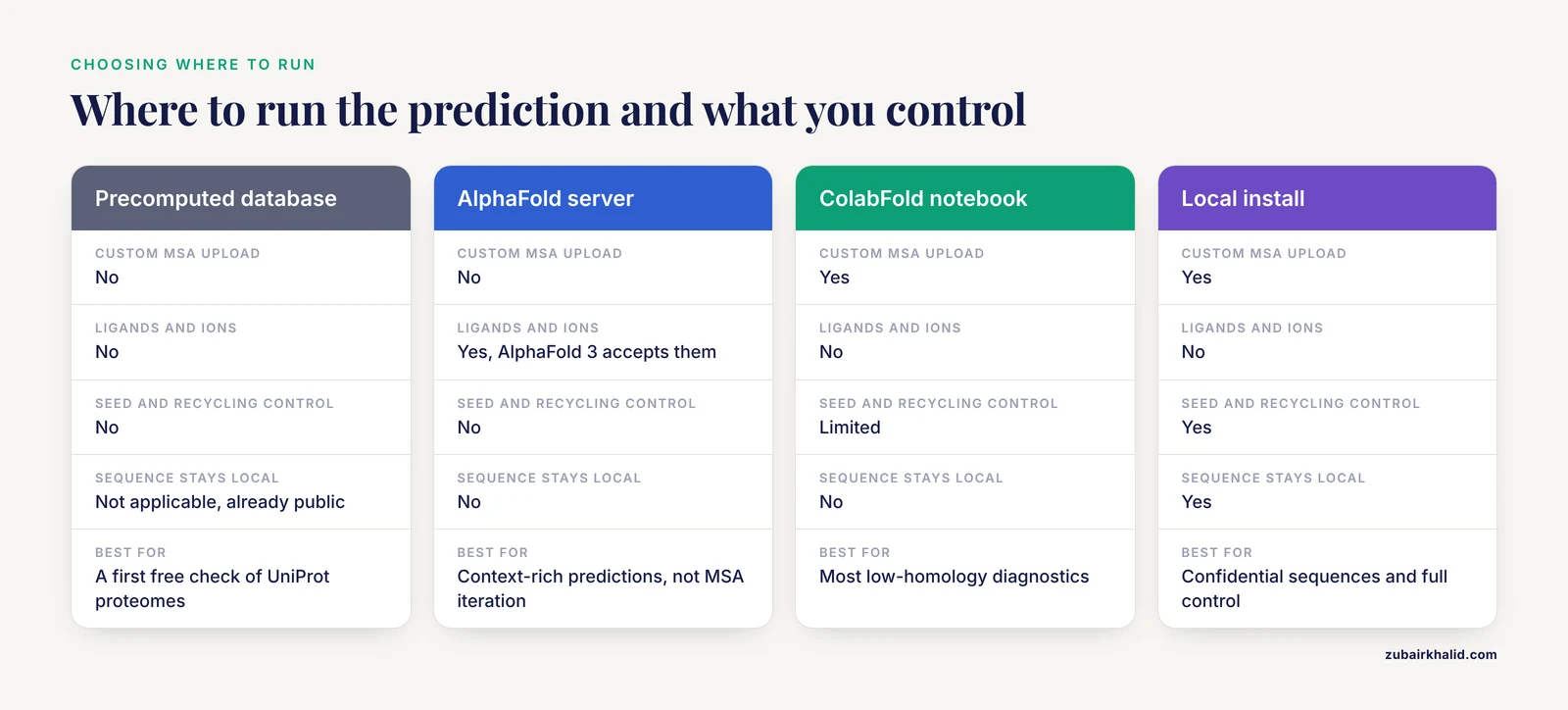
Where you run the prediction determines how much control you have over the fixes above.
AlphaFold Protein Structure Database (EMBL-EBI). Free, precomputed, and the right first stop. Coverage spans UniProt proteomes with chains up to 2,700 residues. No preparation control, but no preparation needed either.
AlphaFold server. DeepMind's web interface to AlphaFold 3 accepts proteins, nucleic acids, ions, ligands, and common modifications, and it is free for non-commercial use under its current terms. The trade-off is control: you cannot supply a custom MSA, fix a seed, or tune recycling. It is excellent for context-rich predictions and poorly suited to iterating on a low-homology MSA.
ColabFold notebook. Free GPU access, MMseqs2 searches, sensible defaults, and a custom MSA upload path. The notebook doubles as a practical tutorial for the standard workflow, and most low-homology diagnostics described here can be run in it. Queue times and session limits are the main constraints.
Local AlphaFold 2 or local ColabFold. Full control over databases, MSAs, templates, seeds, and recycling, and no sequence ever leaves your machine. The costs are setup effort and GPU memory, which scales with chain length.
A data safety note: unpublished and proprietary sequences are sensitive research data. Read the terms of any public server before uploading, and note that commercial use of the AlphaFold server is restricted under its terms. For confidential sequences, use a local install.
For reproducibility, record the tool version, database versions, the random seed, and archive the MSA file with your outputs. A model you cannot regenerate is a model you cannot defend in review.
Prevention: A Sequence Preparation Checklist
Most low-homology failures are preventable at the input stage. Run through this list before every submission:
- Start from the canonical UniProt sequence and verify it against the source genome or publication.
- Validate predicted open reading frames from new genome annotations with BLASTx or ortholog alignment.
- Decide precursor versus mature chain, and document the decision.
- Remove expression tags and cloning artifacts, or model the construct deliberately and say so.
- Screen for non-standard residues, asterisks, gaps, and long low-complexity runs.
- Predict disorder beforehand with a tool such as IUPred2A so low pLDDT does not surprise you.
- BLASTp the sequence against nr to confirm identity and catch annotation errors.
- Check the precomputed AlphaFold database before spending compute.
- Record the MSA depth, seed, and software versions with every run, and archive the MSA.
- Plan at least one independent cross-check, such as a template comparison, a single-sequence model, or enhanced sampling MD, for any low-homology model you intend to use downstream.
Common Mistakes and Limitations
Trusting high pLDDT in low-homology singletons. With no evolutionary restraints, the model can produce a confident wrong structure. Cross-check any low-homology model against templates, a single-sequence language model such as ESMFold, and any experimental data you have.
Reading pLDDT as a distance guarantee. pLDDT is a per-residue confidence measure on the backbone, and side-chain accuracy degrades faster than the score suggests. Two models with similar pLDDT can differ substantially in side-chain placement.
Ignoring the PAE plot. High pLDDT on two domains with high PAE between them means the domains were each modeled well but placed relative to each other on weak evidence. Downstream use that depends on the domain arrangement is built on sand.
Submitting the cloning construct. Tags and linkers generate low-confidence segments that then get misread as disorder. Model the native sequence.
Parameter shopping. Rerunning with different seeds and settings until a model looks right is model selection without a hypothesis. Define acceptance criteria before iterating, and report every run you performed.
Treating disorder as failure. Low confidence in a low-complexity segment is often the model correctly reporting biology. The hydrophobin study reached the same conclusion from the opposite direction, finding that intrinsic disorder explained why one family member resisted both AlphaFold and enhanced sampling MD [1].
The limitations of the method itself also apply. AlphaFold 2 returns a single dominant state, does not model most small molecules, and inherits biases from its training databases. Low-homology models in particular are hypotheses that need experimental validation, whether by crystallography, cryo-EM, NMR, cross-linking data, or functional assays. Individual project decisions, especially for proteins heading into design or drug discovery workflows, benefit from review by an experienced structural biologist before the model is trusted.
Frequently Asked Questions
What does low homology mean in AlphaFold sequence preparation?
It means the query protein has few or no detectable relatives in public sequence and structure databases, so AlphaFold builds a shallow multiple sequence alignment and has little evolutionary information to work with.
Why is my AlphaFold prediction low confidence?
The two most common reasons are a shallow multiple sequence alignment and genuine intrinsic disorder, and the per-residue pLDDT plot together with the MSA depth usually tells you which one you have.
How many sequences does an MSA need for a reliable AlphaFold model?
There is no universal cutoff, but predictions improve sharply as the alignment grows from a handful of sequences to dozens and then hundreds, and the quality and diversity of the sequences matter as much as the raw count.
Can AlphaFold predict a structure with no homologs at all?
It will return a model, but with no evolutionary signal the confidence is usually low and the error rate high, so treat such models as hypotheses and cross-check them with single-sequence tools such as ESMFold.
Should I remove tags and signal peptides before submitting a sequence?
Remove expression and affinity tags because they are cloning artifacts rather than part of the native protein, and decide deliberately whether to model the full precursor or the mature chain, then record that choice.
What is the difference between pLDDT and pAE?
pLDDT is a per-residue confidence score from 0 to 100 that reflects local backbone accuracy, while pAE is a residue-pair error measured in angstroms that tells you how confidently the model places residues, domains, or chains relative to each other.
Can molecular dynamics rescue an unfolded AlphaFold model?
Sometimes, enhanced sampling methods such as accelerated molecular dynamics can fold borderline cases, but a model that stays disordered across methods usually reflects a protein that really is intrinsically disordered rather than a fixable modeling failure.
Is the AlphaFold server different from running AlphaFold locally?
Yes, the AlphaFold server is a web interface to AlphaFold 3 that handles ligands and complexes but offers limited control, while a local AlphaFold 2 or ColabFold setup gives you control over the MSA, templates, seeds, and recycling.