AlphaFold for Proteins with Disordered Regions
By Dr. Zubair Khalid, DVM, MS, PhD ·

You paste a sequence into AlphaFold, wait for the model to finish, and open the result. The folded domain looks fine. Then one end of the protein, or a long stretch in the middle, dangles off the core as an extended ribbon with low pLDDT scores, a poorly packed core, and PAE lines radiating out to everything else. You have just met the most common alphafold disordered protein sequence pattern in the field.
The most likely cause is also the simplest one: that region really is intrinsically disordered. Intrinsically disordered regions (IDRs) are protein segments that lack a stable three-dimensional structure under physiological conditions. Roughly a quarter of the human proteome is intrinsically disordered, and AlphaFold does not hide this from you. It reports it, in the language of confidence scores, whether you know how to read them or not.
The first thing to check is the per-residue pLDDT profile, not the cartoon. If the low-confidence stretch is long, acidic or glycine-rich, and predicted to be solvent-exposed, you are almost certainly looking at a real IDR rather than a prediction failure. A recent systematic comparison of AlphaFold2, OpenFold, ColabFold, and the AlphaFold 3 server found that global scores such as mean pLDDT and pTM often look acceptable while local disagreement is large, which is exactly why you need to inspect per-residue confidence rather than the headline number [1].
This article explains why AlphaFold renders disordered regions the way it does, how to confirm that what you see is disorder rather than a modeling error, and the practical fixes available today, from running the right predictor to modeling the region in complex with its binding partner to simulating the ensemble with coarse-grained methods.
Why AlphaFold Draws Disordered Regions as Extended Loops
AlphaFold2 was trained primarily on structures in the Protein Data Bank, which overwhelmingly contain folded domains. When the model encounters a sequence with no stable fold, it does not return nothing. It returns the least implausible single conformation it can attach to the rest of the protein. In practice, that means IDRs appear as long extended loops that appear to float around the structured core [2].
Three things happen together in an alphafold disordered protein sequence output:
- pLDDT collapses. Per-residue pLDDT drops, typically below 50 for genuinely disordered segments. pLDDT below 50 has long been interpreted as a disorder indicator rather than a modeling error.
- PAE rises. The predicted aligned error between the disordered segment and the rest of the protein climbs, telling you the relative placement of that segment is uncertain.
- The chain sticks out. IDR conformations are often implausibly extended, sometimes resembling polyproline II helices more than anything compact. One survey of AlphaFold models notes that regions lacking experimental structures were frequently modeled as extended PPII conformations, including the disordered C-terminal tails of GroEL and its homologs [3].
AlphaFold3 introduced architectural and training changes, including cross-distillation aimed at reducing structural hallucinations in disordered regions. The hope was that AF3 would represent IDRs more honestly. When Mehdiabadi and colleagues evaluated AF2 and AF3 on the CAID3 disorder benchmark, AF3 did not outperform AF2 at identifying disordered regions. Solvent accessibility remained the most robust and consistent proxy for intrinsic disorder across both models, and the authors concluded that AlphaFold2 remains the preferred choice for identifying IDRs from confidence maps [2].
The mechanism matters because it changes what "fixing" means. If the model fails on a folded domain, you fix the inputs, check the MSA, try orthologs. If the region is genuinely disordered, the correct response is not a better structure. It is a different question: which part of this region folds when it binds, and what experiments or methods will answer that?
The Diagnostic Sequence: Six Steps
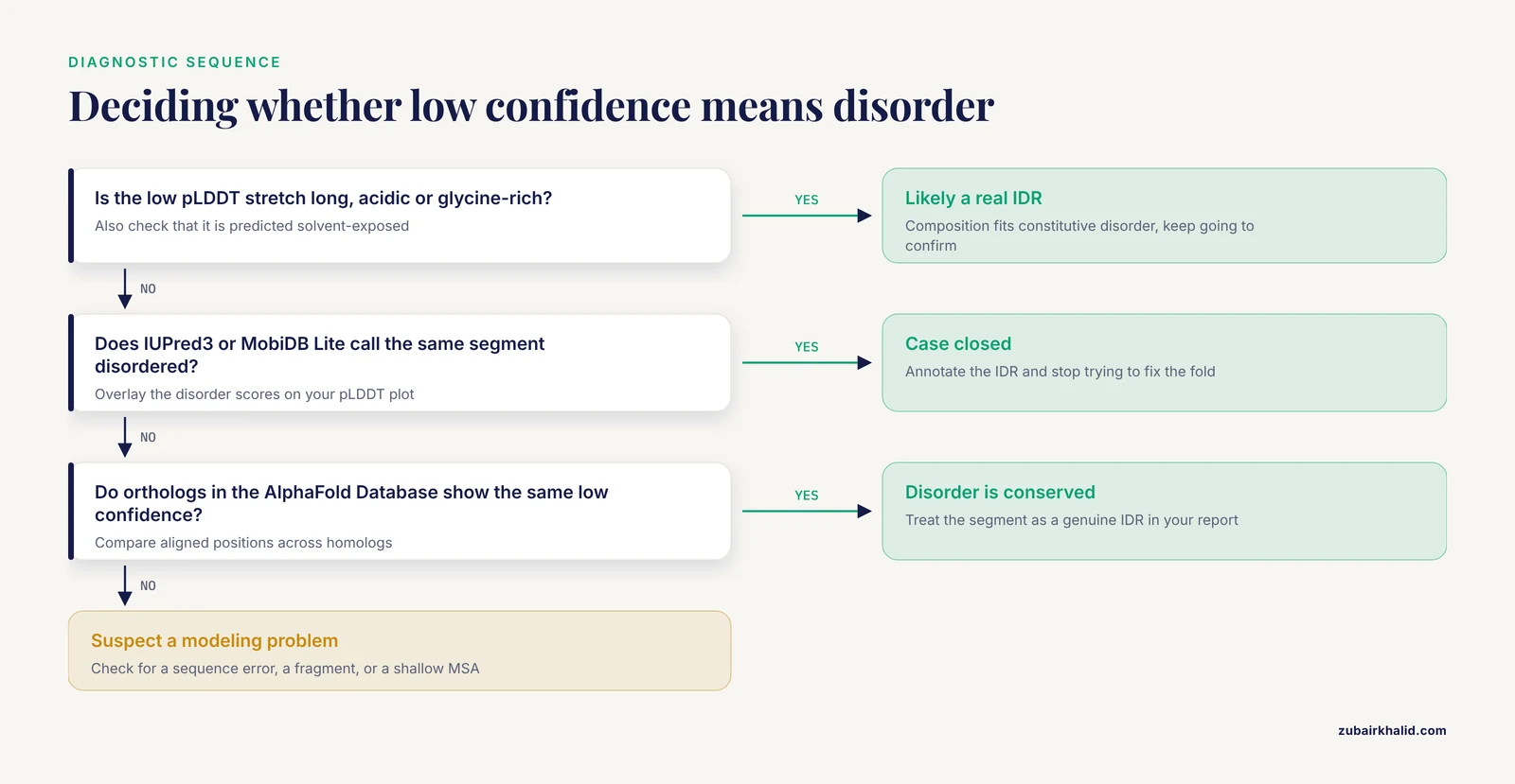
Before changing anything, run this short diagnostic. It separates the four causes described below in under an hour for most proteins.
- Plot pLDDT and PAE together. Open the result in a viewer that shows per-residue confidence. Identify the boundaries of the low-confidence segment. A sharp transition at a residue boundary suggests a domain-disorder junction. A slow decay suggests a gradual loss of packing.
- Check sequence composition. IDRs are enriched in proline, glycine, serine, glutamine, and charged residues, and depleted in hydrophobic core formers. A segment that is 40 percent glutamate is not going to fold on its own. Low-complexity insertions are especially common in some protein families. In malaria parasite type-P5 ATPases, homology modeling detected multiple variable, low-complexity regions that form intrinsically disordered loops, distinct from the conserved core of the ATPase [4].
- Run a dedicated disorder predictor. Use IUPred3, DISOPRED, or MobiDB Lite on the same sequence. If the predictor calls the same segment disordered that AlphaFold calls low confidence, the case is closed. A comparison of AF2 and AF3 against the CAID3 benchmark supports using AlphaFold confidence as one disorder signal among several, with solvent accessibility as a consistent proxy [2].
- Scan for molecular recognition features. Short segments within IDRs that undergo disorder-to-order transitions on binding are called MoRFs. MoRFchibi 2.0 predicts their locations and outperformed all existing MoRF predictors and general IDR binding-site predictors, including top models from CAID rounds 1 through 3 and predictors that use AlphaFold data [5]. A high MoRFchibi score inside your low-pLDDT segment predicts that AlphaFold will only capture that segment in a complex.
- Check homologs in the AlphaFold Database. If orthologs show the same low-confidence pattern at aligned positions, the disorder is conserved and real. If only your sequence is disordered at a position where homologs are confident, suspect a sequence error, a fragment, or a missing MSA.
- Try a partner. If steps 1 through 5 suggest a MoRF or known binding region, model the protein in complex with its partner. Sections below give worked examples, including the LC8 hub protein, where AlphaFold correctly placed binders at the LC8 interface and a threshold scheme on built-in scores separated known binders from nonbinders with an 8 percent false positive rate and a 20 percent false negative rate [6].
Troubleshooting Table: Alphafold Disordered Protein Sequence
| Symptom | Likely cause | How to confirm | Fix |
|---|---|---|---|
| One long tail with pLDDT below 50, predicted solvent-exposed | Genuine constitutive IDR | Disorder predictor agreement, conserved low confidence across orthologs | Do not try to fix the structure. Annotate the IDR and model only interactions for it |
| Low confidence only when the protein is modeled alone | Folding on binding, a MoRF | MoRFchibi 2.0 high score, known partner in the literature | Model with the partner using AlphaFold-Multimer or the AlphaFold server, or use Disobind for interface prediction |
| Disordered region folds into a plausible but unsported compact structure | Hallucination or unjustified packing | No experimental support, contradictory prediction across seeds or implementations | Distrust the compact model. Use ensemble or coarse-grained approaches instead |
| pLDDT fine overall but local stretches disagree between AF2 and AF3 | Implementation-specific local differences | Bland-Altman comparison of per-residue confidence between models | Report per-residue differences, do not rely on mean pLDDT alone |
| Single residue change flips a nearby low-confidence region to ordered | Variant-sensitive local ordering or prediction noise | Repeat with multiple seeds, check experimental disorder data | Treat as hypothesis only. Validate against disorder databases and experiments |
| IDR appears to contact the folded core consistently | Possible intra-chain contact or artifact | Check PAE between the segments, compare across seeds and versions | Low confidence means low confidence. Do not build mechanisms on IDR-core contacts from a single model |
| Whole protein low confidence except a small core | Deep disorder, like LIMCH1's agmatinase-like region | Circular dichroism or sequence-level disorder analysis | Expect AlphaFold to fail. Use biophysics and integrative methods, not single-structure prediction |
Causes, From Most Common to Rarest
Cause 1: The Region Is Constitutively Disordered
This is the default explanation and it is correct most of the time. Constitutive IDRs never fold on their own. They function through dynamics, through transient interactions, and through post-translational modification. AlphaFold renders them as extended, low-confidence loops because that is the honest output for a sequence with no ensemble-preferred state [2].
The LIMCH1 protein shows how far this can go. Its C-terminal agmatinase-like region (ALP) diverges sharply from canonical agmatinases, lacks the conserved residues needed for Mn2+ coordination, and predictive AI algorithms failed to model it reliably. When Reyes and colleagues purified full-length rat LIMCH1 and a truncated ALP variant and measured secondary structure by circular dichroism, both were roughly 60 percent disordered with about 30 percent beta structure, completely unlike the well-defined alpha-beta-alpha sandwich of E. coli agmatinase [7]. No prompt engineering, MSA depth, or model version would have made AlphaFold produce a trustworthy single structure here, because no such structure exists.
Cause 2: The Region Folds Only on Binding
The second most common cause is the disorder-to-order transition. A segment within an IDR is disordered alone and ordered in complex. AlphaFold modeling the chain alone has no partner to bind, so the segment stays a low-confidence tail. This is not a failure of the alphafold disordered protein sequence pipeline. It is a limitation of the question you asked the model.
The LC8 hub protein is the cleanest published demonstration. LC8 binds dozens of intrinsically disordered partners at a shared interface. Walker and colleagues tested whether AlphaFold could predict LC8 binding from sequence alone and found that it places proteins at the correct interface of LC8, and that thresholds on combinations of built-in AlphaFold scores separate known binders from nonbinders with minimal false positives and acceptable false negatives. Their analysis of correlations between binding affinity and AlphaFold scores suggests the model learned an inaccurate but useful energy function [6]. The practical lesson is direct. If your IDR tail contains a binding motif, running AlphaFold with the partner present can turn an unresolvable tail into a well-scored bound conformation.
Cause 3: Missing or Shallow Evolutionary Signal
AlphaFold leans on co-evolution from multiple sequence alignments. IDRs evolve rapidly, so their MSAs are shallow and their columns are poorly conserved. Without co-evolutionary signal, the model has little to grip. Elofsson's review of protein-protein interaction prediction organizes the current failure modes into distinct categories, including interactions that lack sufficient co-evolutionary signal and interactions whose relevance depends on cellular context, dynamics, or disorder [8]. The same taxonomy applies within a single chain. A disordered linker between two domains carries almost no co-evolutionary information, so its conformation is underdetermined.
In specialized protein families this problem compounds. The haemosporidian type-P5 ATPases studied by Wiser contain multiple low-complexity insertions forming disordered loops, and homology modeling did not resolve questions such as substrate specificity for the family [4]. Shallow alignments plus long insertions is a recipe for unreliable local structure even when the core domains model well.
Cause 4: Low-Complexity and Repetitive Sequence
Low-complexity regions, polyprotein tracts, and repetitive motifs confuse the model in a specific way. AlphaFold can assign them regular secondary structure, often extended polyproline II-like conformations, even when no single stable helix exists. The PolyProline Predictor authors observed that AlphaFold modeled several glycine-rich protein families as extended PPII conformations, including mycobacterial PE_PGRS virulence factors and GroEL C-terminal tails, and molecular dynamics supported persistent but heterogeneous PPII helical bundles in some of them [3]. In other words, the AlphaFold conformation can be one physically reasonable member of a broad ensemble, presented with unwarranted precision.
Cause 5: Version and Implementation Differences
The same sequence can look different across AlphaFold2, OpenFold, ColabFold, and the AlphaFold 3 server. The benchmark by Diaz and colleagues compared multiple implementations across 10 proteins and 222 single-amino-acid variants and found that global indicators like mean pLDDT, pTM-score, and RMSD frequently suggested comparable performance while substantial local differences remained hidden. They introduced Bland-Altman agreement analysis of per-residue confidence differences and per-residue profiles to expose them [1]. If your low-confidence segment appears or disappears between runs, that difference is itself information, and it belongs in your report.
How to Fix an Alphafold Disordered Protein Sequence
The word fix needs redefinition. For a constitutive IDR there is no single correct structure to recover, so the fixes below either change the question you ask the model or change the representation of the answer. These are the alphafold disordered protein sequence solutions that current literature supports.
Fix 1: Read Low Confidence as an Annotation
The cheapest fix is interpretive. Treat a continuous pLDDT drop below 50 as a disorder annotation and an extended PAE pattern as evidence that the segment's position relative to the core is undefined. This is standard practice and it is what the CAID3 benchmarking supports: AF2 confidence maps are a reasonable disorder predictor, and solvent accessibility predicted from the models is a robust proxy across AF2 and AF3 [2]. Write the IDR into your figure legend instead of apologizing for the spaghetti.
This interpretation is also the foundation of downstream tools. Hubrich and colleagues combined sequence pattern searches with AlphaFold to structurally annotate 1,300 protein-protein interactions whose interfaces are mediated by short disordered motifs binding to folded domains in partner proteins. That pipeline produced structure-based predictions of deleterious effects for 1,187 missense variants in IDRs, and experimental validation confirmed interfaces and variant effects that AlphaMissense alone had predicted as benign. Context for scale: 37 percent of annotated missense variants fall in the 25 percent of the proteome that is intrinsically disordered [9]. AlphaFold's low-confidence regions, read carefully, become testable structural hypotheses.
Fix 2: Predict the Binding Segment First, Then Model With the Partner
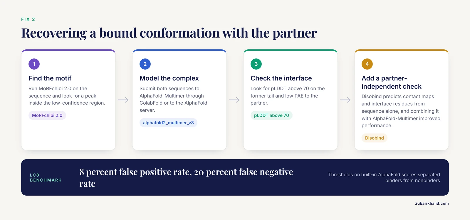
If your IDR contains a binding motif, the sequence-alone alphafold disordered protein sequence problem is solved by adding the partner to the input.
- Find the motif. Run MoRFchibi 2.0, which outperformed all existing MoRF predictors and IDR binding-site predictors, including models built on AlphaFold data and protein language models [5].
- Model the complex. Submit both sequences to AlphaFold-Multimer through ColabFold or to the AlphaFold server. For the LC8 system this strategy correctly localized binders to the LC8 interface and a scoring threshold scheme classified binders against nonbinders with an 8 percent false positive rate and a 20 percent false negative rate [6].
- Add a partner-independent check. Disobind predicts partner-dependent contact maps and interface residues for IDRs from ProtT5 sequence embeddings alone. It outperformed AlphaFold-Multimer and AlphaFold3 at multiple confidence cutoffs, and combining Disobind with AlphaFold-Multimer predictions improved performance further [10]. Because Disobind needs no structures and no MSAs, it works where co-evolution is thin, which is precisely where IDR modeling struggles.
For partner-specific motifs that touch DNA, the same logic holds. AlphaFold 3 modeling of the rice OsDREB1 transcription factors revealed a disordered KRP/RAGR motif adjacent to the AP2 domain, predicted that arginine R40 in that motif contacts the DNA minor groove, and mutational analysis verified the key roles of R40 and E66 [11]. The disordered motif became interpretable only inside a complex model, and the prediction survived experimental testing.
Fix 3: Annotate the IDR Biology You Already Have
Often the question is not the IDR conformation but its function, and modeling can answer function without a single stable structure. For the RNA-binding protein GEMIN5, the C-terminal region contains a TPR-like dimerization module, a non-canonical RNA-binding site, and an IDR. AlphaFold modeling of the TPR-extended protein showed an elongated dimer bound to RNA, and the same RNA-binding surface appeared in the G5C-RNA oligomer model. Mutational work showed that a serine-rich motif and an acidic cluster within the IDR each contributed to RNA binding [12]. Here AlphaFold's role was to organize the folded module and the RNA, while the IDR's contribution was characterized by mutation and binding assays.
For highly disordered proteins where AlphaFold fails outright, biophysics carries the analysis. The Drosophila Polycomb protein is predominantly disordered outside its chromodomain, and structural work required solubility-enhancing MBP fusions, circular dichroism, thermal shift assays, nanoDSF, DLS, SEC-MALS, and cryo-EM with gradient fixation to manage conformational and oligomeric heterogeneity [13]. If your protein resembles this profile, plan the experimental toolkit before the modeling session.
Fix 4: Simulate the Ensemble Instead of Predicting a Snapshot
When you need the disordered region's behavior, an ensemble is the right output format. AF-CALVADOS is a coarse-grained simulation framework that uses AlphaFold 2 to define folded regions and integrates them with the CALVADOS model for disordered chains. It was validated against small-angle X-ray scattering data for more than 400 proteins, performed well across varying ordered and disordered content, and scaled to simulations of 12,483 intracellular human proteins with data made freely available [14]. For a multi-domain protein whose function depends on interplay between folded domains and IDRs, this is currently the most practical route from an alphafold disordered protein sequence to dynamic, experimentally comparable behavior.
Fix 5: Design Disordered Regions on Purpose
If you are engineering a protein and want to add or replace an IDR, structure-prediction tools are the wrong generator. IDiom, an autoregressive protein language model, was trained on 37 million intrinsically disordered region sequences curated from the AlphaFold Database using fill-in-the-middle augmentation. It generates IDR sequences conditioned on surrounding structured context, or fully disordered proteins without context, and post-training with a subcellular localization reward model produced sequences with features consistent with known determinants of compartment-specific localization [15]. This is the design-side mirror of the prediction problem. The same AlphaFold Database confidence maps that frustrate structural biologists became training data for a disordered-region language model.
Fix 6: Integrate Sparse Experimental Restraints
For complexes assembled around IDRs, integrative modeling turns fragmented information into an architecture. In the HDAC2 chromatin remodeling complex, all three components contained extensive IDRs that resisted single-method analysis. Cross-linking mass spectrometry combined with integrative structural modeling showed that the previously uncharacterized C-terminal IDR of HDAC2 acts as an interaction hub engaging both the ELM2 domain of MIER1 and the terminal regions of MHAP1 [16]. If your alphafold disordered protein sequence sits inside a large assembly, XL-MS restraints plus integrative modeling will tell you more than any single-structure predictor.
Alphafold Tutorial: A Worked Workflow for a Multi-Domain Protein
This alphafold tutorial walks through the diagnostic and fix sequence on a realistic case: a 900-residue human protein with a folded kinase domain, a long serine-rich linker, and a C-terminal tail with a reported binding motif. Follow it with your own sequence.
Step 1: Predict With ColabFold and Inspect Confidence
Run a standard monomer prediction with a remote MSA:
colabfold_batch query.fasta results/ --msa-mode mmseqs2_uniref_env --num-recycle 3Open the result. Color the cartoon by pLDDT. Record the residues where pLDDT drops below 50 and below 70. Plot the PAE matrix and note whether the low-pLDDT segment shows high PAE to everything or only to one domain. High PAE to everything indicates free disorder. Localized high PAE to one domain hints at an intra-chain contact worth testing.
Step 2: Confirm Disorder Independently
Run IUPred3 or MobiDB Lite on the same sequence and overlay the disorder scores on your pLDDT plot. Check the predicted solvent accessibility from the AlphaFold output, since solvent accessibility is the most consistent structural proxy for disorder across AF2 and AF3 [2]. Agreement between the confidence drop and the disorder prediction closes the case. A structural alphabet analysis with Protein Blocks can add nuance here, since the entropy-based Neq index distinguishes rigid, flexible, and disordered regions and has been applied to AlphaFold structure analysis [17].
Step 3: Locate Binding Segments
Run MoRFchibi 2.0 on the sequence. A peak inside the low-confidence region marks a candidate disorder-to-order transition [5]. If the literature names a partner, proceed to Step 4. If not, Disobind can nominate interface residues for the IDR given the partner sequence, and its predictions outperformed AlphaFold-Multimer and AlphaFold3 at multiple confidence cutoffs [10].
Step 4: Model the Complex
Append the partner to the FASTA as a second chain and rerun ColabFold with a multimer model:
colabfold_batch complex.fasta results_complex/ --model-type alphafold2_multimer_v3 --num-recycle 3Alternatively, submit both sequences to the AlphaFold server, described in the next section. In the complex model, check whether the previously disordered segment now shows pLDDT above 70 and low PAE to the partner. If yes, you have recovered the bound conformation, exactly the behavior documented for LC8 binders [6]. If the interface score remains ambiguous, treat the model as one hypothesis and look for cross-validation, since interaction prediction degrades sharply when dynamics or disorder dominate [8].
Step 5: Report What You Have
Write up three things separately: the folded-domain model with its confidence, the IDR boundaries as disorder annotations, and any partner-bound conformations with the evidence that motivated them. This reporting discipline prevents the most damaging error in IDR work, which is presenting a single static model of a dynamic region as the structure.
The Alphafold Server Versus Local Installation
The AlphaFold server (alphafoldserver.com) runs AlphaFold 3 from a browser, accepts protein, nucleic acid, and small-molecule inputs, and requires no installation. It suits one-off predictions of complexes and is the fastest way to test whether a partner changes your IDR's confidence profile. The AF3 server was included in the multi-implementation benchmark described above, and the study's headline finding applies to it directly: global scores can look reassuring while per-residue agreement between implementations varies substantially [1].
Local installation through ColabFold or OpenFold gives you control over MSAs, recycling, seeds, and model selection, and it scales to batch jobs. Because AF3 did not outperform AF2 at disorder identification on CAID3, and AF2 confidence remains a solid disorder readout, a local AF2 or ColabFold pipeline is a defensible default for disorder-focused work, with the AF3 server reserved for complex and ligand questions [2]. The benchmark that compared all of these implementations across 222 variants also found that mean pLDDT can mask meaningful local differences between models, so whichever route you choose, compare per-residue confidence across at least two implementations for any IDR-adjacent claim [1].
Common Mistakes and Limitations
- Treating the extended tail as a bug. The single most common error is reporting the IDR conformation as a predicted structure. It is a placeholder for an ensemble.
- Believing a compact low-confidence fold. When AlphaFold packs a disordered region into a plausible-looking fold, the geometry is invented. The LC8 study concluded that AlphaFold learned an inaccurate energy function that is nevertheless useful in the right context, which means its outputs for unbound IDRs deserve calibrated skepticism [6].
- Using mean pLDDT as the quality metric. Mean pLDDT, pTM, and RMSD can agree between two models while per-residue confidence differs substantially, a gap exposed by Bland-Altman analysis across AF2, OpenFold, ColabFold, and the AF3 server [1].
- Assuming AF3 fixes IDRs. Cross-distillation reduced hallucination in disordered regions, but AF3 did not outperform AF2 on the CAID3 disorder benchmark, and interpretations of disorder shift depending on predicted secondary structure and pLDDT [2].
- Predicting IDR interactions without disorder-aware tools. AlphaFold-Multimer and AlphaFold3 can be used to predict IDR binding sites, but they are less accurate at their benchmarked confidence cutoffs, and partner-independent methods such as Disobind beat them at those cutoffs [10].
- Expecting a single structure to explain IDR function. For proteins like LIMCH1's ALP region, roughly 60 percent disordered by circular dichroism, no single model can exist [7]. For assemblies like HDAC2:MIER1:MHAP1, sparse restraints and integrative modeling are the appropriate tools [16].
- Applying variant predictors naively to IDRs. AlphaMissense missed deleterious variants in disordered motifs that combined sequence and AlphaFold-based structural analysis flagged correctly [9]. In clinical domains the same caution applies, since confidence scores may not accurately reflect model reliability for underrepresented protein classes including intrinsically disordered scaffolds [18].
- Ignoring post-translational context. IDR behavior often depends on modification state and cellular compartment, which static prediction does not capture [8].
Prevention Practices
- Screen every new sequence for disorder before modeling. Run IUPred3 or MobiDB in the same session as your first AlphaFold run. Two minutes now prevents a week of chasing a phantom fold.
- Build the IDR annotation into your pipeline output. Report IDR boundaries, MoRF predictions, and confidence-drop intervals alongside any structure files, so downstream users inherit the interpretation rather than the raw spaghetti.
- Standardize per-residue reporting. Export per-residue pLDDT and PAE, not just summary scores, and compare across at least two implementations for any claim touching a disordered segment [1].
- Keep an experimental plan for every IDR you care about. Circular dichroism for secondary structure content, SAXS for ensemble shape, XL-MS for contacts, and NMR where feasible. The GEMIN5 and LIMCH1 studies show how modeling and biophysics divide the labor [12].
- Use ensemble simulation when dynamics matter. AF-CALVADOS provides a validated route from AlphaFold-defined folded regions to proteome-scale IDR dynamics [14].
- When designing IDRs, use a generative model built for them. Structure-based design is not applicable to sequences that lack a stable fold. IDiom is purpose-built for disordered-region design with context conditioning [15].
Frequently Asked Questions
Why does AlphaFold show my protein as an unstructured tail?
Because that segment most likely has no stable structure on its own. AlphaFold2 renders intrinsically disordered regions as long extended loops floating around the structured core, with low pLDDT and high PAE, because no single conformation exists for the model to recover [2].
What pLDDT value indicates disorder?
A continuous per-residue pLDDT drop below 50 is conventionally read as a disorder signal rather than a modeling error. Treat the interval of low pLDDT, together with high PAE to the rest of the chain and predicted solvent exposure, as an IDR annotation [2].
Does AlphaFold3 handle disordered regions better than AlphaFold2?
Not for identification. AlphaFold3 added cross-distillation to reduce hallucinations in disordered regions, but on the CAID3 disorder benchmark it did not outperform AlphaFold2, and AlphaFold2 remains the preferred choice for identifying IDRs from model confidence [2].
Can AlphaFold predict which part of a disordered region binds a partner?
Yes, when the partner is included in the model. For the hub protein LC8, AlphaFold placed binders at the correct interface, and thresholds on built-in scores separated known binders from nonbinders with an 8 percent false positive rate [6]. Partner-independent tools such as Disobind and MoRFchibi 2.0 predict IDR interfaces and MoRFs from sequence and outperform AlphaFold-based approaches at benchmarked cutoffs [10][5].
Should I delete disordered regions before modeling?
For structure-focused questions on the folded domain, truncation is a reasonable and common step. For function, it is usually wrong, because IDRs contribute binding, localization, and regulation, as shown for the acidic cluster and serine-rich motif in GEMIN5's RNA-binding surface [12]. Model the truncated construct for the domain, and treat the IDR with the tools in this article.
How do I get coordinates for a disordered region?
You do not, as a single model. Use ensemble methods instead. AF-CALVADOS combines AlphaFold-defined folded regions with coarse-grained simulation of the disordered parts, was validated against SAXS data for more than 400 proteins, and has been run for 12,483 intracellular human proteins [14].
Is low pLDDT always disorder?
No. Shallow MSAs, low-complexity sequence, and variant-sensitive local effects can also depress confidence. The differential diagnosis is composition analysis, an independent disorder predictor, ortholog comparison, and seed variation. If independent predictors and homologs agree on disorder, accept it. If they disagree, investigate further.
Where can I run AlphaFold without installing anything?
The official AlphaFold server at alphafoldserver.com runs AlphaFold 3 in a browser and accepts protein, nucleic acid, and ligand inputs. For batch work and AF2-based disorder readouts, ColabFold offers a notebook interface and command-line batch prediction. Compare per-residue confidence between implementations for any IDR-adjacent conclusion [1].
Sources
- Comprehensive evaluation of AlphaFold/OpenFold prediction of experimentally unresolved proteins through novel metrics.
- Modeling intrinsically disordered regions from AlphaFold2 to AlphaFold3.
- PolyProline Predictor: A web server for empirical sequence-based prediction of polyproline II helices.
- Homology Modeling of Type-P5 ATPases from the Malaria Parasite: Insight into Their Functions and Evolution, and Implications About the Effect and Role of Intrinsically Disordered Protein Structure.
- Predicting molecular recognition features in protein sequences with MoRFchibi 2.0.
- Successful prediction of LC8 binding to intrinsically disordered proteins sheds light on AlphaFold's black box.
- Structural and Functional Characterization of LIMCH1 and Its Agmatinase-like Region: A Case of Catalysis in a Highly Disordered Protein.
- Structural compatibility is not enough: Current challenges in protein-protein interaction prediction.
- Variant characterization in the intrinsically disordered human proteome.
- Disobind: A sequence-based, partner-dependent contact map and interface residue predictor for intrinsically disordered regions.
- AlphaFold 3-Assisted Deciphering of the DNA Recognition by DREB1 Transcription Factors in Rice.
- Contribution of disordered regions of the essential protein GEMIN5 to RNA binding.
- Comprehensive Biophysical Profiling Evidences Self-Oligomerization of Bacterially Expressed Pc Protein.
- AF-CALVADOS: AlphaFold-guided simulations of multi-domain proteins at the proteome level.
- Generative design of intrinsically disordered protein regions with IDiom.
- Integrative Structural Modeling of Intrinsically Disordered Regions in a Human HDAC2 Chromatin Remodeling Complex.
- A 25-year journey with protein blocks: Unveiling the versatility of a structural alphabet.
- Artificial Intelligence Structural Biology in Nephrology: Powerful Scaffolds, Fragile Certainties.