Moraxella bovis (Pinkeye): Etiology, Pathogenesis, Diagnostics, and Control
By Dr. Zubair Khalid, DVM, MS, PhD ·
Key Takeaways
- Moraxella bovis is the primary bacterial agent of infectious bovine keratoconjunctivitis (IBK), a highly contagious cattle disease causing significant economic losses. Other Moraxella species, including M. bovoculi and M. oculi, are also implicated, and the disease is multifactorial, influenced by environmental stressors and UV radiation.
- The pathogenesis of M. bovis involves type IV pili for adhesion, an RTX cytotoxin (MbxA) that lyses corneal epithelial cells and neutrophils, and a polysaccharide capsule contributing to biofilm formation and immune evasion. Hemolytic strains are strongly associated with virulence.
- Diagnosis relies on a combination of clinical signs, ocular swab culture, and molecular methods. Multiplex real-time PCR and targeted next-generation sequencing (NGS) offer high sensitivity and specificity for direct detection, while MALDI-TOF MS is effective for species identification and genotyping.
- Control strategies include antimicrobial therapy (topical oxytetracycline, tulathromycin, florfenicol), though resistance is a concern, necessitating susceptibility testing. Non-antimicrobial approaches like UV-C light and photodynamic therapy are under investigation.
- Vaccination, using autogenous or commercial vaccines targeting cytotoxin or whole-cell antigens, can reduce IBK incidence and severity, with intranasal cytotoxin vaccines showing promise. Management practices focusing on fly control, shade, and minimizing corneal abrasion are crucial for prevention.
Introduction
Infectious bovine keratoconjunctivitis (IBK), commonly termed pinkeye, is a highly contagious ocular disease of cattle that causes significant economic losses through reduced weight gain, decreased milk production, treatment costs, and premature culling [<a href="#ref-1">1</a>, <a href="#ref-2">2</a>, <a href="#ref-3">3</a>]. The primary etiological agent is Moraxella bovis, a Gram-negative, aerobic, non-motile coccobacillus [<a href="#ref-4">4</a>]. Other Moraxella species, including M. bovoculi and the recently described M. oculi, are also implicated in IBK outbreaks [<a href="#ref-4">4</a>, <a href="#ref-5">5</a>, <a href="#ref-6">6</a>, <a href="#ref-7">7</a>]. The disease is multifactorial, with environmental stressors, ultraviolet radiation, and concurrent infections acting as predisposing factors [<a href="#ref-3">3</a>, <a href="#ref-8">8</a>]. This article provides a detailed, evidence-based review of M. bovis biology, pathogenesis, diagnostic approaches, and control measures, drawing exclusively on peer-reviewed literature from the provided corpus.
Taxonomy and Microbiology
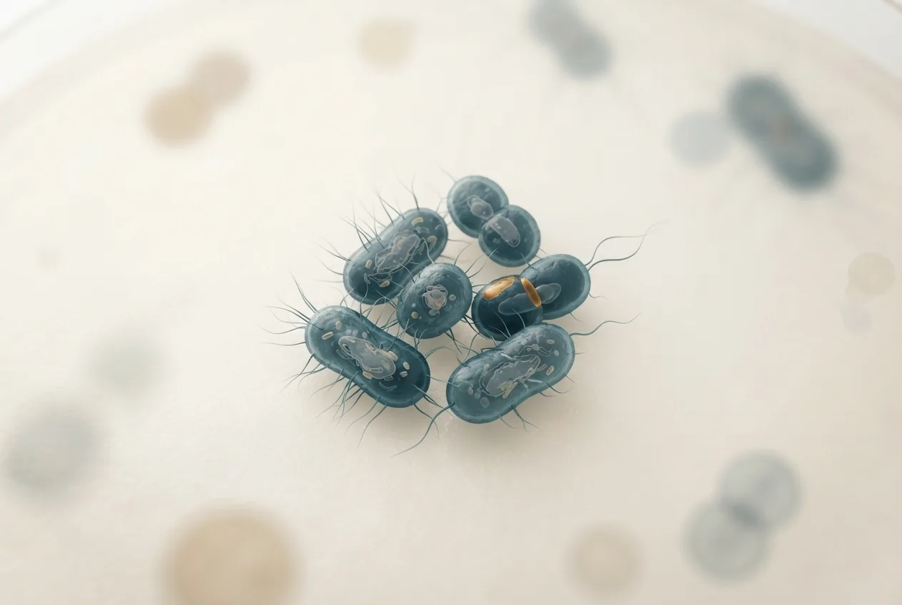
Moraxella bovis belongs to the family Moraxellaceae, order Pseudomonadales. It is a short, plump, Gram-negative rod that often appears in pairs or short chains [<a href="#ref-4">4</a>]. The organism is oxidase-positive, catalase-positive, and non-saccharolytic. On blood agar, hemolytic strains produce a distinct zone of beta-hemolysis, which is a key phenotypic marker associated with virulence [<a href="#ref-5">5</a>, <a href="#ref-9">9</a>]. Non-hemolytic variants are also encountered but are less frequently linked to clinical disease [<a href="#ref-4">4</a>].
Two major genotypes of M. bovis have been identified through whole-genome sequencing and MALDI-TOF MS profiling: genotype 1 and genotype 2 [<a href="#ref-10">10</a>, <a href="#ref-11">11</a>]. These genotypes differ in their genetic determinants, including the presence of specific virulence genes and antimicrobial resistance markers [<a href="#ref-11">11</a>]. The species is closely related to M. bovoculi and M. oculi, which can also cause IBK and are often co-isolated from affected eyes [<a href="#ref-5">5</a>, <a href="#ref-6">6</a>, <a href="#ref-7">7</a>, <a href="#ref-12">12</a>]. A study in the Sahara Desert reported the first simultaneous identification of M. bovoculi and M. bovis in one-humped camels, expanding the known host range [<a href="#ref-13">13</a>].
Pathogenesis
The pathogenesis of M. bovis involves a cascade of virulence factors that enable colonization, immune evasion, and tissue damage.
Adhesion and Colonization
Initial attachment to the corneal and conjunctival epithelium is mediated by type IV pili (fimbriae) [<a href="#ref-4">4</a>]. These filamentous structures are essential for twitching motility and biofilm formation. Pili are antigenically variable, which allows the bacterium to evade host immune responses and contributes to the chronicity of infection [<a href="#ref-4">4</a>]. The bovine ocular microbiome composition, including the presence of commensal bacteria, influences susceptibility to M. bovis colonization [<a href="#ref-14">14</a>, <a href="#ref-15">15</a>, <a href="#ref-16">16</a>]. Genetic factors in the host also affect the ocular microbiome and may predispose certain animals to IBK [<a href="#ref-15">15</a>].
Cytotoxin Production
The major virulence factor of M. bovis is a pore-forming cytotoxin (MbxA) that belongs to the RTX (repeats in toxin) family [<a href="#ref-17">17</a>, <a href="#ref-18">18</a>]. This toxin causes lysis of corneal epithelial cells and neutrophils, leading to corneal ulceration and inflammation [<a href="#ref-18">18</a>]. Heterologous expression of MbxA in human cell lines induces membrane blebbing, confirming its direct cytotoxic activity [<a href="#ref-18">18</a>]. The cytotoxin is immunogenic, and antibodies against it are protective [<a href="#ref-17">17</a>, <a href="#ref-19">19</a>].
Biofilm Formation and Polysaccharides
M. bovis produces a polysaccharide capsule that includes polysialic acid and chondroitin-like polysaccharides [<a href="#ref-20">20</a>]. These surface carbohydrates contribute to biofilm formation and resistance to phagocytosis [<a href="#ref-20">20</a>]. Biofilm production is further enhanced by the presence of other ocular bacteria [<a href="#ref-9">9</a>]. Antimicrobial photodynamic therapy using porphyrins has been shown to disrupt Moraxella biofilms in vitro [<a href="#ref-21">21</a>, <a href="#ref-22">22</a>].
Immune Evasion
The bacterium employs several strategies to subvert host immunity. The antigenic variation of pili and the production of a cytotoxin that kills neutrophils are key mechanisms [<a href="#ref-4">4</a>, <a href="#ref-19">19</a>]. Additionally, the polysaccharide capsule may mask surface antigens from antibody recognition [<a href="#ref-20">20</a>]. The host immune response to M. bovis involves both humoral and cell-mediated components, with IgG1 and IgG2 antibodies playing a role in protection [<a href="#ref-19">19</a>].
Epidemiology
IBK is a worldwide disease with highest incidence in young cattle, particularly calves and heifers [<a href="#ref-1">1</a>, <a href="#ref-2">2</a>]. The disease is more prevalent in summer months when ultraviolet radiation, dust, and fly populations peak [<a href="#ref-3">3</a>]. Face flies (Musca autumnalis) are mechanical vectors that transmit M. bovis from infected to susceptible animals [<a href="#ref-3">3</a>]. Environmental factors such as high stocking density, poor ventilation, and exposure to tall grass or weeds that cause corneal abrasions increase the risk of infection [<a href="#ref-1">1</a>, <a href="#ref-3">3</a>].
A cohort study in dairy heifers under Mediterranean climatic conditions identified several factors associated with IBK incidence, including breed, age, and season [<a href="#ref-1">1</a>]. A cross-sectional study in the same region reported herd-level prevalence and risk factors such as lack of vaccination and presence of other ocular pathogens [<a href="#ref-2">2</a>]. The presence of hemolytic M. bovis and M. bovoculi in dairy herds is strongly correlated with clinical IBK [<a href="#ref-5">5</a>]. In Kazakhstan, the epizootic situation of bovine moraxellosis has been characterized, with multiple economic entities affected [<a href="#ref-23">23</a>].
Clinical Signs and Pathology
The incubation period for IBK is typically 2 to 3 days. Clinical signs progress from mild conjunctivitis and epiphora to severe corneal edema, ulceration, and neovascularization [<a href="#ref-4">4</a>]. In advanced cases, corneal perforation and panophthalmitis may occur, leading to blindness [<a href="#ref-4">4</a>]. The disease is usually unilateral but can become bilateral. Systemic signs such as fever and anorexia are uncommon unless secondary infections occur.
Pathologically, the cornea shows infiltration of neutrophils, epithelial erosion, and stromal necrosis. The cytotoxin of M. bovis is directly responsible for corneal epithelial cell death [<a href="#ref-18">18</a>]. Other bacteria, including Mycoplasma spp., Listeria spp., and Staphylococcus spp., have been isolated from IBK cases and may act as secondary invaders or co-pathogens [<a href="#ref-8">8</a>, <a href="#ref-24">24</a>].
Diagnosis
Accurate diagnosis of M. bovis infection is essential for effective treatment and control. A combination of clinical examination, microbiological culture, and molecular methods is recommended.
Sample Collection and Transport
Ocular swabs (cotton or Dacron) are the preferred sample type. The choice of transport buffer significantly affects the recovery of M. bovis and M. bovoculi; alternative buffers such as Amies with charcoal have been evaluated for their efficacy [<a href="#ref-25">25</a>]. Proper sample handling and rapid transport to the laboratory are critical to maintain bacterial viability.
Culture and Phenotypic Identification
M. bovis grows on blood agar at 37°C under aerobic conditions. Hemolytic colonies are presumptively identified as M. bovis, but confirmation requires biochemical testing or molecular methods [<a href="#ref-9">9</a>]. MALDI-TOF MS has emerged as a rapid and reliable tool for species-level identification and even genotype classification of M. bovis [<a href="#ref-9">9</a>, <a href="#ref-10">10</a>].
Molecular Diagnostics
Polymerase chain reaction (PCR) assays targeting species-specific genes (e.g., rpoB, gyrB, or the cytotoxin gene) are widely used for direct detection from swabs [<a href="#ref-26">26</a>, <a href="#ref-27">27</a>]. A multiplex real-time PCR assay has been developed for the differential diagnosis of Moraxella-induced keratoconjunctivitis in livestock [<a href="#ref-26">26</a>]. Targeted next-generation sequencing (NGS) assays can simultaneously detect M. bovis, M. bovoculi, and M. oculi directly from ocular swabs, providing high sensitivity and specificity [<a href="#ref-27">27</a>]. Whole-genome sequencing has been applied to characterize genetic diversity and antimicrobial resistance profiles of M. bovis strains [<a href="#ref-11">11</a>, <a href="#ref-28">28</a>].
Serology
Serological tests, such as ELISA, can detect antibodies against M. bovis cytotoxin or whole-cell antigens, but they are not routinely used for diagnosis due to cross-reactivity and the time required for seroconversion [<a href="#ref-19">19</a>].
The following Mermaid diagram illustrates a diagnostic decision tree for IBK:
flowchart TD
 A["Clinical signs of IBK"] --> B["Ocular swab collection"]
 B --> C["Transport in appropriate buffer"]
 C --> D{"Diagnostic approach"}
 D --> E["Conventional culture on blood agar"]
 D --> F["Molecular detection"]
 E --> G["Hemolytic colonies?"]
 G -->|"Yes"| H["MALDI-TOF MS or PCR for species confirmation"]
 G -->|"No"| I["Consider non-hemolytic M. bovis or other Moraxella spp."]
 F --> J["Multiplex real-time PCR or targeted NGS"]
 J --> K["Identify M. bovis, M. bovoculi, or M. oculi"]
 H --> L["Antimicrobial susceptibility testing"]
 K --> L
 L --> M["Select appropriate treatment"]
Treatment and Control
Antimicrobial Therapy
Topical administration of antibiotics is the mainstay of treatment. Commonly used agents include oxytetracycline, tulathromycin, and florfenicol, but antimicrobial resistance is a growing concern [<a href="#ref-9">9</a>, <a href="#ref-24">24</a>, <a href="#ref-28">28</a>]. Susceptibility testing should guide therapy, especially in recurrent outbreaks. Systemic administration of long-acting oxytetracycline or ceftiofur is also employed in severe cases.
Non-Antimicrobial Approaches
Due to increasing antimicrobial resistance, alternative therapies are being investigated. Ultraviolet C (UVC) light has demonstrated in vitro antimicrobial activity against M. bovis and may represent a novel treatment modality [<a href="#ref-29">29</a>]. Photodynamic therapy using water-soluble tetra-cationic porphyrins has shown efficacy against Moraxella biofilms and planktonic cells [<a href="#ref-21">21</a>, <a href="#ref-22">22</a>]. A scoping review of non-antimicrobial approaches for IBK prevention and treatment in cow-calf operations identified several promising strategies, including fly control, mineral supplementation, and immunomodulation [<a href="#ref-30">30</a>].
Vaccination
Vaccination is a key component of IBK control. Autogenous and commercial vaccines are available, but their efficacy varies [<a href="#ref-31">31</a>, <a href="#ref-32">32</a>, <a href="#ref-33">33</a>]. A randomized controlled field trial demonstrated that an intranasal M. bovis cytotoxin vaccine reduced the incidence of naturally occurring IBK [<a href="#ref-17">17</a>]. Another five-year randomized controlled trial evaluated the efficacy and antibody responses of a commercial and an autogenous vaccine, showing that vaccination can reduce disease severity but not always prevent infection [<a href="#ref-32">32</a>]. The evidence base for IBK vaccination has been systematically reviewed, highlighting the need for improved vaccine formulations that target multiple virulence factors and account for antigenic diversity [<a href="#ref-33">33</a>]. Bovine immune responses to M. bovis and M. bovoculi following vaccination and natural infection have been characterized, providing insights for vaccine design [<a href="#ref-19">19</a>].
Management Practices
Reducing environmental risk factors is essential for prevention. These include providing shade to reduce UV exposure, controlling face fly populations with insecticides or traps, avoiding dusty or abrasive feed, and maintaining appropriate stocking densities [<a href="#ref-3">3</a>]. Genetic selection for resistance to IBK may also be considered [<a href="#ref-15">15</a>].
FAQ
What is the primary cause of pinkeye in cattle?
The primary cause is Moraxella bovis, a Gram-negative bacterium that produces a cytotoxin causing corneal ulceration [<a href="#ref-4">4</a>]. Other Moraxella species, such as M. bovoculi and M. oculi, are also involved [<a href="#ref-5">5</a>, <a href="#ref-6">6</a>, <a href="#ref-7">7</a>].
How is Moraxella bovis transmitted?
Transmission occurs through direct contact with ocular secretions and via mechanical vectors, primarily face flies (Musca autumnalis) [<a href="#ref-3">3</a>]. Contaminated fomites and aerosols may also play a role.
What are the key virulence factors of Moraxella bovis?
Key virulence factors include type IV pili for adhesion, an RTX cytotoxin (MbxA) that kills corneal epithelial cells, and a polysaccharide capsule that aids biofilm formation and immune evasion [<a href="#ref-4">4</a>, <a href="#ref-18">18</a>, <a href="#ref-20">20</a>].
Which diagnostic methods are most reliable for Moraxella bovis?
Molecular methods such as multiplex real-time PCR and targeted NGS offer high sensitivity and specificity for direct detection from ocular swabs [<a href="#ref-26">26</a>, <a href="#ref-27">27</a>]. MALDI-TOF MS is excellent for species identification and genotyping [<a href="#ref-9">9</a>, <a href="#ref-10">10</a>].
Are there effective vaccines against infectious bovine keratoconjunctivitis?
Yes, vaccines containing M. bovis cytotoxin or whole-cell antigens can reduce disease incidence and severity, but efficacy varies [<a href="#ref-17">17</a>, <a href="#ref-31">31</a>, <a href="#ref-32">32</a>, <a href="#ref-33">33</a>]. Intranasal cytotoxin vaccines have shown promise in field trials [<a href="#ref-17">17</a>].
Can Moraxella bovis infect other species?
Yes, M. bovis has been isolated from one-humped camels with keratoconjunctivitis [<a href="#ref-13">13</a>]. A rare human case of bacteremia and septic arthritis has been reported in an immunocompromised patient, indicating zoonotic potential [<a href="#ref-34">34</a>].
What non-antibiotic treatments are available for pinkeye?
Ultraviolet C light and photodynamic therapy using porphyrins have demonstrated antimicrobial activity against M. bovis in vitro [<a href="#ref-21">21</a>, <a href="#ref-22">22</a>, <a href="#ref-29">29</a>]. These approaches are under investigation for clinical use.