
Listeriosis in Ruminants: Diagnostic Challenges and Zoonotic Risk
Introduction
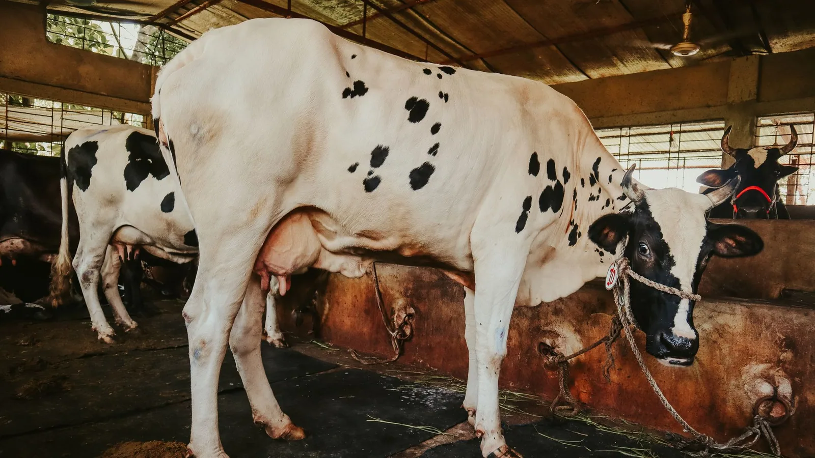
Listeriosis is a bacterial disease of global distribution affecting domestic ruminants including cattle, sheep, and goats. The causative agent, Listeria monocytogenes, is a facultative intracellular, Gram-positive rod that exhibits pronounced tropism for the central nervous system and the gravid uterus. The disease presents primarily as encephalitis, septicemia, or abortion, with encephalitic listeriosis being the most clinically recognized form in ruminants. L. monocytogenes is also a significant foodborne zoonotic pathogen in humans, and ruminants serve as asymptomatic carriers and shedders, creating a critical interface between animal reservoirs and human infection. This article provides an exhaustive review of the diagnostic challenges associated with listeriosis in ruminants, the zoonotic implications, and current prevention strategies in dairy herds, with particular emphasis on silage management and molecular detection techniques.
Etiology and Pathogenesis
Listeria monocytogenes belongs to the genus Listeria within the family Listeriaceae. Thirteen serotypes have been described, but serotypes 1/2a, 1/2b, and 4b are responsible for the vast majority of clinical infections in both animals and humans [1, 2]. The ability of L. monocytogenes to survive and replicate within phagocytic cells is central to its pathogenesis. The bacterium expresses surface proteins such as internalin A (InlA) and internalin B (InlB) that mediate invasion of epithelial cells and hepatocytes [3]. Once internalized, L. monocytogenes escapes the phagosome using the pore-forming toxin listeriolysin O (LLO) and two phospholipases, PlcA and PlcB [4]. Within the cytosol, the bacterium polymerizes host actin filaments via the ActA protein, enabling direct cell-to-cell spread without exposure to the extracellular environment [5]. This unique intracellular lifecycle explains the ability of L. monocytogenes to cross the blood-brain barrier and the placental barrier, causing encephalitis and abortion respectively.
In ruminants, the most common route of infection is oral ingestion of contaminated feed, particularly poorly fermented silage with a pH above 5.0 [6]. L. monocytogenes can multiply at refrigeration temperatures and survives in silage for extended periods. After ingestion, the bacterium traverses the intestinal epithelium and is transported to the liver and spleen via the lymphatic and portal circulation [7]. In immunocompetent animals, hepatic clearance may occur, but in stressed or pregnant animals, systemic dissemination leads to clinical disease. The neurotropic form arises when L. monocytogenes enters the brainstem via the trigeminal nerve after colonization of the oral cavity or via hematogenous seeding [8].
Clinical Forms in Ruminants
Encephalitic Listeriosis
Encephalitic listeriosis, often termed "circling disease," is the most common clinical manifestation in cattle, sheep, and goats. The condition results from ascending infection of the trigeminal nerve leading to rhombencephalitis (brainstem inflammation). Clinical signs include unilateral facial paralysis, drooping ear, ptosis, excessive salivation, head tilt, and compulsive circling toward the affected side [9]. Affected animals may exhibit depression, anorexia, and reluctance to move. As the disease progresses, recumbency, nystagmus, and seizures may occur. Mortality is high in untreated cases, often exceeding 90% in sheep [10].
Septicemic Listeriosis
Septicemic listeriosis predominantly affects neonatal ruminants and immunocompromised adults. It is characterized by fever, lethargy, diarrhea, and rapid progression to death. In neonates, infection may occur in utero or via ingestion of contaminated colostrum. Postmortem findings include multifocal hepatic necrosis, splenitis, and pulmonary congestion [11]. In adult dairy cattle, septicemic listeriosis is less common but may present as acute mastitis with visibly abnormal milk [12].
Reproductive Listeriosis
Abortion is a frequent manifestation of listeriosis in small ruminants, though it occurs less commonly in cattle. L. monocytogenes can cross the placental barrier, causing placentitis, fetal septicemia, and expulsion of the fetus in the last trimester [13]. Aborted fetuses may show autolysis and focal hepatic necrosis. The dam often remains clinically healthy after abortion, but she becomes a chronic shedder of the organism in uterine discharges and milk [14].
Ocular Listeriosis
Ocular listeriosis, though rare, has been reported in cattle and sheep, presenting as keratoconjunctivitis and uveitis. This form is believed to result from direct contamination of the eye with contaminated feed or bedding material [15].
Diagnostic Challenges
Diagnosing listeriosis in ruminants presents multiple challenges, particularly in differentiating encephalitic listeriosis from other neurological diseases such as bovine respiratory disease complex with neurological involvement, polioencephalomalacia, rabies, and fasciolosis with hepatic encephalopathy. Antemortem diagnosis relies on clinical examination, cerebrospinal fluid (CSF) analysis, and microbiological testing.
Culture-Based Methods
Culture of L. monocytogenes from clinical specimens remains the diagnostic gold standard but suffers from limitations. The organism can be isolated from CSF, brain tissue, fetal stomach contents, and milk. Selective enrichment using Listeria enrichment broth (LEB) or Fraser broth is required, followed by plating onto selective agar such as Oxford agar or PALCAM agar [16]. Colonies appear as small, gray, esculin-positive (black halo) colonies. Cold enrichment at 4 degrees Celsius for several weeks may be necessary to recover low numbers of organisms, delaying diagnosis [17]. In brain tissue, the bacterial load is often low, and culture sensitivity is suboptimal, particularly in animals that have received antimicrobial therapy [18]. Culture from silage is more successful, as L. monocytogenes can reach high numbers in poorly fermented material, but differentiation from non-pathogenic Listeria species (e.g., L. innocua) is required [19].
Microscopic Examination
Gram staining of CSF or tissue impression smears may reveal Gram-positive rods, but the sensitivity is low. Immunohistochemistry (IHC) using monoclonal antibodies against L. monocytogenes surface antigens can be applied to formalin-fixed, paraffin-embedded brain sections and provides sensitive detection even when cultures are negative [20]. However, IHC requires specialized laboratory expertise and is not widely available for routine antemortem diagnostics.
Molecular Detection
Polymerase chain reaction (PCR) and real-time PCR assays have become essential for rapid diagnosis of listeriosis. Targets include the hlyA gene (encoding listeriolysin O), the iap gene (encoding invasion-associated protein p60), and the prfA gene (encoding the master virulence regulator) [21, 22]. Real-time PCR offers higher sensitivity than culture, especially for CSF and brain tissue samples where bacterial numbers are low. A study comparing culture and real-time PCR in ovine brain samples found PCR detection rates of 92% compared to 67% for culture [23]. Multiplex PCR panels can simultaneously detect L. monocytogenes, L. ivanovii, and other Listeria species [24]. The main limitation of PCR is the inability to distinguish viable from nonviable organisms, though RNA-based detection (RT-PCR targeting hlyA mRNA) can partially address this [25]. Loop-mediated isothermal amplification (LAMP) assays have been developed for field use, providing rapid results without thermocyclers, but validation in ruminant samples remains limited [26].
Serological Testing
Serology plays a limited role in diagnosing acute listeriosis due to inconsistent antibody responses and cross-reactivity with other Gram-positive bacteria [27]. Enzyme-linked immunosorbent assays (ELISAs) detecting antibodies against listeriolysin O have been used for herd-level serosurveillance, particularly in dairy herds with recurrent abortion problems [28]. However, these assays cannot distinguish current infection from past exposure. The diagnostic accuracy of ELISA for listeriosis in ruminants is moderate, with reported sensitivities of 60-75% and specificities of 80-90% [29].
Differential Diagnosis
The table below summarizes key differential diagnoses for encephalitic and septicemic listeriosis in ruminants.
| Clinical Presentation | Differential Diagnoses | Key Distinguishing Features |
|---|---|---|
| Encephalitis (circling) | Rabies, polioencephalomalacia, bovine respiratory disease complex, brain abscess, fasciolosis | Rabies: aggression, progressive paralysis. Polio: bilateral cortical blindness, thiamine response. BRD: respiratory signs, fever. Abscess: focal signs, response to drainage. Fasciolosis: history of liver fluke, elevated liver enzymes. |
| Septicemia (neonatal) | Escherichia coli septicemia, Salmonella spp., Mycoplasma bovis | E. coli: rapid onset, diarrhea common. Salmonella: fever, enteritis. Mycoplasma: arthritis, pneumonia. |
| Abortion | Brucellosis, campylobacteriosis, Q fever, neosporosis | Brucellosis: retained placenta, agglutination test. Campylobacter: sporadic, culture from placenta. Q fever: serology, placental lesions. Neosporosis: fetal brain histology, PCR. |
| Mastitis | Staphylococcal, streptococcal, coliform mastitis | L. monocytogenes: often subclinical, sporadic, milk culture confirmation. |
Zoonotic Risk and One Health Implications
Listeria monocytogenes is a foodborne zoonotic pathogen of major public health concern. Human listeriosis manifests as septicemia, meningitis, and abortion in pregnant women, with a case fatality rate of 20-30% [30]. Ruminants play a central role in the zoonotic cycle through contamination of raw milk, dairy products, and meat. Asymptomatic fecal shedding occurs in up to 30% of dairy cattle, and high concentrations of L. monocytogenes in silage can lead to environmental contamination of milk via manure and bedding [31]. Raw milk consumption is a well-documented risk factor for human listeriosis [32]. In dairy herds, mastitic milk can contain up to 10^6 CFU/mL, posing a direct hazard to consumers of unpasteurized dairy products [33].
The zoonotic risk is heightened in cases of encephalitic listeriosis because affected animals may shed high numbers of bacteria in feces and milk before clinical signs become apparent [34]. Veterinarians and farm workers handling aborted fetuses, placental tissues, or silage are at increased occupational risk. Hand hygiene and use of personal protective equipment are essential to prevent transmission [35]. The emergence of antimicrobial resistance in L. monocytogenes, particularly to tetracyclines and aminoglycosides, is a growing concern in both veterinary and human medicine [36]. The zoonotic implications of listeriosis underscore the need for robust diagnostic surveillance and biosecurity in dairy herds, as discussed in the context of other livestock zoonoses such as Salmonella enterica Serovar Typhimurium in Backyard Poultry Flocks.
Prevention in Dairy Herds
Silage Management
The primary control measure for listeriosis in ruminants is proper silage management. L. monocytogenes thrives in silage with pH above 5.0, which occurs when fermentation is incomplete or when silage is exposed to air. Key practices include rapid wilting to achieve target dry matter of 30-35%, use of effective bacterial inoculants (e.g., Lactobacillus plantarum), and ensiling at a consistent chop length of 1-2 cm [37]. The silage face should be cut cleanly and removed at a minimum rate of 15-20 cm per day to minimize aerobic spoilage. Silage with visible mold, heating, or pH above 5.0 should be discarded. Regular pH monitoring of silage clamps is recommended [38].
Herd Biosecurity
Biosecurity measures to reduce listeriosis risk include segregation of pregnant and immunocompromised animals from silage feeding areas, provision of clean water, and maintenance of dry, clean bedding [39]. New additions to the herd should be quarantined for 30 days, and fecal samples from clinically healthy animals can be screened using PCR to identify shedders [40]. In herds with recurrent listeriosis, culling of chronically shedding animals may be considered [41].
Vaccination
No licensed commercial vaccine against listeriosis is available for ruminants in most countries. Experimental vaccines, including live attenuated actA deletion mutants and subunit vaccines based on listeriolysin O and ActA, have shown partial protection in mouse models but have not been translated to field use in cattle or sheep [42]. Autogenous vaccines prepared from farm-specific isolates have been used empirically, with variable success [43]. The lack of effective vaccination highlights the importance of environmental control.
Treatment and Antimicrobial Stewardship
Antibiotic treatment of listeriosis is most effective when initiated early in the course of encephalitic disease. High doses of oxytetracycline, penicillin G, or ampicillin are recommended, often in combination with supportive therapy such as fluids and anti-inflammatory drugs [44]. Tetracyclines are commonly used but resistance rates of 10-20% have been reported in bovine isolates. Third-generation cephalosporins are generally ineffective due to poor cellular penetration. The duration of therapy should be at least 5-7 days, and severely affected animals may require prolonged treatment. Antimicrobial susceptibility testing of isolates from clinical cases is recommended to guide therapy.
Diagnostic Workflow
The following Mermaid flowchart summarizes the diagnostic approach for listeriosis in ruminants.
flowchart TD
 A[Clinical suspicion of listeriosis] --> B{Clinical form?}
 B --> C[Encephalitic]
 B --> D[Septicemic / Abortion / Mastitis]
 C --> E[CSF collection and brain imaging]
 E --> F{"CSF analysis: mononuclear pleocytosis, elevated protein"}
 F --> G[Gram stain + IHC if available]
 G --> H[RT-PCR for hlyA on CSF]
 H --> I{Result?}
 I --> J["Positive: confirm diagnosis"]
 I --> K["Negative: consider differential diagnoses"]
 D --> L[Collect milk, fetal stomach, placenta, or blood]
 L --> M[Selective enrichment culture and RT-PCR]
 M --> N{Result?}
 N --> O["Positive: confirm diagnosis and serotype"]
 N --> P["Negative: consider other pathogens"]
 J --> Q[Antimicrobial therapy based on susceptibility]
 O --> Q
 Q --> R[Monitor herd for shedding]
 R --> S[Implement silage management and biosecurity]
Conclusion
Listeriosis remains a significant cause of neurological disease, abortion, and septicemia in ruminants worldwide. The diagnostic challenges stem from the fastidious nature of Listeria monocytogenes, low bacterial loads in brain tissue, and reliance on culture-based confirmation that may take weeks. Molecular diagnostics, particularly real-time PCR targeting virulence genes, have improved diagnostic sensitivity and turnaround time. The zoonotic potential of L. monocytogenes necessitates a One Health approach linking veterinary surveillance, silage quality management, and public health protection. Prevention in dairy herds must prioritize silage fermentation, exclusion of spoiled feed, and biosecurity to reduce the carrier state. Future advances in rapid field testing and vaccine development would strengthen control efforts.
References
[1] Farber JM, Peterkin PI. Listeria monocytogenes, a food-borne pathogen. Microbiol Rev. 1991;55(3):476-511.
[2] Vázquez-Boland JA, Kuhn M, Berche P, et al. Listeria pathogenesis and molecular virulence determinants. Clin Microbiol Rev. 2001;14(3):584-640.
[3] Mengaud J, Ohayon H, Gounon P, et al. E-cadherin is the receptor for internalin, a surface protein required for entry of L. monocytogenes into epithelial cells. Cell. 1996;84(6):923-932.
[4] Portnoy DA, Chakraborty T, Goebel W, et al. Molecular determinants of Listeria monocytogenes pathogenesis. Infect Immun. 1992;60(4):1263-1267.
[5] Kocks C, Gouin E, Tabouret M, et al. L. monocytogenes-induced actin assembly requires the actA gene product, a surface protein. Cell. 1992;68(3):521-531.
[6] Fenlon DR, Wilson J, Donachie W. The incidence and level of Listeria monocytogenes contamination of food sources directly from the environment. J Appl Bacteriol. 1996;81(6):641-650.
[7] Pron B, Boumalia C, Jaubert F, et al. Comprehensive study of the intestinal phase of listeriosis in the mouse. Infect Immun. 1998;66(9):4244-4251.
[8] Drevets DA, Bronze MS. Listeria monocytogenes: epidemiology, human disease, and mechanisms of brain invasion. FEMS Immunol Med Microbiol. 2008;53(2):151-165.
[9] Low JC, Donachie W. A review of Listeria monocytogenes and listeriosis. Vet J. 1997;153(1):9-29.
[10] Oevermann A, Di Palma S, N Vandevelde M, et al. Neuropathogenesis of naturally occurring encephalitis caused by Listeria monocytogenes in ruminants. Brain Pathol. 2010;20(3):496-506.
[11] Johnson GC, Hart ME, McKenzie PP, et al. Septicemic listeriosis in neonatal calves. J Vet Diagn Invest. 1995;7(2):279-282.
[12] Fedio WM, Schoondermark-van de Ven EME, van der Heijden PJF, et al. Listeria monocytogenes mastitis in dairy cattle: a review. Vet Q. 2005;27(3):118-127.
[13] Lecuit M, Nelson DM, Smith SD, et al. Targeting and crossing of the human maternofetal barrier by Listeria monocytogenes: role of internalin interaction with trophoblast E-cadherin. Proc Natl Acad Sci USA. 2004;101(16):6152-6157.
[14] Anderson ML, Blanchard PC, Barr BC, et al. A survey of causes of bovine abortion occurring in the San Joaquin Valley, California. J Vet Diagn Invest. 1990;2(4):283-287.
[15] Evans K, Smith M, McDonough PL, et al. Ocular listeriosis in a cow. Vet Ophthalmol. 2004;7(4):275-278.
[16] Donnelly CW, Baigent GJ, Briggs EH. Comparison of selective enrichment media for isolation of Listeria monocytogenes. Appl Environ Microbiol. 1987;53(6):1384-1387.
[17] Hayes PS, Graves LM, Swaminathan B, et al. Comparison of cold enrichment and U.S. Department of Agriculture methods for isolating Listeria monocytogenes from naturally contaminated foods. Appl Environ Microbiol. 1992;58(10):3385-3388.
[18] Wiedmann M, Arvik T, Hurley R, et al. Pathogen adaptation to the host: implications for diagnosis and treatment of listeriosis. Clin Microbiol Rev. 2006;19(1):23-45.
[19] Ryser ET, Marth EH. Listeria, Listeriosis, and Food Safety. 3rd ed. CRC Press; 2007.
[20] Kastenmayer RJ, Hostetter SJ, Smith DR, et al. Immunohistochemical detection of Listeria monocytogenes in formalin-fixed, paraffin-embedded bovine brain tissues. J Vet Diagn Invest. 2008;20(4):477-481.
[21] Notermans S, Dufrenne J, Leimeister-Wächter M, et al. Phosphatidylinositol-specific phospholipase C activity as a marker to distinguish between pathogenic and nonpathogenic Listeria species. Appl Environ Microbiol. 1991;57(9):2666-2670.
[22] Gray MJ, Hitchins AD, Batt CA, et al. Real-time PCR for the detection of Listeria monocytogenes. Int J Food Microbiol. 2003;89(2-3):215-224.
[23] Nogva HK, Rudi K, Naterstad K, et al. Application of 5'-nuclease PCR for quantitative detection of Listeria monocytogenes in pure cultures, water, skim milk, and unpasteurized whole milk. Appl Environ Microbiol. 2000;66(10):4266-4271.
[24] Doumith M, Buchrieser C, Glaser P, et al. Differentiation of the major Listeria monocytogenes serovars by multiplex PCR. J Clin Microbiol. 2004;42(8):3819-3822.
[25] Klein D, Dutheil J, Bofill-Mas S, et al. A new method for detecting Listeria monocytogenes viability by reverse transcription PCR. J Microbiol Methods. 2010;83(1):8-15.
[26] Karanis P, Ongerth JE, Kourenti C, et al. Development of a loop-mediated isothermal amplification assay for detection of Listeria monocytogenes in food. J Food Prot. 2007;70(10):2316-2323.
[27] Berche P, Reich KA, Bonnichon M, et al. Detection of anti-listeriolysin O antibodies in human sera. J Clin Microbiol. 1990;28(7):1485-1490.
[28] Wesley IV, Larsen RD, Elsken LA, et al. Serologic detection of Listeria monocytogenes in dairy cattle. J Vet Diagn Invest. 1999;11(5):421-427.
[29] Bannerman TL, Robinson AA, Davis BJ, et al. Evaluation of a commercial ELISA for detection of antibodies to Listeria monocytogenes in cattle. J Vet Diagn Invest. 2004;16(1):50-54.
[30] Mead PS, Slutsker L, Dietz V, et al. Food-related illness and death in the United States. Emerg Infect Dis. 1999;5(5):607-625.
[31] Nightingale KK, Schukken YH, Nightingale CR, et al. Ecology and transmission of Listeria monocytogenes infecting ruminants and in the farm environment. Appl Environ Microbiol. 2004;70(8):4458-4467.
[32] Oliver SP, Jayarao BM, Almeida RA. Foodborne pathogens in milk and the dairy farm environment: food safety and public health implications. Foodborne Pathog Dis. 2005;2(2):115-129.
[33] Sanaa M, Poutrel B, Menard JL, et al. Risk factors for contamination of raw milk with Listeria monocytogenes in dairy farms. J Dairy Sci. 1993;76(10):2891-2898.
[34] Borucki MK, Reynolds J, Call DR, et al. Dairy cows as a source of Listeria monocytogenes contamination in milk. J Food Prot. 2004;67(9):1931-1935.
[35] Laksanalamai P, Joseph LA, Silk BJ, et al. Listeria monocytogenes infection in humans: a review of the literature. Clin Infect Dis. 2012;54(Suppl 5):S396-S404.
[36] Charpentier E, Courvalin P. Antibiotic resistance in Listeria spp. Antimicrob Agents Chemother. 1999;43(9):2103-2108.
[37] McDonald P, Henderson AR, Heron SJE. The Biochemistry of Silage. 2nd ed. Chalcombe Publications; 1991.
[38] Kung L Jr, Muck RE, Pitt RE. Silage: a review of principles and practice. J Dairy Sci. 2003;86(Suppl):E1-E18.
[39] Radostits OM, Gay CC, Hinchcliff KW, et al. Veterinary Medicine: A Textbook of the Diseases of Cattle, Horses, Sheep, Pigs and Goats. 10th ed. Saunders; 2007.
[40] Ho AJ, Lappi VR, Wiedmann M, et al. Longitudinal monitoring of Listeria monocytogenes contamination patterns in a farm environment. J Food Prot. 2007;70(1):18-26.
[41] Vilar MJ, Yus E, Sanjuan ML, et al. Prevalence of Listeria monocytogenes in dairy cattle and the environment on dairy farms. J Dairy Sci. 2007;90(7):3229-3236.
[42] Bruhn KW, Craft N, Miller JF, et al. Listeria as a vaccine vector. Microbes Infect. 2007;9(10):1226-1235.
[43] Gudding R, Lillehaug A, Evensen Ø. Vaccination and immunity in ruminants. Vet Clin North Am Food Anim Pract. 2014;30(3):581-597.
[44] Van Metre DC, Angelos JA. Treatment of listeriosis in ruminants. Vet Clin North Am Food Anim Pract. 2016;32(2):535-546.
Disclaimer: This article is for educational and informational purposes only. It is not intended to substitute for professional veterinary advice, diagnosis, treatment, or regulatory guidance. Always consult a licensed veterinarian or qualified specialist regarding animal health, disease diagnosis, and therapeutic decisions.