Erysipelothrix rhusiopathiae: A Comprehensive Veterinary Reference on Etiology, Epidemiology, Diagnostics, and Control
By Dr. Zubair Khalid, DVM, MS, PhD ·
Key Takeaways
- Erysipelothrix rhusiopathiae is a Gram-positive, facultative intracellular bacterium causing significant economic losses in swine (septicemia, arthritis, diamond skin lesions) and high mortality in poultry.
- The pathogen exhibits remarkable host plasticity, with recent reemergence in Arctic wildlife (muskoxen, beluga whales, polar bears) highlighting its ecological adaptability and potential for cross-species transmission.
- Diagnostic confirmation relies on conventional bacteriology (culture, H2S production) and increasingly on molecular methods like PCR for rapid detection, with whole-genome sequencing providing high-resolution epidemiological typing.
- Penicillins and ceftiofur remain the drugs of choice due to intrinsic susceptibility, but resistance to tetracyclines and macrolides is emerging, often mediated by mobile genetic elements.
- Effective control strategies include vaccination with killed bacterins in swine and poultry, with ongoing research into recombinant and probiotic approaches; biosecurity and rodent control are crucial in poultry operations.
- The surface protein SpaA is a critical virulence factor involved in host cell adhesion and serves as a primary target for serological diagnostics and vaccine development.
Introduction
Erysipelothrix rhusiopathiae is a Gram-positive, rod-shaped, facultative intracellular bacterium that causes erysipelas in many domestic and wild animal species [<a href="#ref-1">1</a>, <a href="#ref-2">2</a>]. The organism is of major economic importance in swine production, where it manifests as acute septicemia, chronic arthritis, and characteristic diamond skin lesions, and in poultry, where it can produce acute fatal disease with high mortality [<a href="#ref-3">3</a>, <a href="#ref-4">4</a>]. Over the past decade, renewed attention has focused on this pathogen due to its reemergence in Arctic wildlife populations, including muskoxen, beluga whales, and polar bears, highlighting its ecological plasticity and potential for cross-species transmission [<a href="#ref-1">1</a>, <a href="#ref-2">2</a>, <a href="#ref-5">5</a>, <a href="#ref-6">6</a>].
This article provides a comprehensive, citation-grounded reference for veterinary practitioners, diagnostic microbiologists, and computational biologists. It synthesizes findings from recent molecular epidemiological studies, genomic analyses, vaccine efficacy trials, and antimicrobial resistance surveillance, drawing exclusively on peer-reviewed literature provided in the reference list.
Taxonomy and Morphology
Erysipelothrix rhusiopathiae belongs to the family Erysipelotrichaceae within the phylum Firmicutes [<a href="#ref-1">1</a>, <a href="#ref-7">7</a>]. The genus Erysipelothrix comprises several species, including E. rhusiopathiae, E. tonsillarum, E. piscisicarius, and E. inopinata [<a href="#ref-7">7</a>, <a href="#ref-8">8</a>]. E. rhusiopathiae is the primary pathogen in terrestrial mammals and birds, while E. tonsillarum and E. piscisicarius are more commonly associated with aquatic hosts [<a href="#ref-7">7</a>, <a href="#ref-8">8</a>, <a href="#ref-9">9</a>].
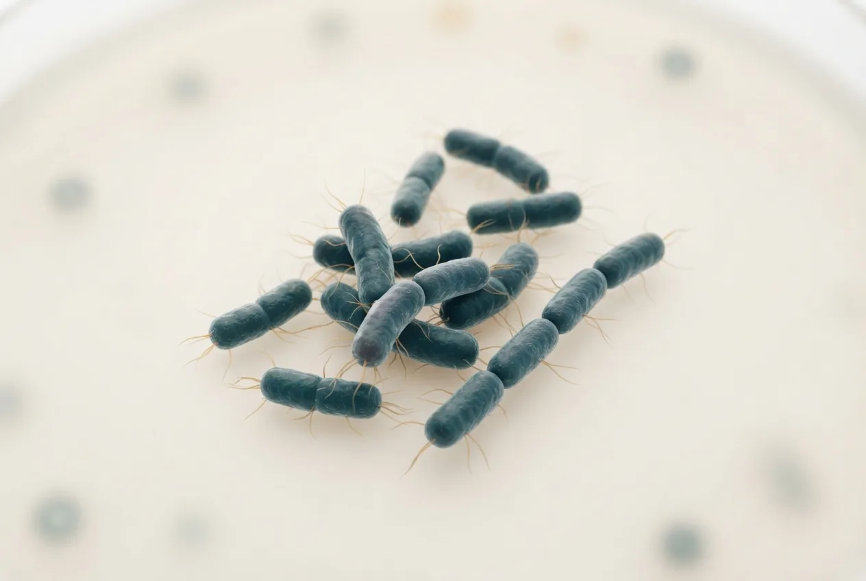
The organism appears as slender, straight or slightly curved rods, 0.2-0.4 µm in width and 1.0-2.5 µm in length, and is non-motile, non-spore-forming, and capsulated in some strains [<a href="#ref-10">10</a>, <a href="#ref-11">11</a>]. Gram staining is variable; older cultures may stain Gram-negative [<a href="#ref-10">10</a>]. On blood agar, colonies are small (0.5-1.5 mm), smooth, translucent, and may produce a narrow zone of alpha-hemolysis, which can be confused with viridans streptococci [<a href="#ref-10">10</a>, <a href="#ref-11">11</a>].
Genomics and Virulence Factors
The genome of E. rhusiopathiae is a single circular chromosome of approximately 1.8-1.9 Mb with a G+C content of 36-38% [<a href="#ref-7">7</a>, <a href="#ref-12">12</a>]. Comparative genomic studies have identified a set of core virulence factors that are conserved across Erysipelothrix species [<a href="#ref-7">7</a>, <a href="#ref-12">12</a>]. The major protective antigen is surface protein SpaA (surface protective antigen A), which mediates adhesion to host cells and is a key target for vaccine development [<a href="#ref-13">13</a>]. Other virulence-associated determinants include neuraminidase (which facilitates tissue invasion), hyaluronidase, and a polysaccharide capsule [<a href="#ref-11">11</a>]
Several strains harbor prophage elements and genomic islands that carry antimicrobial resistance genes. For example, prophage phi Er670 and genomic island GI_Er147 have been identified as carriers of tetracycline and macrolide resistance determinants in swine and poultry isolates [<a href="#ref-12">12</a>]. The presence of mobile genetic elements suggests that horizontal gene transfer contributes to the dissemination of resistance among E. rhusiopathiae strains [<a href="#ref-12">12</a>, <a href="#ref-14">14</a>].
Host Range and Epidemiology
E. rhusiopathiae has an exceptionally broad host range that encompasses swine, poultry (chickens, turkeys, ducks, geese), sheep, cattle, horses, dogs, cetaceans, pinnipeds, and numerous wildlife species [<a href="#ref-3">3</a>, <a href="#ref-4">4</a>, <a href="#ref-5">5</a>, <a href="#ref-6">6</a>, <a href="#ref-9">9</a>, <a href="#ref-15">15</a>]. In swine, disease is most common in grower, finisher pigs aged 8-24 weeks; persistent carriage in tonsils and gut-associated lymphoid tissue sustains within-herd transmission [<a href="#ref-4">4</a>]. In laying hens and turkey breeder flocks, erysipelas frequently emerges after peak production and can cause acute mortality reaching 10-50% within days [<a href="#ref-3">3</a>, <a href="#ref-4">4</a>].
A notable recent phenomenon is the reemergence of a highly virulent clone of E. rhusiopathiae associated with multi-year mass mortality events in high Arctic muskoxen (Ovibos moschatus) in Canada [<a href="#ref-1">1</a>, <a href="#ref-2">2</a>]. Whole-genome sequencing of isolates from these outbreaks revealed high clonality, suggesting a single introduction event followed by environmental persistence [<a href="#ref-2">2</a>]. Similarly, endangered Cook Inlet beluga whales (Delphinapterus leucas) have experienced fatal infections, with E. rhusiopathiae isolated from multiple organs [<a href="#ref-5">5</a>]. Serological surveys have demonstrated antibodies in Beaufort Sea polar bears (Ursus maritimus), with seroprevalence linked to ringed seal demographics, indicating a marine transmission cycle [<a href="#ref-6">6</a>].
In wild boar populations in Poland, seroprevalence ranged from 5% to 15%, confirming that feral swine serve as reservoirs for spillover to domestic herds [<a href="#ref-16">16</a>]. Poultry erysipelas in waterfowl has been increasingly reported in Poland and Hungary, with incidence rates rising over the past decade [<a href="#ref-15">15</a>].
Pathogenesis and Clinical Manifestations
Swine
Acute erysipelas in swine is characterized by sudden onset of fever (40-42°C), depression, anorexia, and cutaneous erythematous lesions that progress to characteristic rhomboid (diamond) urticarial plaques [<a href="#ref-4">4</a>]. These lesions are pathognomonic when present. Septicemic disease may lead to death within 24-48 hours. Subacute and chronic forms include vegetative endocarditis, polyarthritis, and lymphadenitis [<a href="#ref-4">4</a>]. Arthritis typically involves the carpal, tarsal, and stifle joints, with proliferative synovitis and periarticular fibrosis [<a href="#ref-4">4</a>].
Poultry
In chickens, turkeys, and waterfowl, erysipelas presents as peracute septicemia with high mortality, often without premonitory signs [<a href="#ref-3">3</a>, <a href="#ref-4">4</a>]. Gross lesions include generalized congestion, petechial hemorrhages on serosal surfaces, splenomegaly, hepatomegaly, and hemorrhagic enteritis [<a href="#ref-3">3</a>, <a href="#ref-4">4</a>, <a href="#ref-15">15</a>]. Chronic valvular endocarditis is less common in birds than in swine [<a href="#ref-3">3</a>].
Wildlife and Aquatic Mammals
Muskoxen infected with the reemerging clone show severe fibrinous pneumonia, pericarditis, and splenomegaly [<a href="#ref-2">2</a>]. In cetaceans, infection can be peracute (sudden death) or chronic, with meningoencephalitis, pleuritis, and dermatitis [<a href="#ref-5">5</a>, <a href="#ref-9">9</a>].
Diagnostics
Conventional Bacteriology
Isolation of E. rhusiopathiae from liver, spleen, kidney, or joint fluid is the traditional gold standard [<a href="#ref-3">3</a>, <a href="#ref-10">10</a>]. Samples are inoculated onto 5% sheep blood agar or selective media containing antibiotics (e.g., crystal violet, sodium azide) to inhibit contaminants [<a href="#ref-10">10</a>]. After 24-48 h at 37°C in 5% CO2, characteristic small, alpha-hemolytic colonies appear. Identity is confirmed by Gram stain, catalase negative, oxidase negative, and production of hydrogen sulfide in triple sugar iron agar [<a href="#ref-10">10</a>, <a href="#ref-17">17</a>].
Molecular Methods
Polymerase chain reaction (PCR)-based assays have largely replaced culture for rapid detection. A novel multiplex PCR assay has been developed that simultaneously detects E. rhusiopathiae, Streptococcus suis, and Staphylococcus hyicus in swine samples, offering high sensitivity and specificity [<a href="#ref-18">18</a>]. Additionally, pulsed-field gel electrophoresis (PFGE) and random amplified polymorphic DNA (RAPD)-PCR have been used for strain typing in epidemiological investigations [<a href="#ref-2">2</a>, <a href="#ref-17">17</a>]. Whole-genome sequencing provides the highest resolution for outbreak tracing and antimicrobial resistance gene profiling [<a href="#ref-1">1</a>, <a href="#ref-2">2</a>, <a href="#ref-7">7</a>, <a href="#ref-10">10</a>].
Serology
Enzyme-linked immunosorbent assays (ELISAs) targeting the SpaA protein are available for detection of antibodies in swine and poultry [<a href="#ref-16">16</a>, <a href="#ref-19">19</a>]. Commercial or in-house ELISAs have been used to monitor vaccine responses and to determine herd seroprevalence [<a href="#ref-16">16</a>, <a href="#ref-19">19</a>, <a href="#ref-20">20</a>]. In wildlife, serological assays based on whole-cell antigens have been validated for polar bears and belugas [<a href="#ref-5">5</a>, <a href="#ref-6">6</a>].
Diagnostic Workflow
Below is a Mermaid diagram summarizing the recommended diagnostic approach for suspected erysipelas in livestock and poultry.
flowchart TD
 A["Clinical suspicion: fever, diamond lesions, sudden death"] --> B{"Postmortem examination"}
 B --> C["Collect liver, spleen, kidney, joint fluid"]
 C --> D["Gram stain & culture on blood agar\n("24-48h, 5% CO2")"]
 D --> E["Acridine orange stain or\ncatalase/oxidase tests"]
 D --> F["Genus-specific PCR or\nmultiplex PCR"]
 F --> G["Positive: SpaA sequencing\nor whole genome sequencing"]
 E --> H["Alpha-hemolytic, H2S+\nConfirm: API Coryne or MALDI-TOF"]
 H --> I["Antimicrobial susceptibility\n("disc diffusion/MIC")"]
 G --> J["Epidemiological typing:\nPFGE, RAPD, WGS"]
 J --> K["Data submission to public\ndatabases"]
Antimicrobial Susceptibility and Resistance
E. rhusiopathiae is intrinsically susceptible to penicillins, ceftiofur, and other beta-lactam antibiotics, which remain the drugs of choice for treatment [<a href="#ref-14">14</a>, <a href="#ref-21">21</a>]. Resistance to tetracyclines, macrolides, and lincosamides has been documented in up to 30% of isolates from poultry and waterfowl [<a href="#ref-12">12</a>, <a href="#ref-14">14</a>, <a href="#ref-21">21</a>]. Resistance determinants (e.g., tet(M), erm(B)) are frequently carried on mobile genetic elements, including prophages and genomic islands [<a href="#ref-12">12</a>, <a href="#ref-14">14</a>]. In a study of waterfowl isolates from Poland, multidrug resistance (resistance to three or more antibiotic classes) was found in 6.5% of strains [<a href="#ref-14">14</a>].
Surveillance in Hungary (2022-2023) of waterfowl isolates detected high susceptibility to amoxicillin, penicillin, and florfenicol, but reduced susceptibility to enrofloxacin in 10% of isolates [<a href="#ref-21">21</a>]. These findings underscore the need for routine antimicrobial susceptibility testing to guide therapy and minimize selection for resistance.
Vaccination and Control
Swine Vaccines
Commercial bacterins (killed whole-cell vaccines) are widely used in swine to prevent acute erysipelas and reduce the incidence of chronic arthritis [<a href="#ref-20">20</a>, <a href="#ref-22">22</a>]. A field comparison of two vaccine protocols in different swine breeds in Spain demonstrated that a single-dose regimen provided adequate seroconversion, although two-dose protocols improved duration of immunity in high-risk herds [<a href="#ref-20">20</a>]. Self-vaccination strategies (administered by farm personnel) have been evaluated in combination with vaccines against influenza A virus, Mycoplasma hyopneumoniae, and Lawsonia intracellularis and were shown to be safe and immunogenic [<a href="#ref-22">22</a>].
Poultry Vaccines
Vaccination of laying hens with a single dose of erysipelas bacterin has been investigated in Sweden, showing that specific antibody responses are sustained over the production period and that vaccinated flocks had lower mortality during natural outbreaks [<a href="#ref-19">19</a>]. In waterfowl, autogenous vaccines are sometimes used due to the limited availability of licensed commercial vaccines [<a href="#ref-4">4</a>, <a href="#ref-15">15</a>].
Recombinant and Novel Vaccines
Recombinant approaches using Bacillus subtilis as a delivery vector have been explored. A recombinant strain expressing SpaA and CbpB (collagen-binding protein B) of E. rhusiopathiae elicited protective immunity in a mouse model, offering a platform for future live oral vaccines for swine and poultry [<a href="#ref-13">13</a>]. Probiotic candidates, such as Bacillus strains, have been evaluated in vitro for antagonistic activity against E. rhusiopathiae, though in vivo efficacy remains to be confirmed [<a href="#ref-23">23</a>].
Control in Wildlife
Vaccination of captive cetaceans is practiced in some facilities using killed bacterins, though vaccine reactions (including anaphylaxis) have been reported [<a href="#ref-9">9</a>]. For free-ranging Arctic wildlife, no feasible vaccination strategy exists; management focuses on reducing stressors and monitoring population health [<a href="#ref-2">2</a>, <a href="#ref-5">5</a>].
Frequently Asked Questions (FAQ)
What is the primary host species for Erysipelothrix rhusiopathiae?
Swine are considered the primary reservoir and most economically important host, but the bacterium infects many mammals, birds, and fish [<a href="#ref-1">1</a>, <a href="#ref-3">3</a>, <a href="#ref-4">4</a>].
How is erysipelas transmitted among livestock?
Transmission occurs through oral ingestion of contaminated feed or water, direct contact with infected animals, and environmental contamination; the bacterium can survive in soil and organic matter for months [<a href="#ref-4">4</a>].
What are the characteristic clinical signs in pigs?
Acute disease presents with high fever, anorexia, and rhomboid skin lesions (diamonds); chronic disease manifests as arthritis and endocarditis [<a href="#ref-4">4</a>].
Which diagnostic sample is best for PCR detection?
Liver, spleen, or kidney from necropsied animals yield high bacterial loads; joint fluid and blood are also suitable [<a href="#ref-3">3</a>, <a href="#ref-18">18</a>].
Is E. rhusiopathiae susceptible to penicillin?
Yes, penicillin remains the first-line therapeutic agent; resistance to beta-lactams is extremely rare [<a href="#ref-14">14</a>, <a href="#ref-21">21</a>].
Can erysipelas be prevented by vaccination?
Yes, killed bacterins are effective in swine and poultry; recombinant vaccines are under development [<a href="#ref-13">13</a>, <a href="#ref-19">19</a>, <a href="#ref-20">20</a>].
Does E. rhusiopathiae cause disease in wildlife?
Yes, it has caused mass mortality in muskoxen and infections in beluga whales, polar bears, and other Arctic species [<a href="#ref-1">1</a>, <a href="#ref-2">2</a>, <a href="#ref-5">5</a>, <a href="#ref-6">6</a>].
How is antimicrobial resistance monitored in this organism?
Resistance is assessed by broth microdilution or disk diffusion; genomic surveillance targets resistance genes carried on mobile elements [<a href="#ref-12">12</a>, <a href="#ref-14">14</a>].
What is the role of SpaA in pathogenesis?
SpaA is a surface protein involved in adhesion and is the major immunogen; it is used in serological tests and vaccine design [<a href="#ref-13">13</a>].
Are there effective biosecurity measures for poultry farms?
Strict biosecurity, rodent control, and vaccination of replacement flocks are recommended; the bacterium can be introduced via contaminated equipment or feed [<a href="#ref-3">3</a>, <a href="#ref-4">4</a>].
Conclusion
Erysipelothrix rhusiopathiae remains a significant pathogen in livestock, poultry, and wildlife systems. Advances in genomics have clarified virulence mechanisms and resistance dissemination, while molecular diagnostics have improved outbreak detection and strain tracking. Continued surveillance, prudent antimicrobial use, and vaccine development are essential for sustainable control. Future work should focus on understanding the ecology of the bacterium in diverse environments and on developing cross-protective vaccines applicable to multiple host species.
References
[1] Wooten MT, Forde TL, Roberto-Charron A, et al. Correction: Erysipelothrix rhusiopathiae clone reemergence in association with a multi-year mass mortality event in high Arctic muskoxen (Ovibos moschatus). Sci Rep. 2026. URL: https://pubmed.ncbi.nlm.nih.gov/42265220/ [2] Wooten MT, Forde TL, Roberto-Charron A, et al. Erysipelothrix rhusiopathiae clone reemergence in association with a multi-year mass mortality event in high Arctic muskoxen (Ovibos moschatus). Sci Rep. 2025. URL: https://pubmed.ncbi.nlm.nih.gov/41345185/ [3] Eriksson H, Soderlund R, Wattrang E, et al. Poultry erysipelas - a case series from 1998-2023, highlighting diagnostic features, farmer interventions and molecular epidemiology. Avian Pathol. 2026. URL: https://pubmed.ncbi.nlm.nih.gov/41984874/ [4] Eriksson H, Wattrang E, Soderlund R, et al. Erysipelas-A Review of an Emerging Disease in Layers. Avian Dis. 2025. URL: https://pubmed.ncbi.nlm.nih.gov/40249592/ [5] Rouse N, Burek Huntington K, Goertz CEC, et al. Erysipelothrix in Cook Inlet, Alaska, USA: an emerging bacterial pathogen of the endangered Cook Inlet beluga whale. Dis Aquat Organ. 2025. URL: https://pubmed.ncbi.nlm.nih.gov/40607532/ [6] Biddlecombe BA, Pilfold NW, Richardson ES, et al. Seroprevalence of Erysipelothrix rhusiopathiae in Beaufort Sea Polar Bears (Ursus maritimus) is Linked to Ringed Seal (Pusa hispida) Demographics. J Wildl Dis. 2025. URL: https://pubmed.ncbi.nlm.nih.gov/39471827/ [7] Bumgarner JD, Scala C, Older CE, et al. Genomic analysis of an Erysipelothrix tonsillarum isolate from Orcinus orca indicates conservation of major Erysipelothrix virulence factors. Vet Microbiol. 2026. URL: https://pubmed.ncbi.nlm.nih.gov/42114452/ [8] Petri FM, Nogueira GDS, Simao GMR, et al. Pathogenic Potential of Erysipelothrix piscisicarius in Pigs and Its Implications for Surveillance in Brazil. Transbound Emerg Dis. 2025. URL: https://pubmed.ncbi.nlm.nih.gov/40951670/ [9] Lacave G, Cox E. Erysipelothrix rhusiopathiae occurrence, epidemiology and vaccine reactions in cetaceans: a thirty-year retrospective based on two global surveys. Dis Aquat Organ. 2025. URL: https://pubmed.ncbi.nlm.nih.gov/40810335/ [10] Kerek A, Tornyos G, Kaszab E, et al. Comparative Phenotypic and Genotypic Analysis of Erysipelothrix rhusiopathiae Strains Isolated from Poultry. Antibiotics (Basel). 2025. URL: https://pubmed.ncbi.nlm.nih.gov/41594049/ [11] Zhao D, Hu Y, Wu H, et al. Phenotypic and Genotypic Characterization of a Highly Virulent Erysipelothrix rhusiopathiae Strain. Transbound Emerg Dis. 2024. URL: https://pubmed.ncbi.nlm.nih.gov/40303131/ [12] Dec M, Zomer AL, Broekhuizen-Stins MJ, et al. Prophage phiEr670 and Genomic Island GI_Er147 as Carriers of Resistance Genes in Erysipelothrix rhusiopathiae Strains. Int J Mol Sci. 2025. URL: https://pubmed.ncbi.nlm.nih.gov/41516125/ [13] Cheng Z, Huang H, Cao S, et al. Construction of a recombinant Bacillus subtilis strain expressing SpaA and CbpB of Erysipelothrix rhusiopathiae and evaluation of the strain immunogenicity in a mouse model. Sheng Wu Gong Cheng Xue Bao. 2024. URL: https://pubmed.ncbi.nlm.nih.gov/39722513/ [14] Dec M, Nowak T, Webster J, et al. Serotypes, Antimicrobial Susceptibility, and Potential Mechanisms of Resistance Gene Transfer in Erysipelothrix rhusiopathiae Strains from Waterfowl in Poland. Int J Mol Sci. 2024. URL: https://pubmed.ncbi.nlm.nih.gov/39596258/ [15] Nowak T, Wódz K, Kwiecinski P, et al. Incidence of erysipelas in waterfowl in Poland - clinical and pathological investigations. Br Poult Sci. 2024. URL: https://pubmed.ncbi.nlm.nih.gov/39373393/ [16] Czyzewska-Dors EB, Augustyniak A, Piekutowska-Nóżka E, et al. Serological evidence of Erysipelothrix rhusiopathiae antibodies in wild boars (Sus scrofa) in Poland. J Vet Res. 2024. URL: https://pubmed.ncbi.nlm.nih.gov/39776684/ [17] Bobrek K, Gawel A. Phenotypic Characterization and [Pulsed-Field Gel Electrophoresis](/knowledge/diagnostics/molecular/pulsed-field-gel-electrophoresis) and Random Amplified Polymorphic DNA-PCR Profiling of Erysipelothrix rhusiopathiae Isolated from Erysipelas in Domestic Geese in Poland (2008-2018). Vet Sci. 2025. URL: https://pubmed.ncbi.nlm.nih.gov/41472181/ [18] Milton AAP, Khan S, Srinivas K, et al. Novel multiplex PCR approach for the detection of Erysipelothrix rhusiopathiae, Streptococcus suis, and Staphylococcus hyicus in swine. Front Vet Sci. 2025. URL: https://pubmed.ncbi.nlm.nih.gov/40654511/ [19] Wattrang E, Naslund K, Tamminen LM, et al. Immune parameters monitored during the production period of laying hens managed with or without single-dose vaccination against erysipelas. BMC Vet Res. 2026. URL: https://pubmed.ncbi.nlm.nih.gov/42063043/ [20] Sanchez-Tarifa E, Alonso C, Perez I, et al. A field comparison study of two vaccine protocols against Erysipelothrix rhusiopathiae in two types of swine breeds in Spain. BMC Vet Res. 2024. URL: https://pubmed.ncbi.nlm.nih.gov/39394571/ [21] Kerek A, Szabo A, Jerzsele A. Antimicrobial Susceptibility Profiles of Erysipelothrix rhusiopathiae and [Riemerella anatipestifer](/knowledge/bacteria/avian-bacteria/duck-disease-riemerella-anatipestifer-waterfowl-clinical-overview-management) Isolates from Clinical Cases of Waterfowl in Hungary Between 2022 and 2023. Antibiotics (Basel). 2025. URL: https://pubmed.ncbi.nlm.nih.gov/40426544/ [22] Spetic da Selva LC, Robbins R, Archer C, et al. Efficacy of a Self-Vaccination Strategy for Influenza A Virus, Mycoplasma hyopneumoniae, Erysipelothrix rhusiopathiae, and [Lawsonia intracellularis in Swine](/knowledge/bacteria/livestock-bacteria/lawsonia-intracellularis-swine-porcine-proliferative-enteropathy). Vaccines (Basel). 2025. URL: https://pubmed.ncbi.nlm.nih.gov/40266075/ [23] Ayala DI, Evans NP, Wilson D, et al. In vitro Evaluation of Candidate Bacillus Strains Against erysipelothrix rhusiopathiae from Erysipelas Outbreaks in Layer Flocks. Avian Dis. 2025. URL: https://pubmed.ncbi.nlm.nih.gov/40249593/Related Clinical & Scientific Guides
- Duck Diseases: A Comprehensive Overview for Veterinary Practitioners
- Salmonella Dublin in Cattle: Emerging Pathogen, Diagnostic Challenges, and Public Health Impact
- Mycoplasma Infections in Poultry: Vaccination Strategies and Control Programs
- Erysipelothrix rhusiopathiae: Swine Erysipelas, Arthritis, Diamond Skin Lesions, and Vaccination