
Brucellosis in Cattle: Abortion Storms and Serological Surveillance Strategies
1. Introduction and Etiology
Brucellosis in cattle, caused primarily by Brucella abortus, remains one of the most economically consequential bacterial diseases of livestock globally. The pathogen is a Gram-negative, facultative intracellular coccobacillus that exhibits a marked tropism for placental trophoblasts, fetal lymphoid tissues, and the male reproductive tract [1, 2]. The hallmark clinical manifestation in pregnant cattle is late-term abortion, typically occurring after the fifth month of gestation, often clustered as an acute onset of multiple abortions within a herd. This phenomenon, termed an abortion storm, results from the preferential replication of B. abortus within the erythritol-rich environment of the bovine placenta [3, 4]. Bovine erythritol is a key metabolic substrate that B. abortus uses via the erythritol catabolic pathway, a virulence factor absent in Brucella species affecting non-erythritol-accumulating hosts [5].
The economic burden of bovine brucellosis stems not only from direct reproductive losses but also from reduced milk yield, prolonged calving intervals, and trade restrictions [6, 7]. Control and eradication programs rely heavily on serological surveillance, and the selection of appropriate diagnostic assays is critical for detecting infected animals at the herd and individual levels. This article examines the clinical presentation of brucellosis in cattle, the pathophysiological basis of abortion storms, and the principles, performance characteristics, and applications of core serological surveillance strategies including the Rose Bengal test and enzyme-linked immunosorbent assays (ELISAs), with a specific focus on dairy herd management.
2. Pathogenesis and Abortion Storm Dynamics
2.1 Intracellular Survival and Trophoblast Tropism
Following oral or conjunctival exposure, B. abortus is internalized by phagocytic cells at mucosal surfaces and traffics to regional lymph nodes [8]. The pathogen survives within macrophages by inhibiting phagolysosomal fusion and replicating within a Brucella-containing vacuole (BCV) that acquires endoplasmic reticulum-derived membranes [9, 10]. The type IV secretion system VirB is essential for intracellular replication and the establishment of chronic infection [11]. During pregnancy, bacteria are released into the bloodstream and exhibit a pronounced tropism for placental trophoblasts. The molecular basis for this tropism involves the high concentration of erythritol in the bovine placenta, which B. abortus catabolizes more efficiently than other Brucella species, conferring a selective growth advantage [4, 12].
2.2 Mechanisms of Abortion
Within the placenta, B. abortus replicates extensively in the chorioallantoic trophoblasts, leading to severe placentitis, necrosis, and disruption of fetal-maternal exchange [13, 14]. The resultant fetal hypoxia, combined with endotoxin release and inflammatory cytokine cascades (including tumor necrosis factor alpha and interferon gamma), triggers premature parturition [15]. The expelled fetus, placenta, and uterine fluids contain extraordinarily high bacterial loads, often exceeding 10^10 colony-forming units per gram, making the abortus and fetal membranes the primary sources of environmental contamination [16].
Abortion storms occur when a naive herd, lacking protective immunity, experiences simultaneous exposure to a virulent B. abortus field strain. The synchronized timing of late-gestation exposure leads to a wave of abortions within a narrow time window of days to weeks [17]. Risk factors for abortion storm development include newly introduced infected animals, use of community pastures, and breakdowns in vaccination coverage [6].
2.3 Pathological Findings
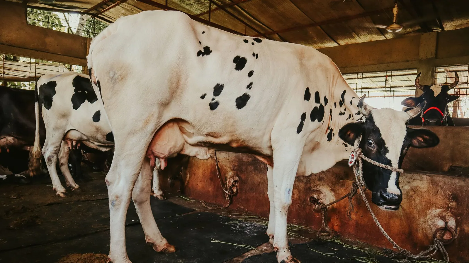
Gross pathological lesions in aborted fetuses include fibrinous pleuritis, peritonitis, and bronchopneumonia [18]. The placenta exhibits the characteristic reddish-brown, edematous appearance with a leathery texture and cotyledonary necrosis. Histologically, the chorionic epithelium shows extensive necrosis with infiltration of neutrophils and macrophages, and the presence of numerous intracytoplasmic coccobacilli within trophoblasts [19]. In adult cows, the udder may become infected, leading to intermittent shedding of B. abortus in milk, a critical route for both vertical transmission to calves and zoonotic exposure via unpasteurized dairy products [20]. In bulls, orchitis and epididymitis with seminal shedding contribute to venereal transmission [21].
3. Serological Surveillance: Core Assays and Diagnostic Principles
Given the chronic and often subclinical nature of B. abortus infection in non-pregnant cattle, serological testing is the backbone of surveillance and eradication programs worldwide [22]. The selection of appropriate serological tests depends on the desired sensitivity and specificity, the prevalence of infection, and the stage of the eradication program.
3.1 Rose Bengal Test (RBT)
The Rose Bengal test is a simple, rapid, and inexpensive agglutination assay used as a primary screening tool. It uses B. abortus whole-cell antigens stained with Rose Bengal dye at an acidic pH (3.65-3.85). This low pH enhances the agglutination of specific immunoglobulins, particularly IgG1, while reducing non-specific agglutination by IgM [23, 24].
Protocol and Interpretation: The test is performed by mixing equal volumes (typically 25 microliters) of serum and rose bengal antigen on a white tile or glass plate, followed by gentle rocking for 4 minutes at room temperature. Any visible agglutination (clumping) constitutes a positive result [25]. The test is highly sensitive (95-99%) for detecting infected animals, but specificity can be compromised in vaccinated populations due to cross-reacting antibodies [26]. False positives can also arise from infection with Yersinia enterocolitica O:9, Escherichia coli O:157, or Salmonella serotypes sharing O-polysaccharide epitopes [27].
Limitations: The RBT is qualitative, subjective in interpretation, and unsuitable for high-throughput automation. Its sensitivity declines in chronic, low-antibody-titer infections, and it cannot reliably distinguish infected from vaccinated animals [28].
3.2 Enzyme-Linked Immunosorbent Assay (ELISA)
ELISA offers objective, quantitative results suitable for large-scale automation. Two principal formats are used for bovine brucellosis: the indirect ELISA (iELISA) and the competitive ELISA (cELISA). The detection of anti-Brucella antibodies is performed using antigen-coated microtiter plates. For technical correlates in other livestock diagnostic platforms, see the article on Enzyme-Linked Immunosorbent Assay (ELISA) for Feline Leukemia Virus: p27 Antigen Detection and Diagnostic Interpretation, which illustrates analogous principles of antibody and antigen capture in a different pathogen system.
Indirect ELISA (iELISA): This format uses purified B. abortus lipopolysaccharide (LPS) or whole-cell sonicate as coating antigen. Bovine serum antibodies bind to the immobilized antigen and are detected using an anti-bovine IgG conjugate labeled with an enzyme (e.g., horseradish peroxidase) [29]. Optical density (OD) values are measured at 450 nm and compared to cut-off thresholds established using reference negative and positive sera. iELISA exhibits superior sensitivity compared to the RBT, particularly in low-titer and chronically infected animals, and is the preferred confirmatory test in many national programs [30].
Competitive ELISA (cELISA): This assay uses a monoclonal antibody specific for Brucella smooth LPS epitopes. The test serum competes with the monoclonal antibody for binding to the antigen. A reduction in signal indicates the presence of specific antibodies in the test serum. The cELISA is highly specific (greater than 99.5%) because the monoclonal antibody targets epitopes unique to Brucella smooth LPS, minimizing cross-reactivity with Y. enterocolitica O:9 [31]. This makes cELISA the preferred confirmatory test in vaccinated populations and in OIE/WHO reference laboratories [32].
Performance Comparison: Table 1 summarizes the comparative performance characteristics of the primary serological assays.
Table 1: Comparative Performance of Core Serological Assays for Bovine Brucellosis
| Feature | Rose Bengal Test (RBT) | Indirect ELISA (iELISA) | Competitive ELISA (cELISA) |
|---|---|---|---|
| Format | Agglutination | Plate-based enzymatic | Plate-based competitive |
| Sensitivity (infected cattle) | 95-98% | 97-99% | 95-98% |
| Specificity (brucellosis-free) | 97-99% | 98-99.5% | >99.5% |
| Specificity (vaccinated / cross-reacting) | 85-95% | 90-97% | 99-100% |
| Objective quantification | No | Yes (OD ratio) | Yes (% inhibition) |
| Throughput | Low (manual) | High (automated) | High (automated) |
| Distinction infected vs. vaccinated | Poor | Moderate | Excellent |
| Cost per test | Low | Moderate | Moderate-High |
3.3 Complement Fixation Test (CFT)
The complement fixation test was historically the reference confirmatory test but has been largely supplanted by ELISA due to its technical complexity, requirement for fresh complement, prozone effects, and anticomplementary activity in certain sera [33]. CFT remains a prescribed test for international trade by some regulatory frameworks [34].
3.4 Milk Ring Test (MRT)
The milk ring test is a rapid, low-cost screening test for bulk tank milk in dairy herds. It detects antibodies in milk fat globules using Brucella whole-cell antigen stained with hematoxylin [35]. The test is performed by mixing fresh milk with stained antigen and observing for the formation of a blue ring at the cream layer. MRT is sensitive for detecting infected herds with multiple lactating animals but is less sensitive in small herds or when using preserved milk. A positive MRT triggers individual animal testing with RBT and ELISA to identify reactors [36].
4. Serological Surveillance Strategies in Dairy Herds
Effective surveillance strategies must integrate testing frequency, test combinations, and interpretive algorithms to optimize sensitivity and specificity while managing the cost of false-positive reactions.
4.1 Screening and Confirmatory Testing Algorithm
A two-tier testing algorithm is standard practice. The first tier uses a high-sensitivity test (RBT or iELISA) to minimize false negatives. All positive or suspicious samples proceed to a second-tier confirmatory test (cELISA or a second iELISA) to maximize specificity [37]. This algorithm is illustrated in Figure 1.
flowchart TD
 A[Individual Serum Sample] --> B{"Tier 1: Screening Test"}
 B -->|RBT or iELISA: Negative| C[Report Negative - No Further Action]
 B -->|RBT or iELISA: Positive / Suspicious| D{"Tier 2: Confirmatory Test"}
 D -->|cELISA or iELISA: Negative| E[Report Negative - Consider False Positive]
 D -->|cELISA or iELISA: Positive| F[Report Positive - Infected Animal]
 D -->|cELISA or iELISA: Inconclusive| G[Repeat Sampling in 4-6 Weeks]
 G --> B
Figure 1. Two-tier serological testing algorithm for bovine brucellosis in dairy herds.
4.2 Herd-Level Surveillance for Dairy
For dairy herds, surveillance strategies differ based on herd infection status:
Brucellosis-Free Herds: Annual or biennial bulk tank milk testing using MRT or milk ELISA is recommended for continuous surveillance [38]. Individual animal testing may be conducted at the time of sale, introduction of new animals, or clinical suspicion.
Enzootic Herds / Eradication Phase: In known infected herds, whole-herd serological testing (RBT + cELISA on all animals over 12-24 months of age) is repeated at 30-90 day intervals. Test-positive animals are immediately isolated and culled. Vaccination with B. abortus strain 19 or RB51 may be used as an adjunct, but serological differentiation of infected from vaccinated animals (DIVA) is critical. The cELISA offers the best DIVA capability in RB51-vaccinated herds because RB51 lacks smooth LPS, so infected animals produce anti-smooth LPS antibodies detectable by cELISA [39].
4.3 Interpreting Serological Results in Context
Many factors influence serological test interpretation: the stage of gestation, vaccination history, presence of cross-reacting infections, and the immunological status of the animal (individual variability, pregnancy-associated immunosuppression). A single positive serological test alone does not always confirm active infection; however, in a herd with clinical abortion storms, positive serology is highly predictive of B. abortus infection [40]. The positive predictive value of a test decreases as disease prevalence decreases. In low-prevalence or brucellosis-free regions, confirmatory testing and epidemiologic investigation are mandatory before declaring an animal infected. The pathophysiological correlation between abortion storms and serological reactivity reinforces the understanding that an infected animal may seroconvert before, during, or after abortion events, so repeated testing of suspect animals is recommended [41].
5. Control Programs and Eradication Strategies
National bovine brucellosis control programs are built upon three pillars: test-and-slaughter, vaccination, and movement restrictions.
5.1 Vaccination
Vaccination reduces the incidence of abortion and decreases bacterial shedding, thereby lowering environmental contamination and transmission risk. Two vaccines are widely used:
Strain 19: A live, attenuated smooth strain of B. abortus. It is highly effective at preventing abortion when administered to heifers between 4-12 months of age. However, strain 19 induces persistent serological responses that interfere with standard serological tests, making the cELISA essential for DIVA [42]. Strain 19 can also cause abortion in pregnant animals and is pathogenic for humans.
RB51: A live, rough mutant strain lacking smooth LPS. It does not induce antibodies against smooth LPS, so vaccinated animals test negative on standard RBT and iELISA, but positive on the cELISA is indicative of field infection [43]. RB51 is safe for use in pregnant cattle and has lower human pathogenicity, making it the vaccine of choice in many eradication programs [44].
5.2 Test-and-Slaughter
In regions with low prevalence, the test-and-slaughter strategy aims to eliminate infected animals from the herd. It relies on highly sensitive and specific serological testing at regular intervals, combined with prompt culling of reactors. This approach is economically viable only when prevalence is low (typically less than 2%) and is often combined with strict biosecurity and movement controls [45].
5.3 Movement Restrictions and Biosecurity
Infected herds are placed under quarantine, restricting the movement of animals, milk (if unpasteurized), and semen. Biosecurity measures include closed herd management, testing of introduced animals, disinfection of calving areas, and proper disposal of aborted fetuses and placentas [46]. Control of wildlife reservoirs, particularly in areas where B. abortus circulates in bison or elk populations, is an additional challenge [47]. The intersection of cattle farming with wildlife interface presents analogous concerns to those described in Tick-Borne Parasites in White-Tailed Deer: Babesia and Theileria Prevalence, PCR-Based Surveillance, and Impact on Livestock Interface, emphasizing the importance of multi-species serological surveillance.
5.4 Eradication Programs
Successful eradication programs, such as those implemented in the United States, Canada, Australia, and many European countries, have relied on sustained application of the test-and-slaughter strategy combined with vaccination of replacement heifers. Surveillance at slaughter (testing blood samples from slaughtered cattle) provides an additional monitoring tool to detect residual infection [48]. Mathematical modeling has demonstrated that surveillance testing at routine intervals can achieve eradication within a defined time horizon when test sensitivity exceeds 95% and culling is immediate [49].
6. Emerging Diagnostic Technologies and Future Directions
While serology remains the cornerstone of surveillance, molecular diagnostic tools are increasingly employed for confirmation and genotyping. Real-time PCR targeting IS711, bcsp31, and per genes offers high sensitivity (detecting as few as 10-100 CFU) and can be applied to fetal stomach contents, placenta, milk, and vaginal swabs [50]. PCR is especially valuable for diagnosing abortion storms when serological results are equivocal or in early infections before seroconversion. For a discussion of molecular diagnostic principles in bacterial livestock diseases, refer to the article on Mycoplasma bovis in Feedlot Cattle.
The integration of PCR with serology in a multi-tier algorithm could further enhance surveillance sensitivity. Additionally, the development of recombinant antigens and synthetic peptide-based ELISAs may improve specificity and enable DIVA capability across both smooth and rough strain vaccinated populations. Whole genome sequencing of B. abortus field isolates provides high-resolution epidemiological data for outbreak tracing and source attribution, supporting targeted biosecurity interventions.
7. Conclusions
Brucellosis in cattle remains a major threat to reproductive efficiency and international trade. The pathophysiology of abortion storms is rooted in the unique metabolic and intracellular survival capabilities of B. abortus within the bovine placenta. Effective surveillance relies on a combination of sensitive screening tests (RBT and iELISA) and highly specific confirmatory assays (cELISA), deployed within a structured two-tier algorithm. For dairy herds, bulk tank milk testing and individual animal serology form the basis of ongoing monitoring. Control programs that incorporate vaccination with DIVA-compatible tests, test-and-slaughter policies, and strict movement controls have successfully eradicated brucellosis from many regions. Continued advances in molecular diagnostics and serological assay refinement will further strengthen global efforts to control this economically devastating zoonotic pathogen of cattle.
References
[1] Corbel MJ. Brucellosis: an overview. Emerg Infect Dis. 1997;3(2):213-221.
[2] Moreno E, Moriyón I. Brucella melitensis: a nasty bug with hidden credentials for virulence. Proc Natl Acad Sci USA. 2002;99(1):1-3.
[3] Keppie J, Williams AE, Witt K, Smith H. The role of erythritol in the tissue localization of the brucellae. Br J Exp Pathol. 1965;46:104-108.
[4] Petersen E, Rajashekara G, Sanakkayala N, Eskra L, Harms J, Splitter G. Erythritol triggers expression of metabolic and virulence genes in Brucella abortus. Infect Immun. 2013;81(11):4056-4064.
[5] Barbier T, Collard F, Zúñiga-Ripa A, et al. Erythritol feeds the pentose phosphate pathway via three new isomerases leading to D-erythrose-4-phosphate in Brucella. Proc Natl Acad Sci USA. 2014;111(49):17815-17820.
[6] McDermott JJ, Grace D, Zinsstag J. Economics of brucellosis impact and control in low-income countries. Rev Sci Tech. 2013;32(1):249-261.
[7] Singh BB, Dhand NK, Gill JP. Economic losses occurring due to brucellosis in Indian livestock populations. Prev Vet Med. 2015;119(3-4):211-215.
[8] Pizarro-Cerda J, Moreno E, Gorvel JP. Invasion and intracellular trafficking of Brucella abortus in nonphagocytic cells. Microbes Infect. 2000;2(7):829-835.
[9] Celli J, de Chastellier C, Franchini DM, Pizarro-Cerda J, Moreno E, Gorvel JP. Brucella evades macrophage killing via VirB-dependent sustained interactions with the endoplasmic reticulum. J Exp Med. 2003;198(4):545-556.
[10] Starr T, Ng TW, Wehrly TD, Knodler LA, Celli J. Brucella intracellular replication requires trafficking through the late endosomal/lysosomal compartment. Traffic. 2008;9(5):678-694.
[11] Boschiroli ML, Ouahrani-Bettache S, Foulongne V, et al. The Brucella suis virB operon is induced intracellularly in macrophages. Proc Natl Acad Sci USA. 2002;99(3):1544-1549.
[12] Smith H, Keppie J, Pearce JH, Fuller R, Williams AE. The chemical basis of the virulence of Brucella abortus. I. Isolation of B. abortus from bovine foetal tissue. Br J Exp Pathol. 1961;42:631-637.
[13] Anderson TD, Cheville NF, Meador VP. Pathogenesis of placentitis in the goat inoculated with Brucella abortus. II. Ultrastructural studies. Vet Pathol. 1986;23(2):229-236.
[14] Samartino LE, Enright FM. Pathogenesis of abortion of bovine brucellosis. Comp Immunol Microbiol Infect Dis. 1993;16(2):95-101.
[15] Giambartolomei GH, Delpino MV, Cahanovich ME, et al. Diminished production of T helper 1 cytokines correlates with increased levels of interleukin-10 in human brucellosis. Infect Immun. 2001;69(6):3671-3675.
[16] Crawford RP, Williams JD, Huber JD, Nicoletti PL. Detection of Brucella abortus in milk of infected cattle. Am J Vet Res. 1986;47(6):1280-1282.
[17] Olsen SC, Palmer MV. Advancement of knowledge of Brucella over the past 50 years. Vet Pathol. 2014;51(6):1076-1089.
[18] López A, Hitos F, Pérez A, Navarro-Fierro RR. Lung lesions in bovine fetuses aborted by Brucella abortus. Can J Comp Med. 1984;48(3):275-277.
[19] Meador VP, Deyoe BL. Intracellular localization of Brucella abortus in bovine placenta. Vet Pathol. 1989;26(5):408-415.
[20] Higgins JL, González-Juarrero M, Bowen RA. Evaluation of shedding of Brucella abortus in milk from infected cattle. J Am Vet Med Assoc. 2005;226(10):1685-1689.
[21] Olsen SC. Brucellosis in the bull. Vet Clin North Am Food Anim Pract. 1997;13(2):361-371.
[22] Godfroid J, Scholz HC, Barbier T, et al. Brucellosis at the animal/ecosystem/human interface at the beginning of the 21st century. Prev Vet Med. 2011;102(2):118-131.
[23] Blasco JM, Garin-Bastuji B, Marin CM, et al. Efficacy of different Rose Bengal and complement fixation antigens for the diagnosis of Brucella melitensis in sheep and goats. Vet Rec. 1994;134(16):415-420.
[24] Díaz R, Moriyón I. Laboratory techniques in the diagnosis of human brucellosis. In: Young EJ, Corbel MJ, eds. Brucellosis: Clinical and Laboratory Aspects. CRC Press; 1989:73-83.
[25] OIE Manual of Diagnostic Tests and Vaccines for Terrestrial Animals. Chapter 3.1.4 Bovine brucellosis. Paris: World Organisation for Animal Health; 2018.
[26] MacMillan AP. The serological diagnosis of bovine brucellosis. Vet Rec. 1990;127(7):167-170.
[27] Nielsen K, Smith P, Widdison J, et al. Serological relationship between cattle exposed to Brucella abortus, Yersinia enterocolitica O:9 and Escherichia coli O157:H7. Vet Microbiol. 2004;100(1-2):87-95.
[28] O'Leary S, Sheahan M, Sweeney T. Brucella abortus detection by PCR in milk from dairy cows. J Dairy Res. 2006;73(1):87-92.
[29] Nielsen K, Gall D, Jolley M, et al. A homogeneous fluorescence polarization assay for detection of antibody to Brucella abortus. J Immunol Methods. 1996;195(1-2):161-168.
[30] Godfroid J, Käsbohrer A. Brucellosis in the European Union and EFTA at the turn of the millennium. Vet Microbiol. 2002;90(1-4):43-58.
[31] Nielsen K, Smith P, Yu WL, et al. Competitive enzyme immunoassay for detection of antibodies to Brucella abortus and Brucella melitensis. J Vet Diagn Invest. 2005;17(1):26-31.
[32] Nielsen K, Smith P, Yu WL, et al. Validation of a competitive ELISA for detection of antibodies to Brucella abortus in cattle sera. J Vet Diagn Invest. 2005;17(2):145-150.
[33] Alton GG, Jones LM, Pietz DE. Laboratory Techniques in Brucellosis. 2nd ed. World Health Organization; 1975.
[34] Godfroid J, Cloeckaert A, Liautard JP, et al. From the discovery of the Malta fever's agent to the discovery of a marine mammal reservoir: brucellosis has continuously been a re-emerging zoonosis. Vet Res. 2005;36(3):313-326.
[35] Nicoletti P. The epidemiology of bovine brucellosis. Adv Vet Sci Comp Med. 1980;24:69-98.
[36] Milian G, Rojas X, León EA. Comparison of the Rose Bengal test and the milk ring test for detection of Brucella abortus in dairy cattle. J Vet Diagn Invest. 2011;23(4):791-793.
[37] Godfroid J, Nymo IH, Larsen AK, et al. A new era for brucellosis surveillance: the importance of laboratory confirmation. Rev Sci Tech. 2014;33(1):221-232.
[38] Veling J, van Zijderveld FG, van Maanen C, et al. Evaluation of three ELISA for the detection of antibodies to Brucella abortus in milk. Vet Microbiol. 2002;89(1):39-49.
[39] Jensen AE, Cheville NF, Thoen CO, MacMillan AP, Miller WG. Genomic fingerprinting and development of a PCR-based method to differentiate Brucella abortus strains. J Clin Microbiol. 1999;37(3):736-741.
[40] Palmer MV, Olsen SC, Cheville NF. Safety and immunogenicity of Brucella abortus strain RB51 vaccine in pregnant cattle. Am J Vet Res. 1997;58(5):472-477.
[41] Olsen SC. Recent developments in livestock and wildlife brucellosis vaccination. Rev Sci Tech. 2013;32(1):207-217.
[42] Stevens MG, Olsen SC, Cheville NF. Comparative analysis of immune responses in cattle vaccinated with Brucella abortus strain 19 or strain RB51. Vet Immunol Immunopathol. 1995;44(3-4):223-235.
[43] Schurig GG, Sriranganathan N, Corbel MJ. Brucellosis vaccines: past, present and future. Vet Microbiol. 2002;90(1-4):479-496.
[44] Olsen SC. Immune responses in cattle and elk following vaccination with Brucella abortus strain RB51. Vaccine. 2010;28(Suppl 5):F54-F59.
[45] Robinson A. Guidelines for coordinated human and animal brucellosis surveillance. FAO Animal Production and Health Paper 156. Food and Agriculture Organization; 2003.
[46] Ragan VE. The Animal and Plant Health Inspection Service's (APHIS) brucellosis eradication program in the United States. Vet Microbiol. 2002;90(1-4):11-18.
[47] Kamper MA, Cross PC, Bienen L, et al. Bison and elk brucellosis in Yellowstone National Park: modeling the impact of a vaccination program. Ecol Appl. 2016;26(2):479-493.
[48] Salman MD, Meyer ME. Epidemiology of bovine brucellosis in the United States. J Am Vet Med Assoc. 1984;184(8):975-979.
[49] O'Brien DJ, Schmitt SM, Fierke JS, et al. A simulation model of the dynamics of Brucella abortus in white-tailed deer. J Wildl Dis. 2002;38(4):747-758.
[50] Bricker BJ, Halling SM. Differentiation of Brucella abortus bv. 1, 2, and 4, Brucella melitensis, Brucella ovis, and Brucella suis bv. 1 by PCR. J Clin Microbiol. 1994;32(11):2660-2666.
Disclaimer: This article is for educational and informational purposes only. It is not intended to substitute for professional veterinary advice, diagnosis, treatment, or regulatory guidance. Always consult a licensed veterinarian or qualified specialist regarding animal health, disease diagnosis, and therapeutic decisions.