
Brachyspira hyodysenteriae and Swine Dysentery: Bloody Mucoid Diarrhea and Diagnosis
Swine dysentery (SD) is a highly contagious mucohemorrhagic colitis of pigs, caused predominantly by the anaerobic spirochete Brachyspira hyodysenteriae. The disease is characterized by severe, often bloody, mucoid diarrhea that leads to significant economic losses in grow-finish pig populations due to morbidity, mortality, reduced feed efficiency, and increased medication costs [1, 88]. This reference article provides a detailed examination of the pathogen, its mechanisms of disease, clinical and pathological presentation, and the full spectrum of diagnostic approaches, with a focus on molecular methodologies.
Pathogen Biology and Taxonomy
B. hyodysenteriae is a Gram-negative, anaerobic, helically shaped spirochete. It is classified within the genus Brachyspira, family Brachyspiraceae, order Spirochaetales. The organism is distinguished from other Brachyspira species by its strong beta-hemolysis on blood agar and its unique genetic and antigenic profile. Other pathogenic Brachyspira species that can cause SD-like disease include Brachyspira hampsonii and Brachyspira suanatina, though B. hyodysenteriae remains the most globally significant agent [2, 3, 91, 97]. The bacterium is highly fastidious, requiring specialized anaerobic culture conditions and enriched media for primary isolation.
The genome of B. hyodysenteriae is approximately 3.1 Mb in size, with a low G+C content of around 27% [4, 44]. Genomic analyses have revealed a core set of virulence-associated genes, including those encoding for hemolysins, lipoproteins, and enzymes involved in mucin degradation [5, 58, 73]. Multilocus sequence typing (MLST) and whole-genome sequencing have demonstrated substantial genetic diversity among global isolates, with specific sequence types (STs) showing distinct geographical distributions and antimicrobial resistance profiles [5, 6, 7, 50, 77]. For instance, ST196 has been reported as a predominant lineage in Switzerland, often associated with macrolide-lincosamide resistance. The population structure of B. hyodysenteriae is dynamic, with evidence for the emergence of weakly hemolytic but genetically distinct variants in Europe.
Pathogenesis and Host Interaction
The pathogenesis of SD is a complex, multifactorial process that begins with oral ingestion of the spirochete and its colonization of the large intestine, specifically the cecum and spiral colon [88, 93]. Colonization is mediated by adhesion to the colonic mucus layer and underlying epithelium. The bacterium exhibits species-specific avidity to colonic mucins, a process facilitated by recognition of sialic acid residues and glycosphingolipids on host cells [8, 45, 71]. Following adherence, B. hyodysenteriae invades the colonic crypts, where it proliferates in the mucus layer, causing disruption of the epithelial barrier [52, 93].
The primary virulence mechanisms involve the secretion of hemolysins and other cytotoxins that damage colonic epithelial cells, leading to inflammation, necrosis, and hemorrhage [58, 88]. In vivo transcriptomic profiling of the colonic mucosa during infection has revealed a profound host immune response, including the upregulation of pro-inflammatory cytokines such as IL-1alpha, IL-8, and IL-17, and the activation of p38 MAPK signaling pathways [9, 10]. This inflammatory cascade contributes to the impairment of electrolyte transport mechanisms. Specifically, infection downregulates the expression of key ion transporters, including the downregulated in adenoma (DRA; SLC26A3) and the sodium-hydrogen exchanger 3 (NHE3) [10, 63]. The decreased expression of DRA is driven by a p38-dependent IL-1alpha response, leading to a loss of electroneutral sodium chloride absorption and contributing to secretory diarrhea [10, 63].
B. hyodysenteriae infection also induces significant changes in the composition and function of the colonic microbiota. Dysbiosis is characterized by a reduction in beneficial bacteria, such as Lactobacillus and Prevotella, and an increase in potentially pathogenic taxa, which can exacerbate disease severity [11, 12, 13, 99]. The disease alters the colonic mucin environment; infection results in a reduction of Mucin 2 (MUC2) and an increase in Mucin 5AC (MUC5AC) expression, along with changes in glycosylation patterns [14, 52]. These mucin alterations impair the protective barrier function and may promote further bacterial adherence and invasion [14, 52]. Metabolomic analyses of the infected colon have demonstrated shifts in purine and amino acid metabolism, reflecting the host's metabolic response to cellular damage and inflammation [15].
Clinical Signs and Pathology
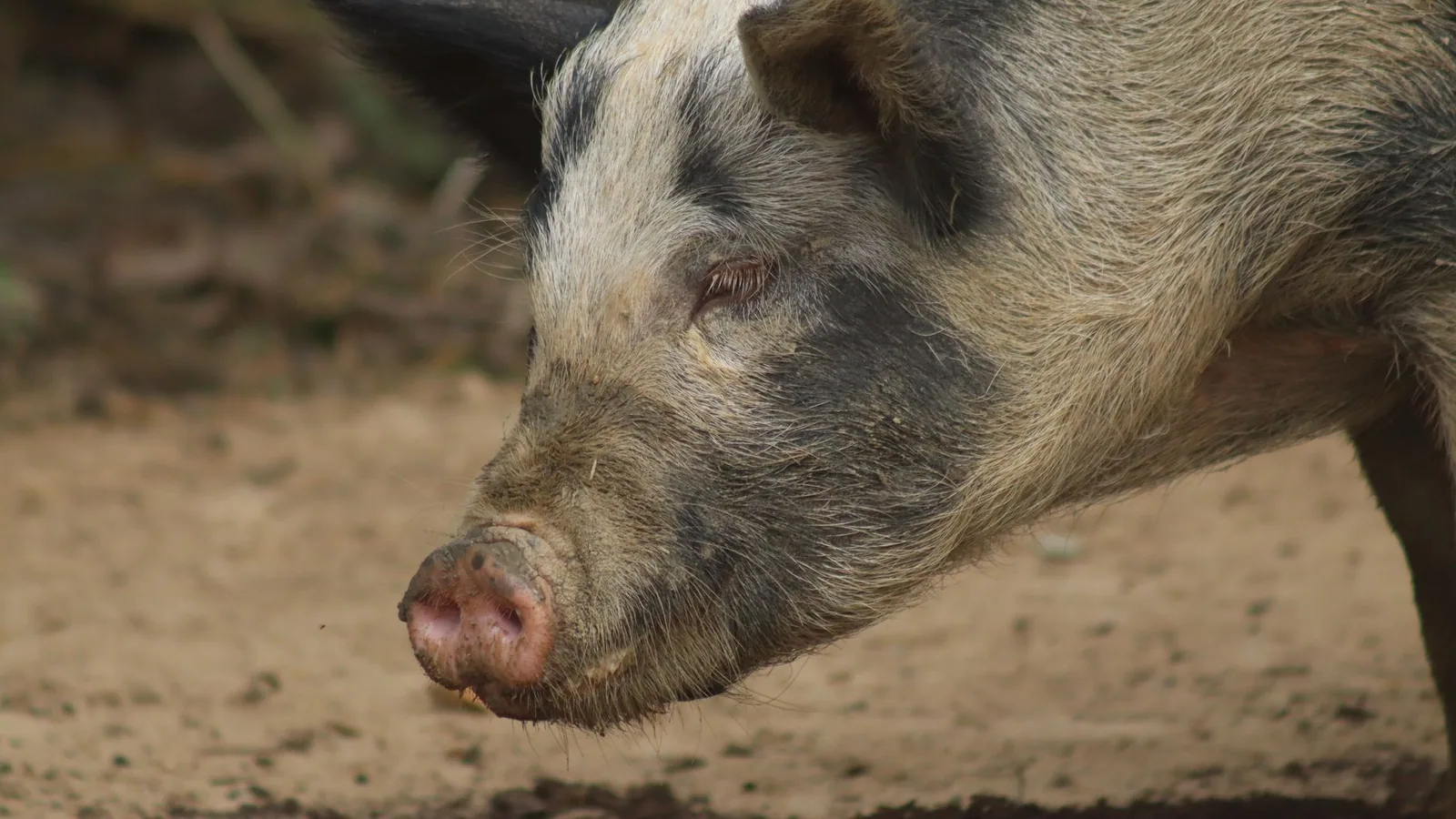
The incubation period for SD typically ranges from 4 to 14 days following oral exposure, but can be longer depending on the infective dose, strain virulence, and host susceptibility [16, 17, 91]. The hallmark clinical sign is the onset of mucoid diarrhea that progresses to contain fresh blood and mucus, giving the feces a characteristic red to dark brown, "bloody mucoid" appearance [18, 88, 93]. Affected pigs are often febrile, anorexic, and dehydrated, with poor growth rates and reduced feed intake [18, 1, 89]. In acute outbreaks, morbidity can approach 100%, with mortality rates varying from 10% to 50% if untreated.
Gross pathological lesions are confined to the large intestine [19, 88]. The cecum and colon exhibit serosal edema and a thickened, corrugated mucosal surface. The luminal contents are often blood-tinged and mucoid [19, 93]. The mucosa is covered by a fibrinous, blood-flecked pseudomembrane, and the underlying tissue is hyperemic and necrotic [19, 89]. Microscopically, lesions include superficial necrosis of the colonic epithelium, goblet cell depletion, crypt elongation with increased mitotic figures, and a mixed inflammatory cell infiltrate comprising neutrophils and lymphocytes in the lamina propria [19, 52, 88]. The spirochetes are most abundant within the crypt lumina, often forming a dense mat on the epithelial surface [52, 93].
Diagnosis
Accurate and timely diagnosis of SD is critical for implementing effective control and treatment measures. The diagnostic approach integrates clinical observation, necropsy findings, and a range of laboratory techniques, with molecular methods now serving as the gold standard. A multi-modal diagnostic strategy is recommended to differentiate SD from other causes of colitis in pigs, such as infection with Lawsonia intracellularis, Salmonella enterica, or Trichuris suis [20, 21, 49, 74].
Traditional Diagnostic Methods
Historically, diagnosis relied on the microscopic detection of spirochetes in Gram-stained fecal smears or colonic scrapings, coupled with anaerobic culture [62, 91]. The characteristic morphology of B. hyodysenteriae (large, helically coiled spirochetes) observed under dark-field or phase-contrast microscopy provides a preliminary indication. For culture, fecal or mucosal samples are plated on selective media (e.g., trypticase soy agar with 5% bovine blood and spectinomycin) and incubated under strict anaerobic conditions for 3 to 7 days. The identification of strong beta-hemolysis around colonies is a key phenotypic feature [58, 70]. However, culture is time-consuming, has low sensitivity, and can be confounded by overgrowth of other Brachyspira species or contaminants. Biochemical tests and older serological methods have largely been superseded by molecular techniques.
Molecular Diagnostics
Polymerase chain reaction (PCR) based assays, particularly real-time quantitative PCR (qPCR), are the preferred methods for direct detection of B. hyodysenteriae in feces, oral fluids, and tissues [22, 23, 24, 25, 89]. These assays typically target species-specific genes, most commonly the NADH oxidase (nox) gene [22, 24, 89]. qPCR offers high sensitivity and specificity, allowing for the detection of low levels of the pathogen even in the presence of a complex background of commensal bacteria [22].
Several multiplex qPCR panels have been developed for the simultaneous detection of B. hyodysenteriae alongside other major enteric pathogens. For example, triplex and quadruplex assays now exist for the concurrent detection of B. hyodysenteriae, L. intracellularis, and Clostridium perfringens [22], or for B. hyodysenteriae, L. intracellularis, and porcine epidemic diarrhea virus [25]. These multiplex formats are highly advantageous for routine diagnostic surveillance, as co-infections are common in herds with diarrheal disease [20, 21]. Oral fluid samples have also been validated as a practical, non-invasive sample type for herd-level monitoring using qPCR, providing a convenient tool for population screening [23].
Advanced Genomic Methods
Whole-genome sequencing (WGS) and long-read sequencing technologies are increasingly applied for detailed characterization of B. hyodysenteriae isolates [5, 26, 46]. WGS provides comprehensive data on the genetic determinants of antimicrobial resistance (AMR) and virulence, and facilitates high-resolution epidemiological tracking of strains [5, 26, 46]. The identification of specific AMR genes, such as those conferring resistance to pleuromutilins (e.g., tva(A)), macrolides, and lincosamides, is crucial for guiding antimicrobial therapy and understanding resistance dynamics within a population [69, 75, 80, 86]. Bioinformatics analysis of core genome single nucleotide polymorphisms (cgSNPs) allows for the differentiation of closely related sublineages, which is invaluable for outbreak investigations [5, 27].
Serological and Immunological Tests
Enzyme-linked immunosorbent assays (ELISAs) for the detection of antibodies against B. hyodysenteriae have been developed, but their use in routine diagnostics is less common compared to molecular methods, mainly due to variable sensitivity and the inability to distinguish current infection from past exposure. However, for large-scale herd surveillance or eradication program monitoring, serology can play a supporting role. The detection of fecal biomarkers such as calprotectin has been explored as a non-specific indicator of intestinal inflammation in SD, but its utility as a diagnostic tool is limited for pathogen-specific confirmation.
Diagnostic Decision Framework
The following Mermaid diagram outlines a recommended diagnostic workflow for a herd presenting with clinical signs consistent with swine dysentery.
flowchart TD
 A["Clinical Signs: Bloody Mucoid Diarrhea"] --> B{Collect Antemortem Samples}
 B --> C[Feces or Oral Fluids]
 B --> D[Rectal Swabs]

 C --> E["Perform Multiplex qPCR: B. hyodysenteriae, L. intracellularis, C. perfringens"]
 D --> E

 E --> F{qPCR Result}
 F -->|Positive for B. hyodysenteriae| G[Confirms Swine Dysentery]
 F -->|Negative for B. hyodysenteriae| H[Consider Other Etiologies]

 G --> I["Optional: Culture & AST"]
 G --> J["Optional: WGS for AMR & Epidemiology"]

 H --> K["Investigate for Other Pathogens: Salmonella, Brachyspira hampsonii, Brachyspira pilosicoli, Trichuris suis"]

 I --> L[Inform Treatment Protocol]
 J --> M[Refine Biosecurity & Surveillance]
Table 1 summarizes the principal laboratory diagnostic methods for B. hyodysenteriae.
| Method | Target | Principle | Sensitivity | Specificity | Turnaround Time | |:-, |:-, |:-, |:-, |:-, |:-, | | Anaerobic Culture | Viable spirochetes | Isolation on selective agar under anaerobic conditions | Low | Moderate | 5-10 days | | qPCR (nox gene) | B. hyodysenteriae DNA | Amplification of species-specific DNA sequences | High | High | 2-4 hours | | Multiplex qPCR | Multiple pathogens | Simultaneous detection of distinct DNA targets | High | High | 2-4 hours | | Whole-Genome Sequencing | Entire genome | High-throughput nucleotide sequencing of isolates | N/A (characterization) | N/A (characterization) | 1-3 days | | ELISA | Host antibodies | Detection of serum or fluid antibodies against B. hyodysenteriae antigens | Moderate | Moderate | 1-2 days |
AST: antimicrobial susceptibility testing. AMR: antimicrobial resistance.
Diagnostic Considerations
The presence of weakly hemolytic or atypical B. hyodysenteriae strains can complicate diagnosis, as these strains may be missed by culture-based methods relying on strong hemolysis [58, 70, 73]. Genotypic methods, such as qPCR targeting the nox gene, are essential for detecting such strains [22, 89]. The existence of other strongly hemolytic spirochetes, such as B. hampsonii and B. suanatina, requires that molecular assays be designed to differentiate these species to ensure an accurate etiological diagnosis [2, 3, 53, 91, 97].
Conclusion
Swine dysentery caused by Brachyspira hyodysenteriae remains a significant challenge for global swine production. The pathogenesis involves a complex interplay of bacterial adhesion, toxin production, host inflammatory responses, and disruption of electrolyte transport, leading to the characteristic bloody mucoid diarrhea. Diagnosis has evolved from traditional microscopic and culture-based methods toward highly sensitive and specific molecular techniques. Multiplex qPCR assays are now essential tools for rapid and accurate detection of the pathogen in clinical samples, while genomic methods provide detailed insights into strain diversity and antimicrobial resistance. A comprehensive diagnostic strategy, incorporating clinical, pathological, and molecular approaches, is paramount for effective disease management, control, and eradication programs.
References
[1] Lee GI, Skou Hedemann M, Borg Jensen B, et al. Influence of infection with Brachyspira hyodysenteriae on clinical expression, growth performance, and digestibility in growing pigs fed diets varying in type and level of fiber. J Anim Sci. 2022. URL: https://pubmed.ncbi.nlm.nih.gov/35255495/
[2] Cybulski P, Strutzberg-Minder K, Michalik E, et al. First molecular detection of Brachyspira hampsonii on pig farms in Poland. Acta Vet Hung. 2024. URL: https://pubmed.ncbi.nlm.nih.gov/38896488/
[3] Cybulski P, Strutzberg-Minder K, Michalik E, et al. First molecular detection of Brachyspira suanatina on pig farms in Poland. J Vet Res. 2023. URL: https://pubmed.ncbi.nlm.nih.gov/37786846/
[4] Rohde J, Jarek M, Goethe R. Complete genome sequences of Brachyspira hyodysenteriae strains B204 and JR80. Microbiol Resour Announc. 2025. URL: https://pubmed.ncbi.nlm.nih.gov/40243424/
[5] Hakimi M, Ye F, Saxena A, et al. Genomic insights into the population structure, antimicrobial resistance, and virulence of Brachyspira hyodysenteriae from diverse geographical regions. Microbiol Spectr. 2025. URL: https://pubmed.ncbi.nlm.nih.gov/40272172/
[6] Hakimi M, Ye F, Stinman CC, et al. Antimicrobial susceptibility of U.S. porcine Brachyspira isolates and genetic diversity of B. hyodysenteriae by multilocus sequence typing. J Vet Diagn Invest. 2024. URL: https://pubmed.ncbi.nlm.nih.gov/37968893/
[7] Sato JPH, Daniel AGS, Leal CAG, et al. Diversity and potential genetic relationships amongst Brazilian Brachyspira hyodysenteriae isolates from cases of swine dysentery. Vet Microbiol. 2022. URL: https://pubmed.ncbi.nlm.nih.gov/35176606/
[8] Quintana-Hayashi MP, Zalem D, Lindén S, et al. Porcine intestinal glycosphingolipids recognized by Brachyspira hyodysenteriae. Microb Pathog. 2023. URL: https://pubmed.ncbi.nlm.nih.gov/36581306/
[9] Pérez-Pérez L, de Toro M, Zaldívar-López S, et al. In vivo transcriptomic profiling of colonic mucosa during Brachyspira hyodysenteriae infection in pigs. Vet J. 2026. URL: https://pubmed.ncbi.nlm.nih.gov/41724371/
[10] Challa N, Enns CB, Keith BA, et al. Decreased expression of DRA (SLC26A3) by a p38-driven IL-1alpha response contributes to diarrheal disease following in vivo challenge with Brachyspira spp. Am J Physiol Gastrointest Liver Physiol. 2024. URL: https://pubmed.ncbi.nlm.nih.gov/39104321/
[11] Pérez-Pérez L, Arguello H, Cobo-Díaz JF, et al. From predisposition to recovery: field evidence of interactions between the gut microbiota and Brachyspira hyodysenteriae infection. Vet Res. 2026. URL: https://pubmed.ncbi.nlm.nih.gov/41618383/
[12] Pérez-Pérez L, Galisteo C, Sanjuán JMO, et al. Severity of Brachyspira hyodysenteriae colitis correlates to the changes observed in the microbiota composition and its associated functionality in the large intestine. Anim Microbiome. 2025. URL: https://pubmed.ncbi.nlm.nih.gov/41044758/
[13] Fodor CC, Fouhse J, Drouin D, et al. Colonic innate immune defenses and microbiota alterations in acute swine dysentery. Microb Pathog. 2022. URL: https://pubmed.ncbi.nlm.nih.gov/36371065/
[14] Lin SJ, Helm ET, Gabler NK, et al. Acute infection with Brachyspira hyodysenteriae affects mucin expression, glycosylation, and fecal MUC5AC. Front Cell Infect Microbiol. 2022. URL: https://pubmed.ncbi.nlm.nih.gov/36683692/
[15] Pérez-Pérez L, Galisteo C, Castillo-Peinado LLS, et al. Metabolomic signatures of colonic infection by Brachyspira hyodysenteriae. Vet Res. 2026. URL: https://pubmed.ncbi.nlm.nih.gov/42157352/
[16] Parra-Aguirre JC, Nosach R, Fernando C, et al. Improving the consistency of experimental swine dysentery inoculation strategies. Vet Res. 2023. URL: https://pubmed.ncbi.nlm.nih.gov/37328906/
[17] Parra-Aguirre J, Nosach R, Fernando C, et al. Experimental natural transmission (seeder pig) models for reproduction of swine dysentery. PLoS One. 2022. URL: https://pubmed.ncbi.nlm.nih.gov/36166423/
[18] Pérez-Pérez L, Carvajal A, Puente H, et al. New insights into swine dysentery: faecal shedding, macro and microscopic lesions and biomarkers in early and acute stages of Brachyspira hyodysenteriae infection. Porcine Health Manag. 2024. URL: https://pubmed.ncbi.nlm.nih.gov/38951921/
[19] Neis LZ, Rodrigues RO, Bertagnolli AC, et al. Visual and microscopic lesions of enteritis in slaughtered swine: pathogen identification and antimicrobial resistance implications. Vet Res Commun. 2026. URL: https://pubmed.ncbi.nlm.nih.gov/42043640/
[20] Chagas S, Jensen P, Paladino E, et al. Immune Response of Pigs Vaccinated Against Proliferative Enteropathy and Co-Infected with Lawsonia intracellularis and Brachyspira hyodysenteriae. Animals (Basel). 2025. URL: https://pubmed.ncbi.nlm.nih.gov/41514801/
[21] Daniel AGS, Pereira CER, Dorella F, et al. Synergic Effect of Brachyspira hyodysenteriae and Lawsonia intracellularis Coinfection: Anatomopathological and Microbiome Evaluation. Animals (Basel). 2023. URL: https://pubmed.ncbi.nlm.nih.gov/37627402/
[22] Wu W, Wang L, Xie R, et al. Development and application of a triplex real-time PCR method for the detection of Lawsonia intracellularis, Brachyspira hyodysenteriae, and Clostridium perfringens. Microbiol Spectr. 2025. URL: https://pubmed.ncbi.nlm.nih.gov/40366193/
[23] Eddicks M, Reiner G, Junker S, et al. Field study on the suitability of oral fluid samples for monitoring of Lawsonia intracellularis and Brachyspira hyodysenteriae by multiplex qPCR under field conditions. Porcine Health Manag. 2025. URL: https://pubmed.ncbi.nlm.nih.gov/39773664/
[24] Wang H, Sun Y, Chen J, et al. Development and application of a quadruplex TaqMan real-time fluorescence quantitative PCR assay for four porcine digestive pathogens. Front Cell Infect Microbiol. 2024. URL: https://pubmed.ncbi.nlm.nih.gov/39660284/
[25] Ren J, Li F, Yu X, et al. Development of a TaqMan-based multiplex real-time PCR for simultaneous detection of porcine epidemic diarrhea virus, Brachyspira hyodysenteriae, and Lawsonia intracellularis. Front Vet Sci. 2024. URL: https://pubmed.ncbi.nlm.nih.gov/39205809/
[26] Vereecke N, Botteldoorn N, Brossé C, et al. Predictive Power of Long-Read Whole-Genome Sequencing for Rapid Diagnostics of Multidrug-Resistant Brachyspira hyodysenteriae Strains. Microbiol Spectr. 2023. URL: https://pubmed.ncbi.nlm.nih.gov/36602320/
[28] Vangroenweghe FACJ. Successful Brachyspira hyodysenteriae Eradication Through a Combined Approach of a Zinc Chelate Treatment and Adapted Management Measures. Pathogens. 2025. URL: https://pubmed.ncbi.nlm.nih.gov/41598985/
[29] Gómez-Martínez S, Pérez-Pérez L, Ucero-Carrretón A, et al. Exploring the potential for competitive exclusion of commensal probiotic candidates against the insidious swine pathogen Brachyspira hyodysenteriae. Anim Microbiome. 2026. URL: https://pubmed.ncbi.nlm.nih.gov/41559772/
[30] Puentes R, Mirzadzare N, Hannawayya R, et al. A curcumin derivative metalloprotease inhibitor (CMC2.24) mitigates Brachyspira spp.-induced swine dysentery. Int Immunopharmacol. 2025. URL: https://pubmed.ncbi.nlm.nih.gov/40752109/
[31] Wallgren P. Control of swine dysentery at national level in Sweden. Acta Vet Scand. 2024. URL: https://pubmed.ncbi.nlm.nih.gov/39238024/
[32] De Lorenzi G, Gherpelli Y, Luppi A, et al. In vitro susceptibility of Brachyspira hyodysenteriae strains isolated in pigs in northern Italy between 2005 and 2022. Res Vet Sci. 2024. URL: https://pubmed.ncbi.nlm.nih.gov/38219471/
[33] Arnold M, Swam H, Crienen A, et al. Prevalence and risk factors of Brachyspira spp. in pig herds with a history of diarrhoea in six European countries. Prev Vet Med. 2023. URL: https://pubmed.ncbi.nlm.nih.gov/36774781/
[34] Vega C, Pérez-Pérez L, Argüello H, et al. In vitro evaluation of gentamicin activity against Spanish field isolates of Brachyspira hyodysenteriae. Porcine Health Manag. 2022. URL: https://pubmed.ncbi.nlm.nih.gov/36451249/
[35] de Groot N, Meneguzzi M, de Souza B, et al. In Vitro Screening of Non-Antibiotic Components to Mitigate Intestinal Lesions Caused by Brachyspira hyodysenteriae, Lawsonia intracellularis and Salmonella enterica Serovar Typhimurium. Animals (Basel). 2022. URL: https://pubmed.ncbi.nlm.nih.gov/36139216/
[36] Sato JPH, Daniel AGS, Pereira CER, et al. Experimental Infection of Pigs with a ST 245 Brachyspira hyodysenteriae Isolated from an Asymptomatic Pig in a Herd with No History of Swine Dysentery. Vet Sci. 2022. URL: https://pubmed.ncbi.nlm.nih.gov/35737338/
[37] Vidondo B, Cadetg RS, Nathues H, et al. Factors driving pig owners' motivation and satisfaction to perform eradications from Swine dysentery. Prev Vet Med. 2022. URL: https://pubmed.ncbi.nlm.nih.gov/35430446/
[38] Arnold M, Schmitt S, Collaud A, et al. Distribution, genetic heterogeneity, and antimicrobial susceptibility of Brachyspira pilosicoli in Swiss pig herds. Vet Microbiol. 2022. URL: https://pubmed.ncbi.nlm.nih.gov/35429815/
[39] Blunt R, Mellits K, Corona-Barrera E, et al. Carriage of Brachyspira hyodysenteriae on common insect vectors. Vet Microbiol. 2022. URL: https://pubmed.ncbi.nlm.nih.gov/35427991/
[40] EFSA Panel on Animal Health and Welfare (AHAW), Nielsen SS, Bicout DJ, et al. Assessment of listing and categorisation of animal diseases within the framework of the Animal Health Law (Regulation (EU) No 2016/429): antimicrobial-resistant Brachyspira hyodysenteriae in swine. EFSA J. 2022. URL: https://pubmed.ncbi.nlm.nih.gov/35317125/
[41] Giovagnoni G, Tugnoli B, Piva A, et al. Dual Antimicrobial Effect of Medium-Chain Fatty Acids against an Italian Multidrug Resistant Brachyspira hyodysenteriae Strain. Microorganisms. 2022. URL: https://pubmed.ncbi.nlm.nih.gov/35208756/
Disclaimer: This article is for educational and informational purposes only. It is not intended to substitute for professional veterinary advice, diagnosis, treatment, or regulatory guidance. Always consult a licensed veterinarian or qualified specialist regarding animal health, disease diagnosis, and therapeutic decisions.