
Bovine Tuberculosis: Diagnostic Tools and Wildlife Reservoirs
Introduction
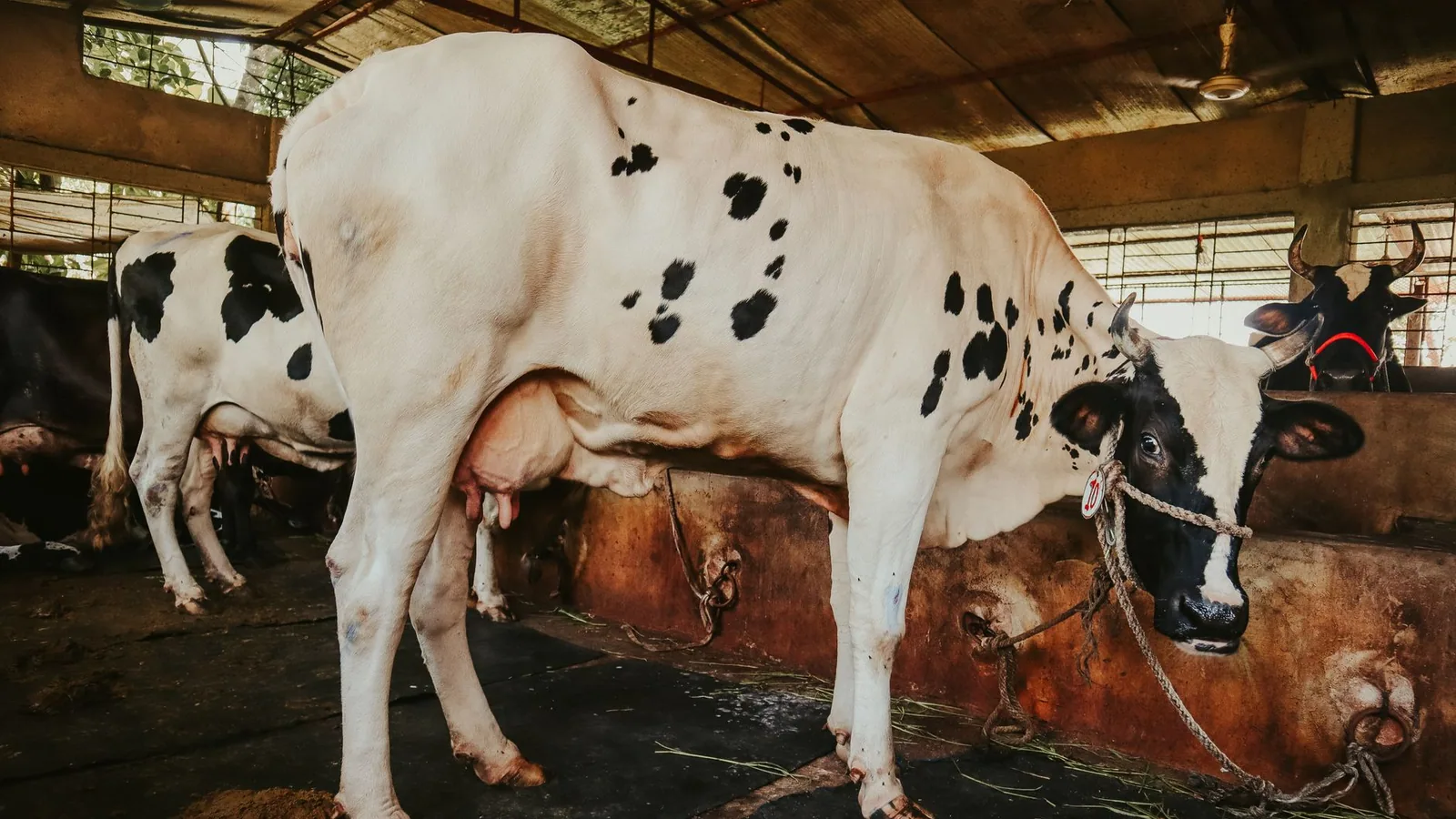
Bovine tuberculosis (bTB) is a chronic infectious disease of cattle caused primarily by Mycobacterium bovis, a member of the Mycobacterium tuberculosis complex. The disease imposes substantial economic burdens on livestock industries worldwide through production losses, trade restrictions, and the costs of surveillance and control programs [1, 2]. Despite decades of eradication efforts in many countries, bTB persists due to the presence of wildlife reservoirs that sustain transmission to domestic cattle [3, 4]. Effective control requires accurate diagnostic tools for ante-mortem detection in cattle and robust surveillance strategies in wildlife populations. This article provides a detailed technical review of the principal diagnostic methods for bTB in cattle, examines the role of key wildlife reservoirs, and discusses the integration of diagnostic data into test-and-cull eradication programs.
Pathogen and Host Range
Mycobacterium bovis is an acid-fast, aerobic, slow-growing bacillus with a lipid-rich cell wall that confers resistance to environmental stressors and host immune defenses [5]. The pathogen primarily causes granulomatous lesions in the respiratory tract of cattle, but can disseminate to lymph nodes, mammary tissue, and other organs [6]. Transmission occurs mainly via aerosolized respiratory secretions, although ingestion of contaminated feed or water can also lead to infection [7]. The host range of M. bovis is exceptionally broad, encompassing domestic livestock (cattle, goats, sheep, pigs), companion animals (dogs, cats), and numerous wildlife species [8]. This wide host tropism complicates eradication because infected wildlife can act as maintenance hosts, reintroducing the pathogen into cleared cattle herds [9].
Diagnostic Tools in Cattle
Ante-mortem diagnosis of bTB in cattle relies on immunological and molecular methods. The two primary immunodiagnostic tests are the tuberculin skin test (TST) and the interferon-gamma (IFN-γ) release assay. Molecular detection via polymerase chain reaction (PCR) is used primarily for confirmatory testing on tissue samples or as a screening tool in certain contexts.
Tuberculin Skin Test (TST)
The TST, also known as the caudal fold test (CFT) or the comparative cervical test (CCT), measures delayed-type hypersensitivity to purified protein derivative (PPD) from M. bovis [10]. A small volume of bovine PPD is injected intradermally, and the increase in skin thickness at the injection site is measured 72 hours later. The CCT uses both bovine and avian PPD to differentiate infection with M. bovis from sensitization due to environmental mycobacteria [11]. The sensitivity of the TST ranges from 68% to 95% depending on the cutoff threshold and the population tested, while specificity is generally high (96% to 99%) in unvaccinated herds [12, 13]. Factors that reduce test performance include desensitization due to repeated testing, concurrent immunosuppression, and early-stage infection before cell-mediated immunity develops [14].
Interferon-Gamma (IFN-γ) Release Assay
The IFN-γ assay (e.g., Bovigam platform) is a whole-blood test that measures the release of IFN-γ from sensitized T lymphocytes after stimulation with M. bovis antigens [15]. Blood samples are incubated with bovine and avian PPD or with specific antigens such as ESAT-6 and CFP-10, which are absent from most environmental mycobacteria and from the BCG vaccine strain [16]. The concentration of IFN-γ in the plasma supernatant is quantified using an enzyme-linked immunosorbent assay (ELISA). The IFN-γ assay has a sensitivity of 81% to 96% and a specificity of 94% to 99% [17, 18]. It is often used as a confirmatory test following a positive TST, or as a parallel test to increase overall detection sensitivity in high-prevalence herds [19]. The assay requires careful sample handling: blood must be processed within 8 to 12 hours of collection, and the stimulation step must be performed under sterile conditions to avoid false-positive results [20].
Polymerase Chain Reaction (PCR)
PCR-based methods detect M. bovis DNA directly from clinical samples such as nasal swabs, bronchoalveolar lavage fluid, milk, or tissue biopsies [21]. Real-time PCR targeting the insertion sequence IS6110 or the mpb70 gene offers high sensitivity (85% to 98%) and specificity (98% to 100%) on tissue samples [22, 23]. However, sensitivity on antemortem samples like nasal swabs is lower (40% to 70%) due to intermittent shedding and low bacterial loads [24]. Multiplex PCR panels can differentiate M. bovis from other members of the M. tuberculosis complex and from nontuberculous mycobacteria [25]. PCR is also used for genotyping to trace transmission chains and identify wildlife sources [26].
Comparative Performance of Diagnostic Tests
The following table summarizes the key characteristics of the three main diagnostic modalities for bTB in cattle.
| Test | Target | Sensitivity (range) | Specificity (range) | Turnaround Time | Advantages | Limitations |
|---|---|---|---|---|---|---|
| Tuberculin Skin Test | Cell-mediated immunity (DTH) | 68% - 95% | 96% - 99% | 72 hours | Low cost, field-deployable, no laboratory required | Operator variability, desensitization with repeated use, requires two visits |
| IFN-γ Release Assay | Cell-mediated immunity (IFN-γ release) | 81% - 96% | 94% - 99% | 24 - 48 hours | Higher sensitivity than TST, can use specific antigens to improve specificity | Requires laboratory processing, short sample stability, higher cost |
| Real-Time PCR | M. bovis DNA | 85% - 98% (tissue) | 98% - 100% | 4 - 6 hours | Rapid, definitive identification, genotyping capability | Lower sensitivity on antemortem samples, requires specialized equipment |
Diagnostic Workflow
The typical diagnostic algorithm for bTB in cattle involves initial screening with the TST, followed by confirmatory testing with the IFN-γ assay or PCR on suspect animals. In eradication programs, parallel testing (both TST and IFN-γ) is sometimes used to maximize sensitivity. The Mermaid diagram below illustrates a common decision tree.
graph TD
 A[Herd Screening] --> B["Caudal Fold Test (CFT)"]
 B -->|Negative| C[No further action]
 B -->|Positive or Inconclusive| D["Comparative Cervical Test (CCT)"]
 D -->|Negative| C
 D -->|Positive| E[IFN-γ Assay]
 E -->|Negative| F[Re-test in 60 days]
 E -->|Positive| G[Slaughter and confirmatory PCR / Culture]
 G --> H[Lesion detection at necropsy]
 H --> I[PCR positive] --> J[Confirmed bTB case]
 H --> K[No lesions] --> L[PCR negative] --> M[Re-evaluate herd status]
Wildlife Reservoirs
Wildlife species that can maintain M. bovis infection independently of cattle are termed maintenance hosts. The two most important wildlife reservoirs for bTB in Europe and North America are the Eurasian badger (Meles meles) and white-tailed deer (Odocoileus virginianus), respectively [27, 28]. Other species, such as wild boar (Sus scrofa), brushtail possum (Trichosurus vulpecula), and African buffalo (Syncerus caffer), play significant roles in specific regions [29, 30].
Eurasian Badger (Meles meles)
In the United Kingdom and Ireland, the Eurasian badger is the primary wildlife reservoir of M. bovis [31]. Badgers become infected through social contact within their setts and can excrete the bacterium in urine, feces, and respiratory secretions [32]. Transmission to cattle occurs when badgers forage in cattle pastures, contaminating grass or water sources [33]. Badger culling has been implemented as a control measure, but its effectiveness is debated due to perturbation effects that can increase disease spread [34]. Vaccination of badgers with an oral BCG vaccine is an alternative strategy that reduces both individual susceptibility and environmental shedding [35]. Diagnostic surveillance in badgers relies on culture of clinical samples (tracheal aspirates, urine, feces) and PCR, as well as serological tests such as the BrockTB Stat-Pak [36].
White-Tailed Deer (Odocoileus virginianus)
In Michigan, USA, and parts of Canada, white-tailed deer are a maintenance host for M. bovis [37]. Deer become infected through direct contact or ingestion of contaminated feed, particularly at supplemental feeding sites that concentrate animals [38]. The bacterium is shed in respiratory secretions, saliva, and feces. Transmission to cattle occurs when deer enter cattle pastures or share water sources [39]. Control measures include banning supplemental feeding, reducing deer density through hunting, and testing hunter-harvested deer using culture and PCR [40]. Oral BCG vaccination of deer is under investigation but not yet widely deployed [41].
Other Wildlife Reservoirs
Wild boar are important reservoirs in the Iberian Peninsula and parts of central Europe [42]. They exhibit high prevalence and can transmit M. bovis to cattle through shared habitats. Brushtail possums are the key reservoir in New Zealand, where they cause significant spillover into cattle and deer herds [43]. African buffalo maintain bTB in southern Africa and pose a threat to livestock and endangered wildlife species [44]. In each case, diagnostic tools adapted for wildlife (e.g., serological tests, PCR on tissue samples) are essential for surveillance and management.
Control Strategies and Eradication Programs
Eradication of bTB requires a multi-pronged approach that combines test-and-cull in cattle with wildlife management.
Test-and-Cull in Cattle
Test-and-cull programs involve regular herd testing using TST and/or IFN-γ assays, followed by slaughter of reactor animals [45]. Compensation is typically paid to farmers to encourage participation. The success of test-and-cull depends on test sensitivity, the frequency of testing, and the removal of all infected animals before they can transmit the disease. In regions with wildlife reservoirs, test-and-cull alone is insufficient because infected wildlife continually reintroduce the pathogen [46].
Wildlife Management
Wildlife management strategies include population reduction (culling), vaccination, and biosecurity measures to reduce contact between wildlife and cattle [47]. Culling of badgers in the UK has been controversial due to ethical concerns and variable efficacy. Vaccination of badgers with oral BCG is now being implemented in some areas. For deer, reducing population density and eliminating artificial feeding are key interventions. Fencing and water trough management can also reduce wildlife-cattle interactions [48].
Vaccination of Cattle
BCG vaccination of cattle has been shown to reduce the severity of disease and bacterial shedding, but it does not prevent infection entirely [49]. A major drawback is that BCG-vaccinated cattle become sensitized to PPD, causing false-positive TST results. The development of DIVA (Differentiating Infected from Vaccinated Animals) tests using specific antigens such as ESAT-6 and CFP-10 allows vaccination to be used in conjunction with immunodiagnostics. However, BCG is not widely used in eradication programs due to concerns about incomplete protection and trade implications.
Challenges and Future Directions
Several challenges impede bTB eradication. The long incubation period and subclinical nature of infection mean that many infected animals are not detected by current tests. The presence of wildlife reservoirs requires coordinated management across large geographic areas. Antimicrobial resistance in M. bovis is rare but has been reported, complicating treatment of valuable animals. Future directions include the development of more sensitive point-of-care tests, such as lateral flow assays for IFN-γ or antigen detection, and the use of genomic epidemiology to trace transmission networks. Computational models integrating diagnostic data, movement records, and wildlife ecology can optimize surveillance and intervention strategies.
Conclusion
Bovine tuberculosis remains a persistent challenge for livestock health and productivity. Accurate diagnosis in cattle relies on the tuberculin skin test, interferon-gamma assays, and PCR, each with distinct strengths and limitations. Wildlife reservoirs, particularly badgers and deer, sustain the pathogen in the environment and necessitate integrated control strategies that combine test-and-cull, wildlife management, and vaccination. Continued research into improved diagnostics and epidemiological modeling is essential for the eventual eradication of this disease.
References
[1] Cousins DV. Mycobacterium bovis infection and control in domestic livestock. Rev Sci Tech. 2001;20(1):71-85.
[2] Pollock JM, Neill SD. Mycobacterium bovis infection and tuberculosis in cattle. Vet J. 2002;163(2):115-127.
[3] Corner LAL. The role of wild animal populations in the epidemiology of tuberculosis in domestic animals: how to assess the risk. Vet Microbiol. 2006;112(2-4):303-312.
[4] Delahay RJ, Smith GC, Barlow AM, et al. Bovine tuberculosis infection in wild mammals in the South-West of England: a survey of prevalence and a semi-quantitative assessment of the relative risks to cattle. Vet J. 2007;173(2):287-301.
[5] Barry CE, Boshoff HI, Dartois V, et al. The spectrum of latent tuberculosis: rethinking the biology and intervention strategies. Nat Rev Microbiol. 2009;7(12):845-855.
[6] Cassidy JP. The pathogenesis and pathology of bovine tuberculosis with insights from studies of tuberculosis in humans and laboratory animal models. Vet Microbiol. 2006;112(2-4):151-161.
[7] Menzies FD, Neill SD. Cattle-to-cattle transmission of bovine tuberculosis. Vet J. 2000;160(2):92-106.
[8] O'Reilly LM, Daborn CJ. The epidemiology of Mycobacterium bovis infections in animals and man: a review. Tuber Lung Dis. 1995;76(Suppl 1):1-46.
[9] Naranjo V, Gortazar C, Vicente J, de la Fuente J. Evidence of the role of European wild boar as a reservoir of Mycobacterium tuberculosis complex. Vet Microbiol. 2008;127(1-2):1-9.
[10] Monaghan ML, Doherty ML, Collins JD, Kazda JF, Quinn PJ. The tuberculin test. Vet Microbiol. 1994;40(1-2):111-124.
[11] de la Rua-Domenech R, Goodchild AT, Vordermeier HM, Hewinson RG, Christiansen KH, Clifton-Hadley RS. Ante mortem diagnosis of tuberculosis in cattle: a review of the tuberculin tests, gamma-interferon assay and other ancillary diagnostic techniques. Res Vet Sci. 2006;81(2):190-210.
[12] Wood PR, Jones SL. BOVIGAM: an in vitro cellular diagnostic test for bovine tuberculosis. Tuberculosis (Edinb). 2001;81(1-2):147-155.
[13] Buddle BM, Livingstone PG, de Lisle GW. Advances in ante-mortem diagnosis of tuberculosis in cattle. N Z Vet J. 2009;57(4):173-180.
[14] Coad M, Clifford D, Rhodes SG, Hewinson RG, Vordermeier HM, Whelan AO. Repeat tuberculin skin testing leads to desensitisation in naturally infected tuberculous cattle which is associated with elevated interleukin-10 and decreased interleukin-1 beta responses. Vet Res. 2010;41(2):14.
[15] Rothel JS, Jones SL, Corner LA, Cox JC, Wood PR. A sandwich enzyme immunoassay for bovine interferon-gamma and its use for the detection of tuberculosis in cattle. Aust Vet J. 1990;67(4):134-137.
[16] Vordermeier HM, Whelan A, Cockle PJ, Farrant L, Palmer N, Hewinson RG. Use of synthetic peptides derived from the antigens ESAT-6 and CFP-10 for differential diagnosis of bovine tuberculosis in cattle. Clin Diagn Lab Immunol. 2001;8(3):571-578.
[17] Gormley E, Doyle MB, Fitzsimons T, McGill K, Collins JD. Identification of risk factors associated with Mycobacterium bovis infection in cattle in Ireland. Vet Rec. 2009;164(15):457-461.
[18] Schiller I, Oesch B, Vordermeier HM, et al. Bovine tuberculosis: a review of current and emerging diagnostic techniques in view of their relevance for disease control and eradication. Transbound Emerg Dis. 2010;57(4):205-220.
[19] Lahuerta-Marin A, Gallagher M, McBride S, et al. Should they stay or should they go? Relative sensitivity of the single intradermal comparative tuberculin test and the gamma-interferon test for detecting bovine tuberculosis in cattle. Vet Rec. 2018;182(10):287.
[20] Ryan TJ, Buddle BM, de Lisle GW. An evaluation of the gamma interferon test for detecting bovine tuberculosis in cattle 8 to 28 days after tuberculin skin testing. Res Vet Sci. 2000;69(1):57-61.
[21] Taylor MJ, Hughes MS, Skuce RA, Neill SD. Detection of Mycobacterium bovis in bovine clinical specimens using PCR and the polymerase chain reaction. Vet Microbiol. 2001;80(3):279-287.
[22] Thacker TC, Harris B, Palmer MV, Waters WR. Improved specificity for detection of Mycobacterium bovis in fresh tissues using IS6110 real-time PCR. BMC Vet Res. 2011;7:36.
[23] Araujo CP, Osorio AL, Jorge KS, et al. Detection of Mycobacterium bovis in bovine tissue samples by real-time PCR. Vet Microbiol. 2014;173(3-4):286-291.
[24] Courcoul A, Moyen JL, Brugère L, et al. Estimation of sensitivity and specificity of bacteriology, histopathology and PCR for the confirmatory diagnosis of bovine tuberculosis using latent class analysis. Vet Microbiol. 2014;171(1-2):127-135.
[25] Reddington K, O'Grady J, Dorai-Raj S, Niemann S, van Soolingen D, Barry T. A novel multiplex real-time PCR for the identification of mycobacteria associated with zoonotic tuberculosis. PLoS One. 2011;6(8):e23481.
[26] Biek R, O'Hare A, Wright D, et al. Whole genome sequencing reveals local transmission patterns of Mycobacterium bovis in sympatric cattle and badger populations. PLoS Pathog. 2012;8(11):e1003008.
[27] Krebs JR, Anderson R, Clutton-Brock T, et al. Bovine tuberculosis in cattle and badgers. London: Ministry of Agriculture, Fisheries and Food; 1997.
[28] Schmitt SM, Fitzgerald SD, Cooley TM, et al. Bovine tuberculosis in free-ranging white-tailed deer from Michigan. J Wildl Dis. 1997;33(4):749-758.
[29] Gortazar C, Ferroglio E, Hofle U, Frolich K, Vicente J. Diseases shared between wildlife and livestock: a European perspective. Eur J Wildl Res. 2007;53(4):241-256.
[30] De Garine-Wichatitsky M, Caron A, Kock R, et al. A review of bovine tuberculosis at the wildlife-livestock-human interface in sub-Saharan Africa. Epidemiol Infect. 2013;141(7):1342-1356.
[31] Cheeseman CL, Wilesmith JW, Stuart FA. Tuberculosis: the disease and its epidemiology in the badger, a review. Epidemiol Infect. 1989;103(1):113-125.
[32] Corner LAL, Murphy D, Gormley E. Mycobacterium bovis infection in the Eurasian badger (Meles meles): the disease, pathogenesis, epidemiology and control. J Comp Pathol. 2011;144(1):1-24.
[33] Garnett BT, Delahay RJ, Roper TJ. Use of cattle farm resources by badgers (Meles meles) and risk of bovine tuberculosis (Mycobacterium bovis) transmission to cattle. Proc Biol Sci. 2002;269(1499):1487-1491.
[34] Donnelly CA, Woodroffe R, Cox DR, et al. Positive and negative effects of widespread badger culling on tuberculosis in cattle. Nature. 2006;439(7078):843-846.
[35] Chambers MA, Rogers F, Delahay RJ, et al. Bacillus Calmette-Guerin vaccination reduces the severity and progression of tuberculosis in badgers. Proc Biol Sci. 2011;278(1713):1913-1920.
[36] Greenwald R, Esfandiari J, Lesellier S, et al. Improved serodetection of Mycobacterium bovis infection in badgers (Meles meles) using multiantigen test formats. J Clin Microbiol. 2003;41(7):3160-3165.
[37] O'Brien DJ, Schmitt SM, Fitzgerald SD, Berry DE, Hickling GJ. Managing the wildlife reservoir of Mycobacterium bovis: the Michigan, USA, experience. Vet Microbiol. 2006;112(2-4):313-323.
[38] Palmer MV, Waters WR, Whipple DL. Shared feed as a means of deer-to-deer transmission of Mycobacterium bovis. J Wildl Dis. 2004;40(1):87-91.
[39] Kaneene JB, Bruning-Fann CS, Granger LM, Miller R, Porter-Spalding BA. Environmental and farm management factors associated with tuberculosis on cattle farms in northeastern Michigan. J Am Vet Med Assoc. 2002;221(6):837-842.
[40] O'Brien DJ, Schmitt SM, Fitzgerald SD, et al. Epidemiology of Mycobacterium bovis in free-ranging white-tailed deer, Michigan, USA, 1995-2000. Prev Vet Med. 2002;54(1):47-63.
[41] Nol P, Palmer MV, Waters WR, et al. Efficacy of oral and parenteral routes of Mycobacterium bovis bacille Calmette-Guerin vaccination against experimental bovine tuberculosis in white-tailed deer (Odocoileus virginianus). Vet Immunol Immunopathol. 2008;124(3-4):235-243.
[42] Gortazar C, Torres MJ, Vicente J, et al. Bovine tuberculosis in Donana Biosphere Reserve: the role of wild ungulates as disease reservoirs in the last Iberian lynx strongholds. PLoS One. 2008;3(7):e2776.
[43] Coleman JD, Cooke MM. Mycobacterium bovis infection in wildlife in New Zealand. Tuberculosis (Edinb). 2001;81(3):191-202.
[44] De Vos V, Bengis RG, Kriek NP, et al. The epidemiology of tuberculosis in free-ranging African buffalo (Syncerus caffer) in the Kruger National Park, South Africa. Onderstepoort J Vet Res. 2001;68(2):119-130.
[45] More SJ, Good M. The tuberculosis eradication programme in Ireland: a review of scientific and policy advances since 1988. Vet Microbiol. 2006;112(2-4):239-251.
[46] Godfray HC, Donnelly CA, Kao RR, et al. A restatement of the natural science evidence base relevant to the control of bovine tuberculosis in Great Britain. Proc Biol Sci. 2013;280(1768):20131634.
[47] Carter SP, Chambers MA, Rushton SP, et al. BCG vaccination reduces risk of tuberculosis infection in vaccinated badgers and unvaccinated badger cubs. PLoS One. 2012;7(12):e49833.
[48] Ward AI, Judge J, Delahay RJ. Farm husbandry and badger behaviour: opportunities to manage badger to cattle transmission of Mycobacterium bovis? Prev Vet Med. 2010;93(1):2-10.
[49] Buddle BM, Wedlock DN, Denis M, Skinner MA. Identification of immune response correlates for protection against bovine tuberculosis. Vet Immunol Immunopathol. 2005;108(1-2):45-51.
Disclaimer: This article is for educational and informational purposes only. It is not intended to substitute for professional veterinary advice, diagnosis, treatment, or regulatory guidance. Always consult a licensed veterinarian or qualified specialist regarding animal health, disease diagnosis, and therapeutic decisions.