Yersinia ruckeri (Enteric Redmouth): Etiology, Pathogenesis, and Diagnostic Advances
By Dr. Zubair Khalid, DVM, MS, PhD ·
Key Takeaways
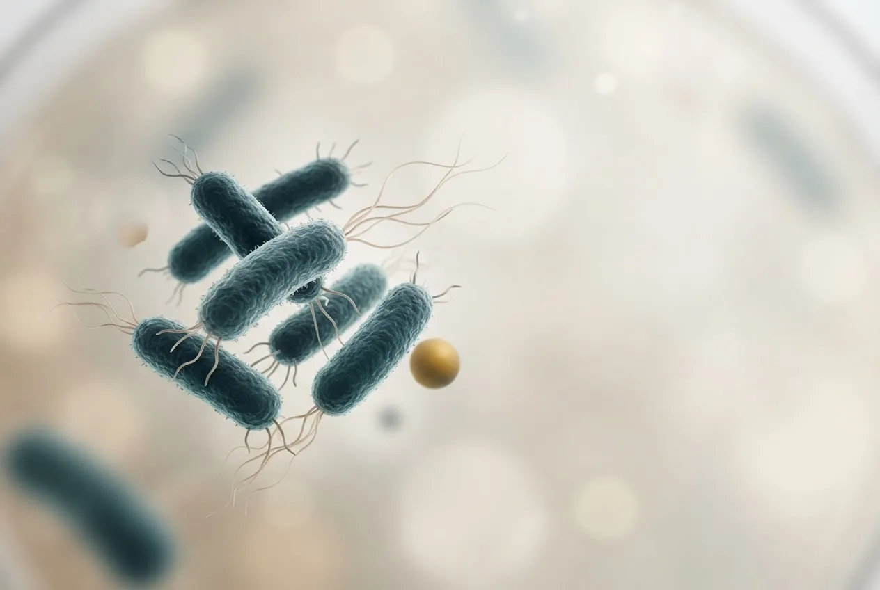
- Yersinia ruckeri is a Gram-negative enterobacterium causing enteric redmouth disease (ERM), a hemorrhagic septicemia with high mortality (30-80%) in salmonid aquaculture, characterized by oral hemorrhages, exophthalmia, and systemic inflammation.
- Virulence is mediated by flagellin C (FliC) interacting with host TLR5, a conserved Ysa type III secretion system (T3SS) regulated by environmental cues, and the ruckerbactin siderophore system for iron acquisition.
- Diagnostic advancements include rapid molecular methods like real-time qPCR targeting gyrA or glnA, field-deployable LAMP assays with high sensitivity, and CRISPR/Cas13a systems for specific nucleic acid detection.
- Host immune responses involve organ-specific transcriptomic shifts in mucosal tissues (tongue, skin, gills, olfactory organ), activation of inflammatory pathways including NLRP3 inflammasome, and local B cell responses.
- Disease management strategies encompass vaccination, dietary immunostimulants (e.g., ginseng polysaccharide, Agaricus bisporus polysaccharides), probiotics, and environmental control, with ongoing surveillance for antimicrobial resistance.
- Pathotyping via MLVA and pan-genome analysis identifies highly virulent lineages (MLVA clonal complexes 1 and 2) and highlights the evolutionary significance of the yrIlm locus, with modern isolates often possessing multiple copies.
Introduction
Yersinia ruckeri is the gram-negative enterobacterium responsible for enteric redmouth disease (ERM), also known as yersiniosis, a hemorrhagic septicemia that causes significant economic losses in global salmonid aquaculture [<a href="#ref-1">1</a>]. The pathogen infects a range of fish species, including rainbow trout (Oncorhynchus mykiss), Atlantic salmon (Salmo salar), channel catfish (Ictalurus punctatus), Chinese sturgeon (Acipenser sinensis), and crucian carp (Carassius carassius) [<a href="#ref-2">2</a>, <a href="#ref-3">3</a>, <a href="#ref-4">4</a>]. The disease is characterized by bilateral petechial hemorrhages of the oral cavity, exophthalmia, darkening of the skin, and systemic inflammation. Outbreaks can result in cumulative mortality rates ranging from 30% to over 80% depending on host age, water temperature, and strain virulence. The bacterium persists in aquatic environments and can survive in biofilms on abiotic surfaces, contributing to recurrent infections in aquaculture facilities [<a href="#ref-5">5</a>].
Taxonomy and Genomic Architecture
Y. ruckeri belongs to the family Enterobacteriaceae and is phylogenetically distinct from the mammalian pathogenic yersiniae such as Y. pestis and Y. enterocolitica. Pan-genomic surveys of the species have revealed a bipartite genome structure comprising a conserved core genome and a variable accessory genome that correlates with virulence potential. The accessory genome includes multiple genomic islands, mobile genetic elements, and plasmid-borne determinants. The inverse-autotransporter invasin locus, designated yrIlm, is the only genetic marker universally present in all virulent strains and absent in putatively avirulent lineages. Highly virulent strains within MLVA clonal complexes 1 and 2 exhibit duplication of the yrIlm locus, with some modern isolates carrying up to three copies. Additional virulence-associated genes include those encoding flagellar components, type III secretion system (T3SS) apparatus proteins, and iron acquisition systems [<a href="#ref-2">2</a>, <a href="#ref-6">6</a>].
Plasmid pYR4, a conjugative element found in some Norwegian outbreak isolates, encodes type 4 pili and a type 4 secretion system. This plasmid is stabilized by a functional HigBA toxin-antitoxin (TA) system that functions as a post-segregational killing mechanism, preventing plasmid loss during cell division. Although pYR4 does not appear to contribute to twitching motility or virulence in Galleria mellonella larval infection models, its conjugative nature and stability mechanisms suggest a role in horizontal gene transfer and bacterial adaptation within aquaculture environments.
Virulence Mechanisms
Flagellin C and Host Recognition
Flagellin C (FliC) is a principal virulence determinant in Y. ruckeri [<a href="#ref-2">2</a>]. Recombinant FliC binds to host Toll-like receptor 5 (TLR5) on intestinal epithelial cells, initiating a signaling cascade that culminates in the nuclear translocation of NF-kappaB and the transcriptional upregulation of pro-inflammatory cytokines including tumor necrosis factor alpha (TNF-alpha), interleukin 1 beta (IL-1-beta), and IL-6 [<a href="#ref-2">2</a>]. Activation of the JAK-STAT signaling pathway further amplifies the inflammatory response [<a href="#ref-2">2</a>]. This FliC-TLR5 interaction has been validated both in cellular models and in vivo in channel catfish, establishing FliC as a key virulence factor responsible for Y. ruckeri-induced intestinal inflammation [<a href="#ref-2">2</a>]. Transcriptomic profiling of the spleen in rainbow trout 24 hours post-infection reveals 2498 differentially expressed genes (DEGs), with 78 immune-related DEGs associated with 20 distinct KEGG pathways including Toll-like receptor, NOD-like receptor, RIG-I-like receptor, and MAPK signaling.
Type III Secretion System
The chromosomal Ysa type III secretion system (T3SS) is a conserved virulence apparatus in Y. ruckeri. Expression of the T3SS structural genes (ysaV, ysaC, ysaJ, and prgH) is differentially regulated across strains and environmental conditions. High salinity (0.3 M NaCl) strongly upregulates T3SS gene expression, whereas iron depletion, low pH, and exposure to fish serum or fish cell culture supernatants produce strain-specific transcriptional responses. These findings suggest that T3SS expression is modulated by environmental cues encountered during host colonization and dissemination within aquatic ecosystems.
Iron Acquisition and the Ruckerbactin System
Y. ruckeri synthesizes a tri-catecholate siderophore named ruckerbactin (Rb) to acquire essential iron during infection [<a href="#ref-6">6</a>]. The biosynthetic gene cluster for ruckerbactin encodes the periplasmic binding protein RupB; however, RupB does not bind Fe(III)-Rb or its hydrolytic di-catecholate (RbDC) or mono-catecholate (RbMC) derivatives with biologically relevant affinities [<a href="#ref-6">6</a>]. Instead, the periplasmic binding protein YiuA, encoded elsewhere on the chromosome, selectively recognizes the 1:2 Fe(III) complex of the mono-catecholate siderophore RbMC, designated Fe(III)-(RbMC)2 [<a href="#ref-6">6</a>]. YiuA is the first characterized periplasmic binding protein that recognizes a mono-catecholate siderophore, and X-ray crystallographic studies have elucidated the structural basis for this selective recognition [<a href="#ref-6">6</a>]. This siderophore-mediated iron uptake system is critical for bacterial proliferation within the iron-limited environment of the fish host.
Biofilm Formation and Regulatory Networks
Y. ruckeri forms robust biofilms on abiotic surfaces, contributing to environmental persistence and recurrent infections [<a href="#ref-5">5</a>]. Biofilm formation is regulated by the RNA chaperone Hfq and its associated small non-coding RNAs (sRNAs) [<a href="#ref-5">5</a>]. The sRNAome of biofilm-forming cells reveals upregulation of conserved sRNAs including RprA, ArcZ, and RybB [<a href="#ref-5">5</a>]. Deletion mutants lacking hfq (delta-hfq) or individual sRNAs (delta-sRNA) exhibit significant alterations in motility, biofilm architecture, and extracellular matrix composition [<a href="#ref-5">5</a>]. Expression analyses demonstrate that these sRNAs modulate transcription of flagellar genes, phosphodiesterases, and key regulatory factors, ultimately influencing intracellular cyclic di-GMP (c-di-GMP) levels, the secondary messenger that controls the planktonic-to-biofilm transition [<a href="#ref-5">5</a>].
Host-Pathogen Interactions
Mucosal Immune Responses
Atlantic salmon alevins exposed to Y. ruckeri mount organ-specific transcriptomic responses at mucosal surfaces [<a href="#ref-3">3</a>]. RNA-Seq analysis reveals distinct basal transcriptomic profiles among skin, gills, and tongue, with gill and tongue showing similarities attributable to anatomical proximity [<a href="#ref-3">3</a>]. Following pathogen exposure, the tongue exhibits peak response at 24 hours with 117 differentially expressed genes (DEGs), while the skin demonstrates the strongest response at 72 hours with 483 DEGs [<a href="#ref-3">3</a>]. Eighteen shared DEGs between these two sites encode acute-phase proteins indicative of activated mucosal inflammation [<a href="#ref-3">3</a>]. The skin shows enrichment of biological processes related to metabolism, gene regulation, and innate immunity at 72 hours, while the tongue at 24 hours enriches processes associated with regulation of lipids, proteins, glucose, and organic acids [<a href="#ref-3">3</a>]. This study provides the first evidence that the teleost tongue contributes to host immunity against bacterial pathogens [<a href="#ref-3">3</a>].
The olfactory organ of Atlantic salmon parr also responds to Y. ruckeri bath exposure, with modulation of cytokines, antibacterial defense genes, and immunoglobulins at 3 and 14 days post-infection [<a href="#ref-4">4</a>]. Embryonic temperature history (4 degrees C versus 8 degrees C) influences disease resistance, as fish reared at 4 degrees C exhibit significantly lower cumulative mortality (approximately 22%) compared to those reared at 8 degrees C (approximately 35%) [<a href="#ref-4">4</a>]. Although embryonic temperature history does not broadly alter immune gene expression in the olfactory organ, interleukin-1-beta (il1b) and tumor necrosis factor-alpha (tnfa) show higher expression in the 8 degrees C group [<a href="#ref-4">4</a>]. Microglial markers (aif1 and cd45) are significantly upregulated in the brain at 14 days post-infection, particularly in the 8 degrees C group, suggesting neuroinflammatory involvement [<a href="#ref-4">4</a>].
Cellular Inflammation and NLRP3 Inflammasome
Y. ruckeri infection of channel catfish induces diffuse acute inflammation through mitochondrial damage and activation of the NLRP3 inflammasome. Transcriptomic analysis of hybrid sturgeon intestine reveals that Y. ruckeri upregulates multiple inflammatory factors including il1-beta, il6, chemokines, casp3, casp8, and multiple tumor necrosis factor family members, resulting in pathological tissue injury. Intestinal microbial composition shifts following infection, with increased relative abundance of Firmicutes and Bacteroidota at the phylum level and decreased abundance of Plesiomonas and Cetobacterium at the genus level.
B Cell Activation in Mucosal Tissues
Rainbow trout mount local B cell responses in skin and gills following Y. ruckeri exposure. Transcriptional analysis of genes related to B cell function supports local differentiation of B cells to plasmablasts and plasma cells in these mucosal surfaces. Plasmablasts secreting specific IgM are detectable as early as 5 days post-exposure, indicating rapid humoral activation at peripheral sites.
Comparative Immune Gene Expression
Rainbow trout infections with Y. ruckeri elicit distinct transcriptional profiles compared to infections with other pathogens including Aeromonas salmonicida, Flavobacterium psychrophilum, Vibrio anguillarum, and Ichthyophthirius multifiliis. Gene expression clustering reveals Th1, Th2, Th17, and innate response signatures that correlate with pathogen invasion routes and immune evasion strategies. Short-term innate immune responses include upregulation of il-1-beta, il-8, il-10, tnf-alpha1, tnf-alpha2, socs3, mmp-9, cath, hsp-70, saa, fer, and pcb within the first 24 hours following infection. Hematological parameters including white blood cell count, hematocrit, neutrophils, monocytes, lymphocytes, and thrombocytes decrease significantly in infected fish, reflecting peripheral leukocyte migration to sites of infection.
Antigen Presentation Pathways
Naturally infected rainbow trout show significant upregulation of the endogenous antigen presentation pathway, including mhc-I, tapasin, and beta-2-microglobulin transcripts and Tapasin protein levels in head kidney, spleen, and skin tissues. In contrast, genes associated with the exogenous antigen presentation pathway are not significantly modulated, suggesting that Y. ruckeri is primarily processed as an intracellular pathogen. Pro-inflammatory cytokines (il1-beta, tnfa, il6, ifng) are significantly upregulated at both transcript and protein levels in infected fish.
Diagnostic Detection
Conventional and Molecular Methods
Traditional diagnosis of ERM relies on bacterial culture from internal organs (spleen, head kidney) on selective media, followed by biochemical identification and serotyping. However, culture-based methods are time-consuming and may lack sensitivity for detecting carrier fish or environmental reservoirs.
Real-time quantitative PCR (qPCR) assays targeting conserved genes such as gyrA or glnA provide rapid, specific detection of Y. ruckeri from tissue samples and water filtrates [<a href="#ref-1">1</a>]. Transcriptomic profiling of spleen tissue at 24 hours post-infection using RNA-Seq has identified 2498 DEGs, providing a molecular signature for infection status. A multiplex RT-qPCR platform for simultaneous detection of Y. ruckeri alongside other salmonid pathogens such as Aeromonas salmonicida and Vibrio anguillarum has been developed, enabling differential diagnosis in polyculture settings.
Loop-Mediated Isothermal Amplification (LAMP)
A field-deployable LAMP assay targeting the glutamine synthetase gene (glnA) of Y. ruckeri has been optimized for detection of environmental DNA (eDNA) from water samples [<a href="#ref-1">1</a>]. The Yr-LAMP assay amplifies the target in under 20 minutes with a limit of detection (LOD) of 0.5 x 10^(-7) ng/microliter, significantly surpassing the LOD of 0.5 x 10^(-4) ng/microliter achieved by conventional PCR [<a href="#ref-1">1</a>]. When applied to environmental water samples spiked with transformed Escherichia coli carrying the Y. ruckeri glnA amplicon, the assay demonstrates an analytical sensitivity of 0.08 cells/microliter [<a href="#ref-1">1</a>]. Cumulative time from sample preparation to amplification is under 1 hour, making the assay suitable for on-site surveillance in aquaculture facilities [<a href="#ref-1">1</a>].
CRISPR/Cas13a-Based Detection
A CRISPR/Cas13a system (SHERLOCK) has been adapted for specific detection of Y. ruckeri nucleic acids from planktonic and biofilm samples. The assay targets the gyrA gene and the small non-coding RNAs MicA and RprA. Nucleic acids are first subjected to recombinase polymerase amplification (RPA) followed by T7 transcription of RPA amplicons. Cas13a collateral RNA cleavage upon target recognition generates a reporter signal measurable by fluorescence or lateral flow readouts. The CRISPR/Cas13a assay achieves sensitivity comparable to qPCR and can distinguish Y. ruckeri from phylogenetically related bacteria.
Antimicrobial Susceptibility Testing
Minimum inhibitory concentration (MIC) testing of Peruvian Y. ruckeri isolates against oxytetracycline and florfenicol reveals wild-type susceptibility profiles for both antimicrobials, with epidemiological cut-off values of less than or equal to 16.0 microgram/mL for florfenicol and less than or equal to 4.0 microgram/mL for oxytetracycline. However, whole-genome sequencing of a Chinese sturgeon isolate (zhx1) identified 135 drug-resistance genes and demonstrated high sensitivity to chloramphenicol and florfenicol but varying degrees of resistance to 18 other antimicrobial drugs. These findings underscore the need for continued antimicrobial resistance surveillance in Y. ruckeri populations.
flowchart TD
 A["Fish exhibiting clinical signs of ERM: oral hemorrhages, exophthalmia, darkening"] --> B["Sample collection: head kidney, spleen, water filtrate"]
 B --> C{"Diagnostic pathway"}
 C --> D["Conventional culture on selective media"]
 D --> E["Biochemical identification and serotyping"]
 C --> F["Molecular detection"]
 F --> G["Real-time qPCR: gyrA, glnA"]
 F --> H["LAMP: glnA gene"]
 F --> I["CRISPR/Cas13a: gyrA, sRNAs"]
 G --> J["Positive identification"]
 H --> J
 I --> J
 C --> K["Antimicrobial susceptibility testing: MIC for florfenicol, oxytetracycline"]
 K --> L["Therapeutic decision making"]
 J --> M["Confirmatory sequencing for virulence markers: yrIlm, pYR4, T3SS genes"]
 M --> N["Epidemiological typing: MLVA, pan-genome analysis"]
Disease Management and Control
Vaccination Strategies
Vaccination remains a cornerstone of ERM control. Oral vaccination combined with dietary supplementation of immunostimulants such as methionine enhances vaccine efficacy in rainbow trout [<a href="#ref-7">7</a>]. Vaccinated fish exhibit reduced bacterial load in gills, posterior gut, and spleen, with more rapid resolution of infection through early production of reactive oxygen species [<a href="#ref-7">7</a>]. Methionine-supplemented vaccinated fish show distinct plasma proteomic profiles at 24 hours post-infection, with increased hemostasis-related proteins [<a href="#ref-7">7</a>]. Reverse vaccinology approaches have identified common antigens including TonB-dependent siderophore receptor, OMP assembly factor BamA, LPS assembly protein LptD, flagellar hook assembly protein FlgD, and flagellar basal body rod protein FlgG. These antigens harbor B cell and T cell epitopes binding to major histocompatibility complex class I and II, offering potential targets for polyvalent or universal vaccines against multiple Gram-negative fish pathogens.
Immunostimulatory Feed Additives
Dietary supplementation with natural immunostimulants enhances resistance against Y. ruckeri. Ginseng polysaccharide (GP) at 200-800 mg/kg improves growth performance, digestive enzyme activities, and intestinal barrier integrity in channel catfish, with significant upregulation of epithelial barrier-associated genes (zo-1, zo-2, occludin) and immunoregulatory mediators (tgf-beta1, tgf-beta2, tgf-beta3, il-10) [<a href="#ref-8">8</a>]. GP supplementation modulates key immune signaling pathways (tlrs, nf-kappaB, Nrf2-Keap1) while suppressing pro-inflammatory cytokines (il-1-beta, il-8, tnf-alpha, nf-kappaB) and enriches beneficial gut microbiota including Lactococcus and Weissella [<a href="#ref-8">8</a>]. Challenge studies demonstrate significantly enhanced survival in GP-treated groups [<a href="#ref-8">8</a>].
Agaricus bisporus polysaccharides (ABPs) at 250 mg/kg increase weight gain, specific growth rate, and activities of immune enzymes (ACP, MDA, T-SOD, AKP, T-AOC, GSH, CAT) in channel catfish. ABP supplementation upregulates immune-related genes (il-1-beta, Hsp70, IgM) in head kidney and promotes expression of intestinal immunity and growth metabolism genes. Protection rates exceed 60% when ABP inclusion is above 125 mg/kg.
Pot marigold (Calendula officinalis) powder at 1.5% dietary inclusion improves growth indices, digestive enzyme activities, antioxidant capacity, and immune parameters in rainbow trout, with the highest survival rate observed following Y. ruckeri challenge. Licorice extract (Glycyrrhiza glabra) at 2 g/kg enhances serum immunoglobulin, lysozyme activity, complement components (C3, C4), and skin mucus bactericidal activity, reducing cumulative mortality from 53.6% in controls to 29.0% in treated fish. Origanum onites essential oil at 0.5 mL/kg combined with vaccination results in no mortality following Y. ruckeri challenge.
Copper nanoparticles (Cu-NPs) at 2 mg/kg combined with vitamin C at 250-500 mg/kg in rainbow trout diets improve weight gain, specific growth rate, protein efficiency ratio, lysozyme activity, and alternative complement activity while modulating oxidative stress enzyme gene expression. Survival rates are significantly enhanced in supplemented groups.
Probiotics and Postbiotics
Enterococcus strains isolated from the gastrointestinal tract of aquatic animals demonstrate anti-Yersinia activity through bacteriocin-like inhibitory substances (BLIS). Enterococcus sp. MA176 and E. thailandicus MA122 exhibit high adhesion to Atlantic salmon intestinal mucus (25% and 98%, respectively) and withstand simulated salmonid gastrointestinal tract conditions of low pH (3.4) and 3% bile salt content. Postbiotics produced by Weissella cibaria significantly reduce Aeromonas salmonicida growth in co-culture and show some inhibitory activity against Y. ruckeri.
Environmental Influences on Disease Susceptibility
Alternative feed sources such as Hermetia illucens (black soldier fly) and Arthrospira platensis (spirulina) do not impair innate immune mechanisms in rainbow trout skin and gills, as assessed by in vitro gill explant infection models. However, microplastic exposure increases susceptibility to Y. ruckeri infection, with altered immune parameters and elevated mortality in rainbow trout. PACAP-38 (pituitary adenylate cyclase activating polypeptide) formulated feeds modulate immune responses, with amidated PACAP-35 exhibiting greater immunological activity across multiple tissues and decreased mortality rates [<a href="#ref-9">9</a>].
Pathotyping and Virulence Determinants
Strain typing is essential for epidemiological surveillance and vaccine development. MLVA typing has resolved multiple clonal complexes, with clonal complexes 1 and 2 representing highly virulent lineages associated with major salmonid outbreaks. The serotype O1-LPS in combination with yrIlm is characteristic of strains causing significant mortalities. Pan-genome analysis reveals that duplication of the yrIlm locus has evolved over time, with modern isolates carrying up to three copies, suggesting ongoing selection for increased invasiveness.
Frequently Asked Questions
What is the primary bacterial species responsible for enteric redmouth disease?
Yersinia ruckeri is the etiological agent of enteric redmouth disease (ERM), a hemorrhagic septicemia affecting primarily salmonid fish species [<a href="#ref-1">1</a>].
Which fish species are most susceptible to Yersinia ruckeri infection?
Rainbow trout (Oncorhynchus mykiss), Atlantic salmon (Salmo salar), channel catfish (Ictalurus punctatus), Chinese sturgeon (Acipenser sinensis), and crucian carp (Carassius carassius) are documented susceptible species [<a href="#ref-2">2</a>, <a href="#ref-3">3</a>, <a href="#ref-4">4</a>].
What are the characteristic clinical signs of enteric redmouth disease?
Clinical signs include bilateral petechial hemorrhages of the oral cavity, exophthalmia, darkening of the skin, and systemic inflammation. Intestinal inflammation and multi-organ dysfunction are observed in severe cases.
How is Yersinia ruckeri transmitted in aquaculture settings?
Transmission occurs horizontally through waterborne exposure, with the bacterium capable of surviving in planktonic and biofilm states on abiotic surfaces [<a href="#ref-5">5</a>]. The point of first susceptibility in rainbow trout is the time of first exogenous feeding (14 days post-hatch).
What is the role of the yrIlm locus in virulence?
The yrIlm locus encodes an inverse-autotransporter invasin and is the only genetic marker universally present in all virulent Y. ruckeri strains; it is absent in putatively avirulent lineages.
Which molecular detection methods are available for Yersinia ruckeri?
Available methods include conventional real-time qPCR, LAMP targeting glnA (limit of detection 0.5 x 10^(-7) ng/microliter), and CRISPR/Cas13a-based detection targeting gyrA and small non-coding RNAs [<a href="#ref-1">1</a>].
What antimicrobials are effective against Yersinia ruckeri?
Florfenicol and chloramphenicol demonstrate high sensitivity in Chinese sturgeon isolates, while oxytetracycline and florfenicol show wild-type susceptibility profiles in Peruvian rainbow trout isolates.
Can dietary supplements reduce mortality from enteric redmouth disease?
Yes. Ginseng polysaccharide, Agaricus bisporus polysaccharides, pot marigold powder, licorice extract, Origanum onites essential oil, and copper nanoparticles combined with vitamin C have all demonstrated reduced mortality in controlled challenge studies [<a href="#ref-8">8</a>].
Does vaccination protect against Yersinia ruckeri infection?
Vaccination reduces bacterial load and mortality. Dietary methionine supplementation enhances vaccine efficacy, and reverse vaccinology approaches have identified common antigens for polyvalent vaccine development [<a href="#ref-7">7</a>].
What is the significance of the T3SS in Yersinia ruckeri?
The Ysa type III secretion system is a conserved virulence apparatus whose gene expression is modulated by environmental cues such as salinity, iron depletion, and pH.
Related Clinical & Scientific Guides
- Duck Diseases: A Comprehensive Overview for Veterinary Practitioners
- Salmonella Dublin in Cattle: Emerging Pathogen, Diagnostic Challenges, and Public Health Impact
- Mycoplasma Infections in Poultry: Vaccination Strategies and Control Programs