Edwardsiella ictaluri: Pathogen Profile of Enteric Septicemia of Catfish and Bacillary Necrosis of Pangasianodon
By Dr. Zubair Khalid, DVM, MS, PhD ·
Key Takeaways
- Edwardsiella ictaluri is a Gram-negative, facultative intracellular bacterium responsible for economically devastating enteric septicemia of catfish (ESC) and bacillary necrosis of Pangasianodon (BNP), with mortality rates approaching 100% in naive populations.
- Critical virulence factors include the type III secretion system effector EseG, which disrupts host cell cytoskeleton and modulates immune responses, and the type VI secretion system structural protein EvpC, both essential for pathogenesis.
- Diagnosis relies on bacterial culture and biochemical tests, but molecular methods such as PCR targeting the gyrB gene and multiplex TaqMan qPCR offer superior specificity and speed.
- Vaccination strategies, including live attenuated vaccines with T3SS/T6SS or hfq gene deletions and nanovaccines, provide high relative percent survival (RPS) in susceptible fish species.
- Multidrug resistance (MDR) is a significant concern, with high prevalence of resistance to florfenicol, trimethoprim/sulfamethoxazole, and tetracyclines, necessitating careful antimicrobial stewardship and exploration of phytochemical alternatives like henna leaf and sappanwood extracts.
- E. ictaluri exhibits significant genomic diversity, with distinct lineages identified in Vietnamese striped catfish and southeastern US channel catfish, impacting vaccine efficacy and epidemiological surveillance.
Introduction
Edwardsiella ictaluri is a Gram-negative, facultative intracellular bacterium belonging to the family Hafniaceae within the order Enterobacterales. This pathogen is the etiological agent of enteric septicemia of catfish (ESC) in channel catfish (Ictalurus punctatus) and hybrid catfish, and of bacillary necrosis of Pangasianodon (BNP) in striped catfish (Pangasianodon hypophthalmus) [<a href="#ref-1">1</a>, <a href="#ref-2">2</a>]. The bacterium causes severe economic losses in aquaculture worldwide, particularly in the southeastern United States and the Mekong Delta region of Vietnam [<a href="#ref-1">1</a>, <a href="#ref-2">2</a>]. Mortality rates in outbreaks can approach 100% in naive populations, making E. ictaluri one of the most significant bacterial pathogens in warmwater finfish aquaculture [<a href="#ref-3">3</a>].
This article provides an exhaustive reference on the bacteriology, genomics, pathogenesis, diagnostics, vaccinology, and antimicrobial resistance of E. ictaluri, with emphasis on the channel catfish and striped catfish production systems.
Taxonomy and Bacteriological Characteristics
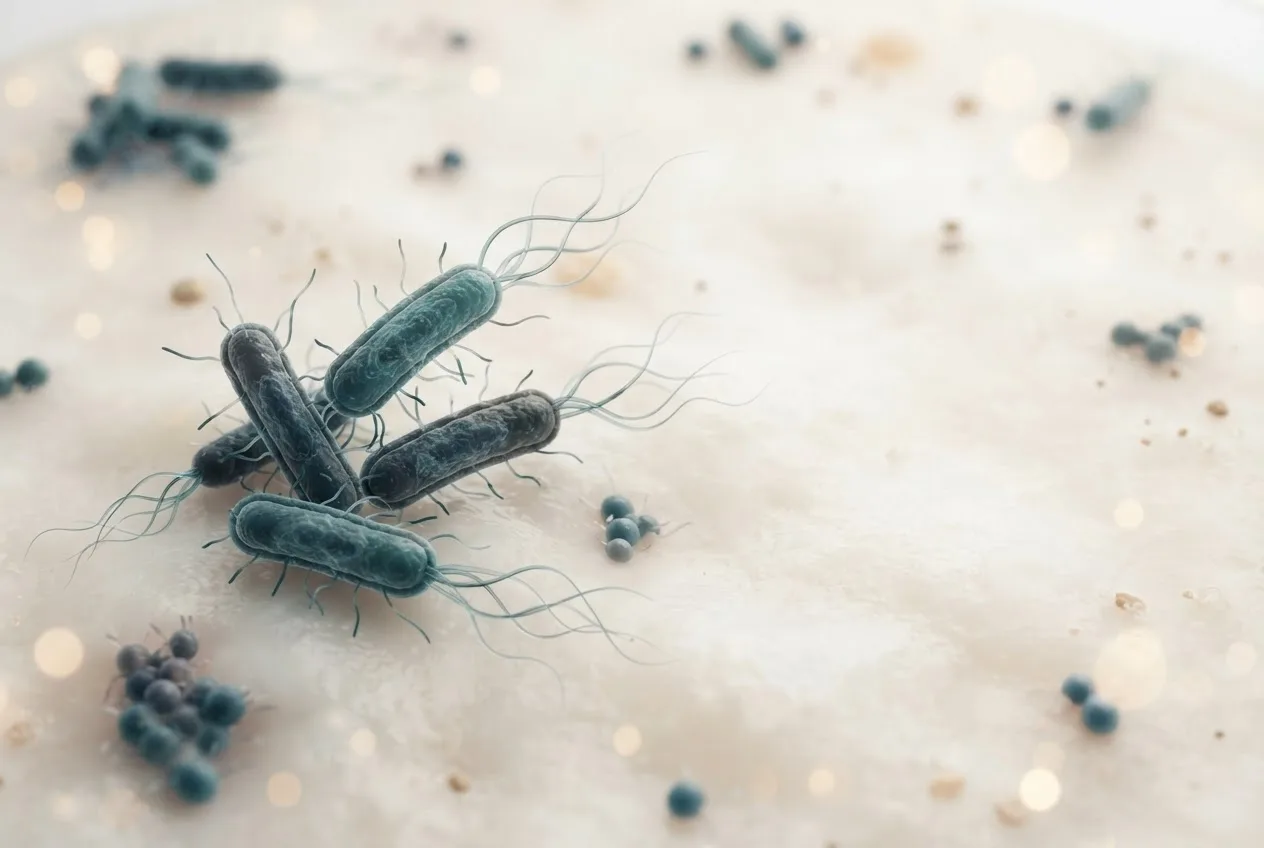
Edwardsiella ictaluri is a non-spore-forming, motile, rod-shaped bacterium measuring approximately 0.5 × 1.0-2.0 μm. The organism is oxidase-negative, catalase-positive, and ferments glucose with gas production. Optimal growth occurs at 25-30°C in tryptic soy broth or brain heart infusion broth supplemented with 0.5% NaCl. Colonies on solid media appear as small, circular, convex, and translucent after 24-48 hours.
The bacterium is highly conserved within the genus Edwardsiella. Multilocus sequence analysis (MLSA) and whole-genome sequencing have revealed distinct lineages. Vietnamese isolates of E. ictaluri from striped catfish cluster into two major clades, with an estimated ancestral origin dating back to the 1950s [<a href="#ref-1">1</a>]. In contrast, isolates from channel catfish in the southeastern USA show genotypic differences compared to ornamental fish isolates, with distinct plasmid profiles and virulence gene content [<a href="#ref-4">4</a>]. All Vietnamese striped catfish isolates belong to sequence type ST-26, differing by a maximum of 90 single nucleotide polymorphisms [<a href="#ref-2">2</a>].
Host Range and Disease Syndromes
Edwardsiella ictaluri infects a growing number of commercially important fish species beyond its original catfish hosts. The primary disease syndromes include:
- Enteric septicemia of catfish (ESC) in channel catfish, blue catfish, and hybrid catfish (USA) [<a href="#ref-5">5</a>].
- Bacillary necrosis of Pangasianodon (BNP) in striped catfish (Vietnam, Malaysia) [<a href="#ref-1">1</a>, <a href="#ref-2">2</a>, <a href="#ref-3">3</a>].
- Emerging edwardsiellosis in tilapia (Oreochromis spp.) in northern Vietnam, with extremely high virulence (LD50 of 42-61 CFU/fish).
- Disease in yellow catfish (Pelteobagrus fulvidraco) in China, causing severe gut dysbiosis and brain pathology [<a href="#ref-6">6</a>, <a href="#ref-7">7</a>, <a href="#ref-8">8</a>].
- Infections in ornamental fish such as zebrafish (Danio rerio) and tiger barb (Puntius tetrazona) [<a href="#ref-4">4</a>].
- Disease in hybrid grouper (Epinephelus fuscoguttatus × E. lanceolatus) [<a href="#ref-9">9</a>].
Coinfections with other bacterial pathogens exacerbate disease severity. Synergistic infection of E. ictaluri with Flavobacterium oreochromis in tilapia results in mortality rates of 65-85%. Similarly, coinfection with Flavobacterium covae in channel catfish elevates cumulative percent mortality to over 90%, with delayed mortality onset when F. covae precedes E. ictaluri exposure.
Pathogenesis and Virulence Factors
Edwardsiella ictaluri employs a suite of virulence determinants to invade, survive, and proliferate within host cells. The bacterium is a facultative intracellular pathogen that survives and replicates within macrophages [<a href="#ref-5">5</a>, <a href="#ref-10">10</a>].
Type III and Type VI Secretion Systems
The type III secretion system (T3SS) and type VI secretion system (T6SS) are critical for E. ictaluri virulence [<a href="#ref-2">2</a>, <a href="#ref-5">5</a>, <a href="#ref-10">10</a>]. The T3SS effector protein EseG is translocated via the chaperone EscB and inactivates the small GTPase RhoA, leading to disassembly of microtubule and actin cytoskeletons [<a href="#ref-10">10</a>]. This disruption impairs phagocytosis, alters cell morphology, and modulates immune responses by reducing expression of pro-inflammatory interleukins and cyclooxygenase-2 (COX-2), thereby decreasing prostaglandin E2 production [<a href="#ref-10">10</a>]. An eseG deletion mutant is significantly attenuated in channel catfish, with reduced persistence in tissues [<a href="#ref-5">5</a>, <a href="#ref-10">10</a>].
The T6SS structural protein EvpC is also essential for full virulence; an evpC mutant shows marked attenuation and provides protective immunity in channel catfish [<a href="#ref-5">5</a>]. The master regulator EsrC controls expression of both T3SS and T6SS genes [<a href="#ref-5">5</a>].
Other Virulence Factors
Plasmids are highly prevalent in E. ictaluri isolates, particularly from Vietnamese striped catfish, where up to four plasmids per genome have been identified [<a href="#ref-1">1</a>]. These plasmids carry diverse virulence genes, including those associated with protein secretion systems [<a href="#ref-1">1</a>]. The RNA chaperone Hfq plays a multifaceted role in virulence: deletion of hfq results in reduced growth rate, decreased biofilm formation, increased motility, impaired survival under oxidative and acidic stress, reduced persistence in catfish macrophages, and complete attenuation in vivo. An hfq mutant confers 100% protection against wild-type challenge in channel catfish.
The bacterium also harbors urease genes (ureA-C), which may contribute to acid tolerance and intracellular survival. Comparative genomics reveals temporal variation in motility and immune modulation genes among Vietnamese isolates [<a href="#ref-1">1</a>].
Clinical Signs and Pathology
Clinical signs vary by host species but generally include anorexia, lethargy, erratic swimming, and exophthalmia. External lesions include cutaneous hemorrhages, petechiae, and ulcerations. Internally, gross pathology commonly includes pale or patchy liver, white nodular spleen, and congested or swollen kidney. In striped catfish, BNP is characterized by multifocal white nodules in the liver, spleen, and kidney [<a href="#ref-2">2</a>]. In yellow catfish, infection leads to severe enteritis, with E. ictaluri dominating the gut microbiota (>99% relative abundance in diseased fish) [<a href="#ref-8">8</a>].
High-dose infection in hybrid grouper induces acute liver injury with cytoplasmic vacuolation, inflammatory cell infiltration, nuclear pyknosis, and hepatocyte apoptosis. Biochemical markers such as glutamate oxaloacetate transaminase (GOT) and glutamate pyruvate transaminase (GPT) are significantly elevated [<a href="#ref-9">9</a>]. Hepatic transcriptomic changes involve dysregulation of steroid hormone biosynthesis, PPAR signaling, and protein digestion pathways.
Central nervous system involvement is observed in yellow catfish, where E. ictaluri increases blood-brain barrier permeability and alters brain immunity [<a href="#ref-6">6</a>]. Both head and trunk kidneys mount robust immune responses, with increased IgM and IgT B cells following infection [<a href="#ref-7">7</a>].
Immune Response and Immunomodulation
The host immune response to E. ictaluri involves both innate and adaptive components. Infection upregulates pro-inflammatory cytokines such as interleukin-6 (IL-6), interleukin-8 (IL-8), tumor necrosis factor alpha (TNF-α), and interleukin-1 beta (IL-1β) [<a href="#ref-9">9</a>]. Simultaneously, anti-inflammatory mediators such as IL-10 and nuclear factor kappa B P65 subunit are downregulated in certain tissues [<a href="#ref-9">9</a>]. The T3SS effector EseG specifically modulates immune responses by reducing COX-2 expression, thereby limiting inflammation [<a href="#ref-10">10</a>].
Systemic immunoglobulin M (IgM) levels in serum and mucus increase following vaccination and infection [<a href="#ref-3">3</a>, <a href="#ref-7">7</a>]. In yellow catfish, the trunk kidney shows higher IgM upregulation than the head kidney after infection [<a href="#ref-7">7</a>].
Dietary immunomodulators such as oligochitosan and trans-cinnamaldehyde (TC) have been investigated for their ability to prime immune responses. Oligochitosan supplementation in hybrid grouper reduces liver injury, normalizes metabolic pathways, and decreases apoptosis [<a href="#ref-9">9</a>]. TC feeding in channel catfish enhances expression of innate and adaptive immune genes, including IgM, in the anterior kidney and spleen following E. ictaluri challenge.
Diagnostic Approaches
Accurate and timely diagnosis is essential for disease management. Conventional identification relies on bacterial culture from affected organs (liver, spleen, kidney, brain) on selective agar, followed by biochemical testing. However, genotypic methods offer superior specificity.
Molecular Diagnostics
Polymerase chain reaction (PCR) targeting the gyrB gene is widely used for species confirmation. A multiplex TaqMan real-time quantitative PCR assay has been developed that simultaneously detects Edwardsiella ictaluri, Aeromonas veronii, and Aeromonas sobria with detection limits as low as 100 copies/μL for E. ictaluri. This assay demonstrated higher detection rates than conventional PCR in clinical samples from yellow catfish.
16S rRNA gene sequencing and multi-locus sequence analysis (MLSA) are employed for phylogenetic characterization and genotyping [<a href="#ref-2">2</a>, <a href="#ref-4">4</a>]. Whole-genome sequencing combined with hybrid assembly provides high-quality reference genomes for epidemiological surveillance [<a href="#ref-1">1</a>].
Serological and Phenotypic Characterization
Serotyping has revealed distinct antigenic profiles between catfish and ornamental fish isolates, indicating that vaccines derived from catfish strains may have limited efficacy against ornamental strains [<a href="#ref-4">4</a>]. Plasmid profiling and antimicrobial susceptibility testing (minimum inhibitory concentration determination) complement molecular characterization [<a href="#ref-2">2</a>, <a href="#ref-4">4</a>].
The following Mermaid flowchart outlines a diagnostic decision tree for suspected E. ictaluri infection:
flowchart TD
 A["Clinical Signs: anorexia, lethargy, exophthalmia, skin lesions"] --> B["Gross Pathology: pale liver, nodular spleen, swollen kidney"]
 B --> C["Collect liver, kidney, spleen, brain tissues"]
 C --> D{"Bacterial Culture on TSA/BHI"}
 D --> E["Gram-negative rods, oxidase-negative, catalase-positive"]
 E --> F["PCR targeting gyrB or 16S rRNA"]
 F --> G{"Multiplex TaqMan qPCR for three pathogens"}
 G --> H["Positive for E. ictaluri"]
 G --> I["Co-infection with Aeromonas spp."]
 H --> J["Confirmed E. ictaluri infection"]
 I --> J
 J --> K["Antimicrobial susceptibility testing (MIC)"]
 K --> L["Select treatment or vaccination strategy"]
Prevention and Control Strategies
Vaccination
Vaccination is the cornerstone of sustainable disease control. Several vaccine platforms have been developed:
- Live attenuated vaccines: Strains with deletions in T3SS/T6SS genes (esrC, eseG, evpC) confer 95-100% relative percent survival (RPS) in channel catfish following single immersion [<a href="#ref-5">5</a>]. An hfq deletion mutant provides 100% protection. Inactivated mutated strains (wzz and aroA deletions) show up to 94% RPS in striped catfish.
- Inactivated whole-cell vaccines: Formalin-killed sonicated cells (FK-SC) and polyvalent formulations with booster regimens enhance immune responses in striped catfish, achieving RPS up to 61.9% when combined with oral and injection routes [<a href="#ref-11">11</a>].
- Nanovaccines: Cationic lipid-based chitosan-coated nanoemulsion (CS-NE) vaccines deliver formalin-killed antigens via immersion, providing higher RPS than conventional FK-SC [<a href="#ref-3">3</a>]. The CS-NE vaccine penetrates skin up to 100 μm within 5 minutes and upregulates immunoglobulin and inflammatory gene expression [<a href="#ref-3">3</a>].
- Combination strategies: Prime-boost protocols combining injection and oral vaccination produce superior protection compared to either route alone.
Antimicrobial Treatment
Antimicrobials remain a common intervention, but resistance is widespread. Florfenicol is the drug of choice for ESC in the USA, but resistance genes such as floR are prevalent among Vietnamese and some US isolates [<a href="#ref-1">1</a>, <a href="#ref-2">2</a>]. Tetracycline resistance via tet(A) or tet(D) is also common [<a href="#ref-2">2</a>]. Extended-spectrum beta-lactamase gene *bla*CTX-M-15 has been detected in 30% of Vietnamese genomes [<a href="#ref-2">2</a>]. The IncA/C plasmid pEIMS-171561 mediates resistance to florfenicol, sulfonamides, and tetracycline, and is stable without selection pressure but does not readily transfer to gut microbiota in vivo.
Phytochemicals and Nutraceuticals
Plant extracts and feed additives offer alternative or adjunct control measures:
- Henna leaf extract (Lawsonia inermis): Strong antibacterial activity against E. ictaluri with inhibition zones up to 16.85 mm at 48% concentration.
- Sappanwood extract (Caesalpinia sappan): Inhibits growth at 5% concentration, comparable to ciprofloxacin.
- Brazilian propolis constituents (vestitol, neovestitol, methylvestitol): Active against Flavobacterium covae but not significantly against E. ictaluri.
- Oligochitosan: Dietary supplementation restores hepatic metabolic homeostasis, reduces apoptosis, and modulates immune gene expression in hybrid grouper [<a href="#ref-9">9</a>].
- Trans-cinnamaldehyde: Dietary supplementation primes innate and adaptive immune responses in channel catfish.
- Combined leaf extracts (Terminalia catappa and Nelumbo nucifera): Reduce oxidative stress and enhance resistance in ornamental fish.
Probiotics and Prebiotics
Probiotic supplementation following florfenicol therapy improves survival against ESC challenge in channel catfish compared to prebiotics or unsupplemented controls, likely by restoring gut microbiota balance. Iron supplementation, conversely, increases susceptibility to E. ictaluri infection in hybrid catfish, possibly through increased haematocrit or pathogen proliferation.
Epidemiology and Genomic Diversity
Edwardsiella ictaluri exhibits an open pan-genome with 4,148 genes and a core genome of 3,060 genes, accounting for over two-thirds of the genome [<a href="#ref-1">1</a>]. Comparative genomics of Vietnamese isolates over 20 years (2001-2021) reveals temporal increases in antimicrobial resistance gene content, while virulence gene numbers remain stable [<a href="#ref-1">1</a>]. Three plasmid replicon types (IncA, p0111, IncQ1) are common in Vietnam [<a href="#ref-2">2</a>]. In the southeastern USA, plasmid profiles are more diverse among catfish and ornamental isolates [<a href="#ref-4">4</a>].
Risk factors for BNP in Malaysian cage-cultured striped catfish include water temperature (strong correlation), ammonia, sulfide, and total suspended solids, with highest prevalence in fish weighing 1-50 g and 150-200 g. Disease spread in Vietnam occurs primarily through contaminated seeds and within the Mekong Delta region [<a href="#ref-2">2</a>].
Antimicrobial Resistance Profile
Multidrug resistance (MDR) is alarming. In Vietnamese striped catfish, 97.3% of isolates are MDR, with 89.3% resistant to 3-6 antimicrobial classes. High resistance rates are observed to trimethoprim/sulfamethoxazole (97.3%), nalidixic acid (93.3%), streptomycin (74.7%), ampicillin (61.3%), and florfenicol (60.0%). In tilapia isolates from Vietnam, 80.8-100% are MDR, resistant to 4-8 antimicrobials including penicillins, macrolides, sulfonamides, amphenicols, and glycopeptides.
The MAR index (0.37-0.48) indicates high antibiotic usage pressure in aquaculture environments. Continued genomic surveillance is essential to monitor resistance trends and inform disease management [<a href="#ref-1">1</a>].
Reference Gene Selection for Gene Expression Studies
Accurate normalization of reverse transcription quantitative PCR (RT-qPCR) data requires stable reference genes. In E. ictaluri, nine genes (aspA, glyA, gyrB, mutS, recP, tkt, atpA, dnaG, rpoS) are most stable during catfish serum exposure, while aspA, g6pd, glyA, gyrB, mdh, mutS, pgm, recA, recP, and tkt are most stable across growth phases. The classical 16S rRNA gene is among the least stable across growth phases and should not be used as a single reference.
Frequently Asked Questions
What is the primary disease caused by Edwardsiella ictaluri in channel catfish?
The primary disease is enteric septicemia of catfish (ESC), a systemic infection causing high mortality in farmed channel catfish and hybrid catfish in the southeastern United States [<a href="#ref-5">5</a>].
Can Edwardsiella ictaluri infect tilapia?
Yes, a highly virulent strain (LD50 42-61 CFU/fish) has been reported in farmed tilapia in northern Vietnam, causing white spots in visceral organs and mortality rates of 30-65%.
What virulence factors are most critical for Edwardsiella ictaluri pathogenesis?
The type III secretion system effector EseG and the type VI secretion system structural protein EvpC are essential for intracellular survival, cytoskeletal modulation, and immune evasion [<a href="#ref-5">5</a>, <a href="#ref-10">10</a>].
How is Edwardsiella ictaluri diagnosed in a laboratory setting?
Diagnosis is confirmed by bacterial culture from internal organs, followed by PCR targeting the gyrB gene or by multiplex TaqMan qPCR that simultaneously detects Edwardsiella ictaluri, Aeromonas veronii, and Aeromonas sobria.
What vaccine types are available for Edwardsiella ictaluri?
Live attenuated vaccines (T3SS/T6SS deletion mutants or hfq deletion) provide 95-100% relative percent survival in channel catfish [<a href="#ref-5">5</a>]; inactivated vaccines and chitosan-coated nanoemulsion immersion vaccines also show high efficacy in striped catfish [<a href="#ref-3">3</a>].
What antibiotics are commonly used to treat Edwardsiella ictaluri infections?
Florfenicol is a primary therapeutic agent, but resistance mediated by floR and tet(A) genes is widespread, especially in Vietnamese isolates [<a href="#ref-1">1</a>, <a href="#ref-2">2</a>].
Are there phytochemical alternatives to antibiotics for Edwardsiella ictaluri?
Henna leaf extract and sappanwood extract demonstrate strong in vitro antibacterial activity, while dietary oligochitosan and trans-cinnamaldehyde enhance immune resistance in vivo.
Does iron supplementation affect susceptibility to Edwardsiella ictaluri?
Excess dietary iron increases haematocrit and potentially decreases disease resistance in hybrid catfish, leading to higher mortality following challenge.
What is the role of the RNA chaperone Hfq in Edwardsiella ictaluri?
Hfq regulates growth, biofilm formation, motility, stress tolerance, intracellular survival, and virulence; deletion of hfq completely attenuates the bacterium and confers protective immunity.
How does Edwardsiella ictaluri alter the gut microbiota of infected fish?
In yellow catfish, infection causes near-complete dominance of the gut microbiota by Edwardsiella ictaluri (99.22%), with disappearance of beneficial genera such as Cetobacterium, Plesiomonas, and Romboutsia [<a href="#ref-8">8</a>].
Related Clinical & Scientific Guides
- Duck Diseases: A Comprehensive Overview for Veterinary Practitioners
- Salmonella Dublin in Cattle: Emerging Pathogen, Diagnostic Challenges, and Public Health Impact
- Mycoplasma Infections in Poultry: Vaccination Strategies and Control Programs