
Poultry Salmonellosis: Control, Diagnosis, and Differentiation from Other Enteric Pathogens
Introduction
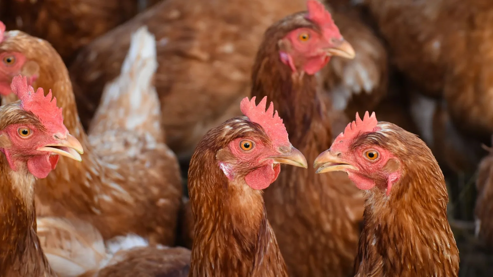
Poultry salmonellosis encompasses a spectrum of diseases caused by infection with members of the genus Salmonella enterica subsp. enterica. These infections range from subclinical carrier states in broilers to acute septicemic pullorum disease and fowl typhoid in chicks and poults, and to persistent reproductive tract colonization in layers [1, 2, 3]. The global economic burden of salmonellosis in poultry production is compounded by the public health implications of foodborne transmission through meat and eggs [4, 5, 6]. In the European Union alone, salmonellosis remains the second most frequently reported zoonosis. The control of Salmonella in poultry therefore requires a multifaceted approach integrating vaccination, biosecurity, feed additives, probiotics, bacteriophages, and rigorous diagnostic surveillance [7, 61]. Crucially, accurate differentiation of Salmonella from other enteric pathogens such as Escherichia coli in Chickens and Poultry Products, Campylobacter spp., Clostridium perfringens Type A in Broilers, and Histomonas meleagridis is essential for targeted intervention [8, 83].
Etiology and Serovar Distribution
The genus Salmonella comprises over 2,600 serovars, but only a limited number are consistently isolated from poultry. Host-restricted serovars such as Salmonella Pullorum (biovars of S. enterica serovar Gallinarum) and S. Gallinarum cause systemic disease in young birds, whereas broad-host-range serovars including S. Enteritidis, S. Typhimurium, and S. Infantis are frequently associated with subclinical intestinal colonization and contamination of carcasses and eggs [1, 3, 9, 62]. Recent genomic surveillance has revealed clonal dissemination of multidrug-resistant S. Enteritidis strains in Thailand and persistent emergence of S. Infantis clone REPJFX01 in the United States poultry supply chain [10]. High-resolution serotyping using CRISPR-SeroSeq has uncovered previously unrecognized serovar diversity in broiler flocks. Pangenome analyses of serovar Hadar further illustrate population structure and zoonotic potential. The dynamic epidemiology of Salmonella serovars is influenced by geographic region, production system, and antimicrobial use [55, 66, 74].
Pathogenesis and Virulence Factors
Salmonella virulence is multifactorial, involving type III secretion systems (T3SS-1 and T3SS-2), flagella, fimbriae, lipopolysaccharide (LPS), and multiple effector proteins [7, 69]. The virulence factor SptP activates the NLRP3/caspase-1 pathway, inducing pyroptosis and amplifying intestinal injury in chicks. Iron-manganese superoxide dismutases contribute to antioxidative defense, antibiotic tolerance, and virulence of S. Pullorum. Biofilm formation is a critical mechanism for persistence on poultry farms and in processing environments [11, 61]. Fimbrial regulators such as FimZ modulate type I fimbriae and can be targeted by phytochemicals like naringenin to reduce virulence. Kinome analysis has identified host signaling pathways that influence resistance to Salmonella colonization. Co-infection with other pathogens, such as Eimeria tenella or Histomonas meleagridis [8], can exacerbate the severity of salmonellosis by disrupting intestinal barrier function and immune responses.
Control Strategies
Vaccination
Vaccination is a cornerstone of Salmonella control in poultry. Live attenuated vaccines, including those based on S. Enteritidis and S. Typhimurium mutants, can reduce colonization and shedding [12, 13, 85]. Novel vaccine candidates include LPS-deficient strains with steD deletions that confer cross-protection against heterologous serovars. Recombinant S. Enteritidis vectors expressing Clostridium perfringens antigens have demonstrated dual protection against salmonellosis and necrotic enteritis [14, 15]. Outer membrane vesicle (OMV) vaccines derived from S. Typhimurium provide cross-protective immunity against multiple serovars in SPF chickens. Protein surface display on bioengineered Saccharomyces cerevisiae has been explored for oral vaccination against S. Pullorum. Conjugate and whole-cell killed vaccines against S. Typhimurium have also been evaluated [16]. High-throughput sequencing (HTS) is now used to monitor the genetic stability of live vaccine strains.
Biosecurity and Management
Biosecurity measures, including cleaning and disinfection of housing, control of rodent and insect vectors, and all-in/all-out production, are essential to prevent Salmonella introduction and spread [2, 7]. The use of contaminated eggshells in poultry feed has been implicated in diffuse outbreaks [6]. Chick paper sampling is a non-invasive surveillance tool during outbreaks linked to backyard poultry.
Feed Additives and Organic Acids
Organic acids (e.g., formic, propionic, butyric acids) exert a dual-action mode by reducing pH in the gut and directly disrupting Salmonella cell membranes [17, 42]. Oregano essential oil has been shown to reduce S. Enteritidis colonization in market-age broilers [18]. Houttuynia cordata extract targets T3SS-1 to protect against Salmonella infection [19]. Dihydromyricetin ameliorates S. Enteritidis-induced pyroptosis via NLRP3 inflammasome modulation. Clove extract protects against intestinal dysfunction through JAK2/STAT3-mediated stem cell activation. Systems pharmacology approaches have been used to elucidate the mechanism of traditional Chinese medicine formulations against avian salmonellosis [20]. Matrine combined with tannic acid protects against S. Typhimurium intestinal infection. Purslane extract improves gut microbiota and growth in S. Pullorum-challenged chicks.
Probiotics, Prebiotics, and Synbiotics
Probiotic strains, particularly Bacillus subtilis [21], Lactiplantibacillus plantarum [22, 81], Lactobacillus salivarius, Lactobacillus rhamnosus, and Lactococcus lactis, have been shown to reduce Salmonella colonization and improve gut health. Competitive exclusion using undefined cecal microbiota from healthy birds impairs Salmonella colonization [24]. Synbiotic combinations of probiotics and prebiotics (e.g., fructo-oligosaccharides) further enhance protection [25, 26, 95]. A commercial synbiotic's effects on pathogen colonization can depend on administration route. Antimicrobial peptides isolated from probiotics offer an alternative to conventional antibiotics.
Bacteriophage Therapy
Bacteriophages represent a promising tool for targeted reduction of Salmonella in poultry. Phage cocktails targeting multiple receptors reduce S. Enteritidis colonization and modulate the cecal microbiome [27]. Single phages such as vB_SalS_KY05 and vB_SalP_NW15 combined with cinnamaldehyde show therapeutic efficacy. Phage treatment significantly reduces S. Enteritidis shedding in layer hens and egg surface contamination. Alginate encapsulation improves gastrointestinal stability of phage cocktails for oral delivery. Phage application in feed and drinking water can reduce S. Pullorum contamination. Phage therapy has also been evaluated for multidrug-resistant S. Pullorum [28] and S. Infantis [29, 48]. Holins from S. Pullorum phages have therapeutic efficacy in chicks. A phage cocktail combined with vaccination induced complete protection against homologous and heterologous Salmonella [30]. Lytic phages isolated from poultry farms in Ecuador show genomic diversity and potential for biocontrol [31].
Diagnostic Approaches
Accurate and timely diagnosis is critical for effective control and differentiation from other enteric pathogens.
Bacteriological Culture and Serotyping
Conventional culture remains the gold standard for Salmonella isolation from poultry samples. Selective enrichment media (e.g., Rappaport-Vassiliadis broth, xylose-lysine-tergitol 4 agar) are used, followed by biochemical and serological confirmation. Serotyping using antisera identifies O and H antigens, but is increasingly supplemented by molecular methods.
Serological Methods
Enzyme-linked immunosorbent assays (ELISAs) are widely used for flock-level surveillance. Blocking ELISAs have been developed for detection of antibodies against S. Enteritidis. Whole-cell and conjugate vaccine evaluation often involves serological monitoring [16].
Molecular Diagnostics
Polymerase chain reaction (PCR) and real-time PCR targeting invasion genes (e.g., invA) are standard for rapid detection. A dual-gene colorimetric loop-mediated isothermal amplification (LAMP) assay enables genus-level detection of Salmonella and specific identification of non-motile S. Gallinarum. High-throughput sequencing (HTS) and whole-genome sequencing (WGS) provide high-resolution genomic epidemiology, antimicrobial resistance profiling, and outbreak tracking [32, 56, 63, 66, 94]. CRISPR-SeroSeq is a targeted amplicon sequencing method for serovar diversity detection in mixed infections. Quantile regression forest models using multi-source surveillance data can provide early warning of Salmonella foodborne risk [4].
Differentiation from Other Enteric Pathogens
Differentiation of salmonellosis from other enteric infections requires integration of clinical signs, gross pathology, histopathology, and laboratory testing. Key differentials include:
Table 1. Differentiation of Salmonella from Common Enteric Pathogens in Poultry
| Pathogen | Key Clinical Features | Diagnostic Method | Distinguishing Characteristics |
|---|---|---|---|
| Salmonella spp. | Diarrhea (yellow, mucoid), septicemia, high mortality in young; carrier state in adults | Culture, PCR, serology, WGS | Invades via M cells; T3SS effectors; often causes hepatitis, splenomegaly, and typhlitis |
| Escherichia coli (APEC) | Omphalitis, airsacculitis, peritonitis, coligramuloma | Culture (MacConkey), PCR (iss, iroN) | Typically associated with secondary infections; lactose-fermenting on MacConkey; no T3SS |
| Campylobacter jejuni / C. coli | Mild enteritis, watery diarrhea (often subclinical in poultry) | Culture (Campy-CVA), mPCR | Microaerophilic; requires special media; no T3SS; flagella-mediated invasion |
| Clostridium perfringens Type A | Necrotic enteritis (focal or diffuse mucosal necrosis), sudden death | Anaerobic culture, toxin ELISA (NetB, TpeL) | Gram-positive rod; toxin production; typically associated with predisposing factors (coccidiosis) |
| Histomonas meleagridis | Cecal cores, liver necrosis ("target lesions"), turkeys more susceptible | Microscopy, PCR (18S rRNA) | Protozoan; causes typhlitis and hepatitis; can be co-infection with Salmonella |
| Eimeria spp. | Bloody diarrhea, cecal cores, intestinal petechiation | Oocyst count (McMaster), PCR | Intracellular apicomplexan; site-specific lesions; no bacterial invasion |
Co-infection scenarios are common. Eimeria tenella infection dose-dependently increases Salmonella colonization. Campylobacter and Salmonella co-inoculation affects cecal microbiota and serum metabolome. Histomonas and Salmonella co-infection in turkeys can worsen histomonosis outcomes [8]. Diagnostic differentiation often relies on molecular panels that simultaneously detect multiple pathogens.
Mermaid Diagram: Diagnostic Decision Tree for Suspect Poultry Enteric Disease
The following diagram outlines a systematic approach to differential diagnosis.
flowchart TD
 A["Clinical signs: diarrhea, depression, mortality"] --> B{Postmortem examination}
 B --> C["Gross lesions: typhlitis, hepatitis, splenomegaly"]
 B --> D[Cecal cores / liver necrosis]
 B --> E[Focal intestinal necrosis]
 B --> F[No specific lesions]
 C --> G[Culture on selective media<br>XLT4, brilliant green agar]
 G --> H["'Salmonella suspected colonies<br>black-centered (H2S+')"]
 H --> I[Biochemical identification<br>and serotyping]
 I --> J[Confirmed Salmonella]
 J --> K[Antimicrobial susceptibility testing<br>and WGS / CRISPR-SeroSeq]
 D --> L[Microscopy of cecal/liver<br>scrapings and PCR for Histomonas]
 L --> M[Histomonas meleagridis detected]
 L --> N[Also culture for Salmonella<br>to rule out co-infection]
 E --> O[Anaerobic culture<br>and NetB toxin ELISA]
 O --> P[Clostridium perfringens]
 F --> Q["Molecular panel: PCR for<br>Salmonella, Campylobacter, Eimeria, Cryptosporidium"]
 Q --> R[Campylobacter positive<br>or Eimeria positive]
 R --> S[Specific treatment<br>and control measures]
Conclusions
Poultry salmonellosis remains a complex challenge requiring integrated control strategies that combine vaccination, biosecurity, feed additives, probiotics, and bacteriophage therapy. Advances in molecular diagnostics, including LAMP, HTS, and CRISPR-SeroSeq, have enhanced our ability to detect, serotype, and differentiate Salmonella from other enteric pathogens quickly and accurately. Continued genomic surveillance is essential to monitor emerging serovars and antimicrobial resistance trends [32, 55, 62, 66]. Differentiation from pathogens such as E. coli, Campylobacter, Clostridium perfringens, and Histomonas is critical for implementing appropriate control measures and reducing flock mortality and food safety risks.
References
[1] Pelyuntha W, Yamik DY, Khongkhai H, et al. Serovar Distribution, Virulence Gene Profiles, and Antimicrobial Resistance of Salmonella Isolated from Chicken Meat from the Broiler Slaughterhouse and Processing Plant in Central Thailand. Foodborne Pathog Dis. 2026.
[2] Ayala Velastegui D, Shariat NW. Investigating the differences in Salmonella serovar transmission within broiler production. Front Vet Sci. 2026.
[3] Lee JB, Lim JH, Park JH, et al. Serotype distribution, genotype, and antimicrobial resistance profiles of Salmonella enterica isolated from retail chicken meat in South Korea. Food Sci Anim Resour. 2026.
[4] Liu W, Zhou Y, Yan R, et al. Quantile regression forest-based early warning of Salmonella foodborne risk using multi-source food safety surveillance data. Food Res Int. 2026.
[5] Bai L, Wang J, Wang Y, et al. Quantitative risk assessment of nontyphoidal Salmonella in retail pork in China. Food Res Int. 2026.
[6] Adriaansens DL, van den Berg OE, Lanzl MI, et al. The role of contaminated eggshells used in poultry feed in a diffuse nationwide outbreak of Salmonella Enteritidis, the Netherlands, 2023 to 2025. Euro Surveill. 2026.
[7] El-Shall NA, Adiguzel MC, Abd El-Ghany WA, et al. Salmonella infection in chickens: pathogen, pathogenesis, and dietary non-drug feed additives as alternatives to antibiotics - a comprehensive review. Folia Microbiol (Praha). 2026.
[8] Rafieian-Naeini HR, Keshavareddy VPR, Katha HR, et al. Does Salmonella co-infection worsen Histomonas meleagridis infection in turkeys? Poult Sci. 2026.
[9] Alsufyani AT, Bin Jaddua R, Alreshoodi FM, et al. Genomic insights into the prevalence and genetic diversity of Salmonella in chicken eggs in Saudi Arabia. Front Microbiol. 2026.
[10] Ford L, Weller DL, Steele MK, et al. Trends in a Persistent Strain of Multidrug-Resistant Salmonella Infantis (REPJFX01) in Humans and Chickens - United States, 2010-2023. J Food Prot. 2026.
[11] Oastler C, La Ragione RM, Chambers MA, et al. Biofilm-forming capability of Salmonella isolates sourced from poultry production and farm environments in Great Britain. J Med Microbiol. 2026.
[12] Filho RCP, Sarabia J, Sesti L, et al. Evaluation of the Efficacy of Different Vaccination Programs Using Two Live Salmonella Vaccines Against Salmonella Enteritidis, Salmonella Typhimurium, and Salmonella Gallinarum in Brown Layer Hens. Avian Dis. 2026.
[13] Quinteros JA, Wilson TB, Anwar A, et al. A vaccination programme combining a live attenuated Salmonella Typhimurium and an autogenous inactivated Salmonella Enteritidis vaccine confers protection and reduces ovarian colonisation with the Salmonella Enteritidis isolate 7A, PT12. Aust Vet J. 2026.
[14] Li W, Li YA, Liu X, et al. Oral immunization with attenuated Salmonella enterica serovar Enteritidis expressing dual-toxin antigen induces protective immunity against avian necrotic enteritis. Vaccine. 2026.
[15] Li W, Li YA, Liu X, et al. Recombinant Attenuated Salmonella Enteritidis Vector Enhances the Immunogenicity of Clostridium perfringens EntB Antigen for Effective Prevention of Avian Necrotic Enteritis. Biomolecules. 2026.
[16] Ahmad A, Yousaf Z, Naeem M, et al. Preparation and comparative evaluation of conjugate and whole-cell killed vaccine candidates against Salmonella enterica serovar Typhimurium. Vaccine. 2026.
[17] Marcu D, Balta I, Gundogdu O, et al. Organic acids - a dual-action mode influencing both host and pathogen integrity to reduce Salmonella infection. Microb Pathog. 2026.
[18] Swaggerty CL, Sasia S, Cabrera MD, et al. Oregano essential oil: a pre-harvest tool to reduce Salmonella enterica serovar Enteritidis in market-age broilers. Poult Sci. 2026.
[19] Wang T, Cai S, Zhang J, et al. Houttuynia cordata extract protects against Salmonella infection by targeting type III secretion system 1. Poult Sci. 2026.
[20] Pu S, Zhang L, Yang H, et al. A systems pharmacology-based in vivo study elucidating the mechanism of Wengxian granules against avian salmonellosis. Front Vet Sci. 2026.
[21] Chen Y, Li H, Zhang X, et al. Dietary Bacillus subtilis Group Reduces the General Infection of Salmonella Pullorum in Broiler Chicken. Antibiotics (Basel). 2026.
[22] Hoang TAP, Nguyen XH, Phan VH. Lactiplantibacillus plantarum-fermented shallot (Allium cepa L.) bulb in drinking water as a potential antibiotic alternative against Salmonella pullorum infection in broilers. Vet Immunol Immunopathol. 2026.
[24] Kolososki IMM, Benevides VP, Rodrigues HLS, et al. Competitive exclusion strategies using healthy caecal microbiota impair Salmonella enterica serovars colonization in broilers. Avian Pathol. 2026.
[25] Salim AA, Mohamed NA, El-Gammal GA, et al. The role of synbiotic in controlling Salmonella infection in broilers. Sci Rep. 2026.
[26] Babot J, Hidalgo V, Fernández M, et al. Preventive effects of synbiotic supplementation against Salmonella Enteritidis infection in broiler chickens. Can J Microbiol. 2026.
[27] Gao D, Hu D, Xu H, et al. A phage cocktail targeting multiple receptors reduces Salmonella Enteritidis colonization in chicks and modulates the cecal microbiome. Vet Microbiol. 2026.
[28] Zhao H, You S, Fu J, et al. Phage therapy: A novel strategy to combat drug-resistant Salmonella Pullorum infection in chickens. Vet Microbiol. 2026.
[29] Wei S, Wu J, Zhou L, et al. Isolation of lytic phage and its inhibition of multidrug-resistant Salmonella and its biofilm. Vet Microbiol. 2026.
[30] Zhang J, Zhang Z, Yang D, et al. Combination of lytic phage Sal-P001 and RAST002 vaccine induces complete protection against homologous and heterologous Salmonella strains. Vaccine. 2026.
[31] Vela-Chauvin MG, Ramirez-Villacis DX, Armijos CE, et al. Characterization and evaluation of a phage cocktail targeting Salmonella enterica in a Turkey farm. BMC Microbiol. 2026.
[32] Yang Q, Zhang J, Chen T, et al. Global prevalence and distribution of bla-harboring Salmonella: A genome-wide association study of cephalosporin resistance mechanisms. Food Microbiol. 2026.
[33] Zhao Y, Li G, Li YA, et al. Identification and protective efficacy characterization of novel immunogenic antigens for Salmonella Enteritidis vaccines. Poult Sci. 2026.
[34] Huo Z, Li Y, Zhong C, et al. Cationic Liposome-Fused Endolysin Lys40 Overcomes Outer Membrane Barriers and Enhances Survival in Salmonella-Infected Chicks. Animals (Basel). 2026.
[35] Ager EO, Nickodem CA, Brown J, et al. Diet-vaccine interactions: SQM Iron and Salmonella vaccination shape poultry gut microbiota. Appl Environ Microbiol. 2026.
[36] Liu K, Wei S, Liu C, et al. Structural rationality and enhanced immunoprotective efficacy of endogenously expressed OmpX-IL-9 fusion protein against Salmonella in broilers. Protein Expr Purif. 2026.
[37] He H, Jiang X, Luo Z, et al. Identification of Salmonella enterica serovar Enteritidis mutants overproducing OMVs as vaccine candidates. Vet Microbiol. 2026.
[38] Chuwatthanakhajorn S, Wedley A, Watts A, et al. Lactobacilli Isolated from the Caecum of Healthy Broilers with Antimicrobial Activity are Probiotic Candidates for Controlling Salmonella. Probiotics Antimicrob Proteins. 2026.
[39] Rahman MA, Abraham R, Abraham S, et al. Application of bacteriophages to control bacterial infections of food animals in the era of antimicrobial resistance: An overview. Vet Microbiol. 2026.
[40] Kerek Á, Szabó Á, Bárdos K, et al. Impact of plant-based antibiotic alternative supplemented feed on the gut microbiota of Bábolna Tetra-SL chickens experimentally infected with Salmonella enterica and Escherichia coli. BMC Vet Res. 2026.
[41] Fenster DA, Qudsieh R, Wang JJ, et al. Effects of a direct fed microbial and xylanase enzyme blend on Salmonella Typhimurium colonization, antioxidant status, and performance in broiler chickens. Poult Sci. 2026.
Disclaimer: This article is for educational and informational purposes only. It is not intended to substitute for professional veterinary advice, diagnosis, treatment, or regulatory guidance. Always consult a licensed veterinarian or qualified specialist regarding animal health, disease diagnosis, and therapeutic decisions.