
A Comprehensive Guide to Poultry Diseases: MSD Manual and Clinical Insights
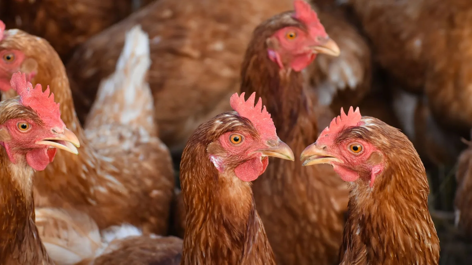
The diagnosis and management of bacterial diseases in commercial poultry flocks require a structured approach that integrates clinical observation, gross pathology, histopathology, and molecular or microbiological confirmation. This reference article synthesizes information from standard veterinary texts, including the MSD Veterinary Manual and Diseases of Poultry, with practical clinical insights for the field veterinarian and diagnostic pathologist. Bacterial infections of poultry span a diverse array of pathogens, each with distinct pathogenesis, tissue tropism, and clinical presentation. Because antimicrobial resistance is an escalating concern, judicious antimicrobial selection guided by culture and susceptibility testing is critical.
Major Bacterial Pathogens of Poultry
Bacterial diseases remain a leading cause of morbidity, mortality, and economic loss in commercial poultry operations. The most clinically relevant bacterial pathogens are summarized in Table 1.
| Pathogen | Primary Diseases | Affected Systems | Key Clinical Signs |
|---|---|---|---|
| Escherichia coli | Colibacillosis, airsacculitis, salpingitis, omphalitis | Respiratory, reproductive, systemic | Pericarditis, perihepatitis, airsacculitis, egg peritonitis |
| Pasteurella multocida | Fowl cholera | Systemic, respiratory | Septicaemia, swollen wattles, diarrhoea, sudden death |
| Gallibacterium anatis | Salpingitis, peritonitis | Reproductive, systemic | Decreased egg production, peritonitis, mortality |
| Mycoplasma synoviae | Infectious synovitis, respiratory disease, eggshell apex abnormalities | Joints, respiratory, reproductive | Lameness, rales, eggshell apex irregularities |
| Mycoplasma gallisepticum | Chronic respiratory disease (CRD) | Respiratory | Coughing, nasal discharge, airsacculitis, reduced growth |
| Mycoplasma meleagridis | Turkey airsacculitis, leg deformities | Respiratory, skeletal | Airsacculitis, leg deformities, poor growth |
| Avibacterium paragallinarum | Infectious coryza | Upper respiratory | Facial oedema, nasal discharge, sneezing, conjunctivitis |
| Clostridium perfringens type A | Necrotic enteritis | Intestinal | Sudden death, diarrhoea, necrotic intestinal lesions |
| Clostridium perfringens type C | Necrotic enteritis (less common) | Intestinal | Haemorrhagic enteritis, high mortality |
| Salmonella spp. | Salmonellosis (pullorum disease, fowl typhoid, paratyphoid) | Systemic, intestinal | White diarrhoea (pullorum), septicaemia, reduced hatchability |
| Staphylococcus aureus | Bumblefoot, osteomyelitis, septicaemia | Skeletal, skin, systemic | Swollen foot pads, lameness, joint abscesses |
| Streptococcus zooepidemicus | Septicaemia, endocarditis | Systemic | Sudden death, cyanosis, valvular endocarditis |
Table 1: Major bacterial pathogens of poultry and their clinical manifestations [1, 2, 3, 4].
Colibacillosis and the Role of Escherichia coli
Escherichia coli is a commensal inhabitant of the avian gastrointestinal tract, but certain serogroups acquire virulence factors that permit extraintestinal infection [1]. Colibacillosis in poultry is characterized by fibrinous polyserositis, including airsacculitis, pericarditis, and perihepatitis [1, 5]. The pathogenesis involves inhalation of contaminated dust or direct penetration of respiratory epithelium following viral infection (e.g., infectious bronchitis virus) [5]. The adhesion of fimbriae (F1, P, and S fimbriae) to host epithelial cells is the critical first step [5]. Invasion of the respiratory epithelium leads to bacteraemia and localization in serosal surfaces [5]. The gross lesion of "pancarditis" refers to the thickened, cloudy pericardial sac, while perihepatitis produces a classic "glazed liver" appearance [1]. Omphalitis in chicks (yolk sac infection) results from contamination of the eggshell with faecal material and subsequent bacterial translocation through the navel [1]. In broilers, colibacillosis manifests as increased mortality and poor weight gain [1]. In layers, salpingitis and egg peritonitis are common sequelae, associated with egg yolk peritonitis and a subsequent drop in production [1]. Diagnosis is confirmed by isolation of E. coli from affected tissues (e.g., pericardial sac, liver, yolk sac) and serotyping or molecular virulence gene detection [5]. Antimicrobial susceptibility testing is essential given the high frequency of multidrug resistance [6]. Control measures emphasize biosecurity, effective hatchery hygiene, vaccination against primary viral pathogens, and proactive litter management [6].
Fowl Cholera: Pasteurellosis in Poultry
Fowl cholera, caused by Pasteurella multocida, is a highly contagious septicaemic disease affecting chickens, turkeys, ducks, and game birds [1, 2]. The bacterium is a Gram-negative coccobacillus that colonizes the upper respiratory tract after inhalation [1]. Capsular serotypes A and D are most commonly associated with disease in poultry [1, 2]. The acute form presents with sudden death, cyanosis of comb and wattles, mucoid diarrhoea, and fever [1, 2]. The chronic form manifests as swollen wattles (wattle oedema), conjunctivitis, lameness, and torticollis [1, 2]. At necropsy, generalized congestion and petechial haemorrhages on the epicardium and serosal surfaces are characteristic [1]. Fibrinous pericarditis and airsacculitis may be present in peracute cases [1]. Diagnosis relies on culture of the organism from blood, liver, or bone marrow on blood agar, with identification via Gram staining, biochemical profiling, or PCR [1, 2]. Differentials include avian influenza infection and infectious coryza [2]. Vaccination with bacterins or live attenuated vaccines is used in endemic areas, and tetracyclines or sulfonamides are effective therapeutic options [2, 7]. However, resistance to tetracyclines has been reported, underscoring the need for susceptibility profiling [7].
Mycoplasma Infections
Mycoplasmas are cell wall-deficient bacteria that cause chronic respiratory disease (CRD) in chickens, infectious synovitis, and eggshell apex abnormalities [1]. Mycoplasma gallisepticum is the most economically significant mycoplasma of chickens and turkeys [1, 3]. The organism colonizes the respiratory mucosa, cilia, and air sacs [1]. Virulence factors include a specialized attachment organelle and production of hydrogen peroxide, which causes oxidative damage to host epithelial cells [3]. Clinical signs include rales, coughing, nasal discharge, and reduced growth rates [1, 3]. Coinfection with Escherichia coli or infectious bronchitis virus exacerbates the severity of airsacculitis [1, 3]. Serological screening via ELISA or hemagglutination inhibition is standard for monitoring breeder flocks [1]. Molecular detection using real-time PCR targeting the 16S rRNA gene or specific adhesin genes provides rapid and sensitive diagnosis [1, 8]. Mycoplasma synoviae causes synovitis, tenosynovitis, and respiratory disease [8]. A hallmark presentation is eggshell apex abnormalities in commercial layers [8]. Mycoplasma meleagridis is primarily a pathogen of turkeys, causing airsacculitis in poults and leg deformities [1]. Control relies on maintaining Mycoplasma-free breeder flocks through rigorous testing and eradication [1]. Antibiotic therapy (tylosin, tiamulin, enrofloxacin) may reduce clinical signs but does not eliminate the carrier state [1].
Diagnostic Pathways in Poultry Bacterial Diseases
Diagnosis of bacterial diseases in poultry follows a systematic workflow. The diagram below illustrates the diagnostic decision tree from initial flock observation to confirmatory laboratory testing.
graph TD
 A["Flock Observation: Increased mortality, drop in production, respiratory signs"] --> B[On-Farm Postmortem Examination]
 B --> C[Gross Lesions Present?]
 C -->|Yes: Fibrinous polyserositis, organ congestion, synovitis| D["Collect Tissue Samples: Liver, Lungs, Joint Fluid, Yolk Sac"]
 C -->|No: No significant gross lesions| E[Collect Blood, Tracheal Swabs, Faecal Samples]
 D --> F["Microbiological Culture: Blood Agar, MacConkey Agar, Mycoplasma Agar"]
 E --> F
 F --> G[Gram Stain & Biochemical ID]
 F --> H["Molecular Detection: 16S rRNA PCR, Species-Specific qPCR"]
 G --> I["Antimicrobial Susceptibility Testing: Disk Diffusion or MIC Determination"]
 H --> I
 I --> J[Interpret Results & Select Therapy]
 J --> K["Implement Flock-Level Interventions: Biosecurity, Vaccination, Litter Management"]
 K --> L["Follow-Up: Monitor Mortality, Lesion Scores, and Production Metrics"]
Figure 1: Diagnostic decision tree for bacterial poultry diseases [1, 5, 9].
Sampling and Culture
Selection of appropriate samples depends on the clinical presentation [1]. For suspected colibacillosis, liver and pericardial swabs are optimal [1]. Mycoplasma culture requires specialized media (e.g., Frey's medium) and reduced oxygen conditions [1, 4]. For necrotic enteritis, the lumen of the small intestine should be examined for gas and necrotic debris, and anaerobic culture on egg yolk agar is recommended [1]. For fowl cholera, bone marrow from the femur provides high-quality isolation material [1, 9]. Culture results should be interpreted in the context of clinical lesions to differentiate commensal flora from pathogens [1].
Molecular and Serological Methods
Polymerase chain reaction (PCR) and real-time PCR assays offer rapid detection of specific pathogens, particularly for mycoplasmas and Avibacterium paragallinarum [1, 10]. ELISA-based serology is routinely employed for flock-level surveillance of Mycoplasma gallisepticum, Mycoplasma synoviae, and Salmonella [1, 10]. The use of pooled PCR testing for Salmonella in faecal samples enhances sensitivity compared with culture alone [1]. For Pasteurella multocida, PCR amplification of the capsular typing genes (hyaD, dcbF, ecbJ) permits serogroup identification critical for vaccine selection [9].
Antimicrobial Resistance and Stewardship
Antimicrobial resistance (AMR) in poultry pathogens is a growing threat to both animal and human health [1, 6]. Escherichia coli isolates from poultry often carry extended-spectrum beta-lactamase (ESBL) genes, rendering third-generation cephalosporins ineffective [5]. Fluoroquinolone resistance in Pasteurella multocida and Mycoplasma gallisepticum has been documented in multiple countries [1, 6]. The cornerstone of AMR mitigation is culture-based susceptibility testing [6]. Disc diffusion methods (Kirby-Bauer) and minimal inhibitory concentration (MIC) determination using broth microdilution are standard [6]. The clinical breakpoints used should be those established by the Clinical and Laboratory Standards Institute (CLSI) for veterinary pathogens [6]. Judicious use of antimicrobials includes restricting prophylactic use, selecting narrow-spectrum drugs where possible, and following guidelines established by veterinary regulatory bodies [6]. Alternatives to antibiotics such as probiotics, prebiotics, organic acids, bacteriophages, and essential oils are under investigation for control of necrotic enteritis and salmonellosis [7].
Flock Health Management and Biosecurity
The cornerstone of bacterial disease prevention is robust biosecurity [1, 2]. Management practices to reduce bacterial disease incidence include [1, 2]:
- All-in/all-out production to break pathogen cycles.
- Adequate ventilation to reduce ammonia and respiratory irritation.
- Litter management to control moisture and pathogen load.
- Vaccination against primary viral agents (infectious bronchitis, Newcastle disease) that predispose to secondary bacterial infections.
- Hatchery sanitation to reduce egg and yolk sac contamination.
Vaccination against Pasteurella multocida, Escherichia coli, and Avibacterium paragallinarum is available in many regions [1, 2, 6]. Autogenous vaccines may be indicated for persistent problems on individual farms [1, 6].
Differential Diagnosis and Integration with Other Pathogens
Many bacterial diseases of poultry mimic viral or parasitic infections [1]. For example, fowl cholera must be differentiated from highly pathogenic avian influenza and Newcastle disease [1, 4]. Respiratory signs of infectious coryza overlap significantly with those caused by Mycoplasma gallisepticum and Avibacterium paragallinarum [4]. The presence of swollen wattles is pathognomonic for chronic fowl cholera but can also be seen in cases of vaccine reactions [1]. Necrotic enteritis caused by Clostridium perfringens must be distinguished from coccidiosis, which often occurs concurrently [1, 3]. The typical coinfections in poultry include E. coli secondary to Mycoplasma gallisepticum, and necrotic enteritis secondary to Eimeria infection [1, 7]. A thorough postmortem examination, including histopathology and targeted PCR, is essential for definitive diagnosis [1].
Conclusions
Bacterial diseases of poultry present a significant diagnostic and therapeutic challenge in commercial production. A systematic approach combining clinical examination, gross pathology, and laboratory confirmation is necessary for accurate diagnosis and effective treatment. The judicious use of antimicrobials guided by susceptibility testing is critical to preserve therapeutic options and minimize AMR. Vaccination, biosecurity, and management practices remain the most sustainable strategies for long-term disease control in poultry flocks.
References
[1] Swayne DE, Boulianne M, Logue CM, McDougald LR, Nair V, Suarez DL, editors. Diseases of Poultry. 14th ed. John Wiley & Sons; 2020.
[2] Merck & Co., Inc. The Merck Veterinary Manual. 11th ed. Merck & Co., Inc.; 2016.
[3] Huff GR, Huff WE, Rath NC, Donoghue AM, Anthony NB, Balog JM. Bacterial infections in turkeys: Pathogenesis and diagnosis. Avian Diseases. 2005;49(3):409-415.
[4] Glisson JR. Bacterial respiratory diseases of poultry. Poultry Science. 1998;77(8):1135-1139.
[5] Dho-Moulin M, Fairbrother JM. Avian pathogenic Escherichia coli (APEC). Veterinary Research. 1999;30(2-3):299-316.
[6] Singer RS, Hofacre CL. Antimicrobial resistance in poultry: Epidemiology and control. Avian Diseases. 2006;50(3):373-380.
[7] Christensen H, Bisgaard M. The genus Gallibacterium: Pathogenesis and clinical significance in poultry. Veterinary Microbiology. 2006;114(3-4):197-207. *** Disclaimer This article is for educational and informational purposes only. It is not intended to substitute for professional veterinary advice, diagnosis, treatment, or regulatory guidance. Always consult a licensed veterinarian or qualified specialist regarding animal health, disease diagnosis, and therapeutic decisions.
[8] Ley DH, Yoder HW. Mycoplasma synoviae infection. In: Swayne DE, editor. Diseases of Poultry. 13th ed. Wiley-Blackwell; 2013. p. 885-903.
[9] Blackall PJ, Miflin JK. Development of a PCR assay for the detection of Pasteurella multocida. Veterinary Microbiology. 2000;72(1-2):79-86.
[10] Kleven SH. Mycoplasmosis in poultry: Diagnosis and control. Avian Pathology. 2008;37(6):591-598. *** Disclaimer: This article is for educational and informational purposes only. It is not intended to substitute for professional veterinary advice, diagnosis, treatment, or regulatory guidance. Always consult a licensed veterinarian or qualified specialist regarding animal health, disease diagnosis, and therapeutic decisions.