
Avian Influenza (HPAI) Spread: Transmission Pathways, Biosecurity, and Clinical Implications
Introduction
Highly pathogenic avian influenza (HPAI) represents a significant threat to global poultry production, wildlife conservation, and food security. The causative agents, primarily influenza A viruses of the H5 and H7 subtypes, have demonstrated a remarkable capacity for sustained circulation, intercontinental spread, and host range expansion [1, 2]. The emergence of clade 2.3.4.4b viruses has fundamentally altered the epizootiological landscape, with unprecedented incursions into naive geographic regions and novel mammalian hosts [3, 4]. Understanding the complex interplay of transmission pathways, biosecurity vulnerabilities, and clinical manifestations is essential for effective surveillance and control.
Virological Basis of Transmission
Viral Shedding and Environmental Persistence
Transmission of HPAI viruses is fundamentally dependent on the shedding of infectious virions from infected hosts. Infected poultry excrete high concentrations of virus via the respiratory tract, conjunctival secretions, and feces [5, 56]. The magnitude and duration of shedding vary by species, viral strain, and host immune status. In experimentally infected blue-winged teal, H5N1 clade 2.3.4.4b viruses demonstrated efficient replication and shedding without clinical disease, highlighting the role of waterfowl as asymptomatic shedders [5]. Viral particles can remain infectious in aquatic environments for extended periods, with persistence influenced by temperature, pH, and salinity [6]. Fecal-oral transmission via contaminated water sources is a primary mechanism for maintaining infection cycles in wild waterfowl populations [54, 56].
Molecular Determinants of Transmission Fitness
Specific molecular signatures within the viral genome govern transmission efficiency. The hemagglutinin (HA) protein's receptor binding specificity is a critical determinant. HPAI viruses typically exhibit preferential binding to alpha-2,3-linked sialic acid receptors, which are abundant in the avian intestinal and respiratory tract [7]. Mutations in the HA receptor binding site can alter tropism. The PB2 gene segment harbors multiple adaptive mutations that enhance replication efficiency in mammalian cells. A triad of PB2 mutations (I147T, K339T, and A588T) has been identified in avian influenza viruses, with implications for host range expansion and zoonotic potential [8]. Mutations in PB1, NP, HA, and NA have been shown to contribute to increased viral fitness in chickens for H5N2 clade 2.3.4.4 viruses [9]. The presence of a multibasic cleavage site in the HA protein is the defining molecular feature of HPAI, enabling systemic replication through ubiquitous furin-like protease activation [7].
Transmission Pathways
Wild Bird Reservoir and Long-Distance Dispersal
Wild aquatic birds, particularly members of the orders Anseriformes (ducks, geese, swans) and Charadriiformes (gulls, terns), constitute the primary natural reservoir for influenza A viruses [2, 10]. Migratory movements facilitate the long-distance dispersal of HPAI viruses across continents and along flyways [54, 62]. Spatiotemporal co-occurrence patterns between migratory birds and virus detection events have been systematically documented, providing a framework for predicting high-risk periods and locations [2]. The introduction of H5N1 clade 2.3.4.4b to remote islands, such as Gough Island in the South Atlantic Ocean, demonstrates the capacity for long-distance oceanic transport via infected avian vectors [3]. Phylogeographic analyses of H5N1 clade 2.3.4.4b in South America revealed converging transmission routes, with independent introductions from both North American and Eurasian lineages [11]. Low pathogenic avian influenza (LPAI) viruses of subtypes H13 and H16 are primarily maintained in gulls and terns, with limited mammalian infectivity, but their role in the broader ecology of influenza A viruses is significant [10].
Direct and Indirect Contact Transmission
Direct contact between infected and susceptible birds is a highly efficient transmission route. This occurs through shared feeding and watering sites, communal roosting, and close confinement in poultry housing. Indirect transmission via fomites is equally important. Contaminated equipment, vehicles, clothing, and footwear can mechanically transfer virus between premises [12, 52]. The movement of live poultry, including broilers, layers, and ducks, through marketing chains and production networks creates extensive opportunities for viral dissemination [13, 14, 15]. In France, live-duck movement networks were identified as a critical pathway for HPAI transmission during the 2016-2017 epizootic [15]. Similarly, analysis of broiler movements in Guangxi, China, demonstrated that trade networks significantly influence the spatial distribution of infection risk [13].
Airborne and Aerosol Transmission
Respiratory aerosols and droplet nuclei generated by coughing, sneezing, and ventilation systems can transmit HPAI virus over short to moderate distances within poultry houses [16]. The physical properties of aerosols, including particle size and composition, influence transport distance and infectivity. Experimental evidence from H9N2 virus detection in air samples from live poultry markets confirms the presence of airborne virus [16]. The relative contribution of aerosol transmission compared to direct contact and fomite routes remains an area of active investigation, but it is considered a significant pathway for within-flock spread.
Vector-Mediated Transmission
Mechanical vectors, including rodents, wild songbirds, and insects, may contribute to local spread. Rodents and wild songbirds can traverse farm perimeters and introduce virus into poultry facilities [17]. The role of ectoparasites, such as the poultry red mite (Dermanyssus gallinae), in mechanical transmission has been hypothesized but requires further empirical confirmation. The potential for transmission via artificial insemination in turkey breeder flocks has been evaluated, with risk assessments indicating that contaminated semen could introduce HPAI virus into naive flocks [18].
Human-Mediated Transmission
Human activities are among the most significant drivers of between-farm transmission. Farm personnel, veterinarians, feed delivery drivers, and catching crews can mechanically transport virus on clothing, boots, and hands [52, 58]. Shared equipment, including tractors, loaders, and egg collection trolleys, represents a high-risk fomite. The movement of manure and litter from infected farms for land application or disposal poses a risk for environmental contamination and subsequent introduction to susceptible flocks [19]. Social network analysis has demonstrated that the structure of human-mediated contacts, including service provider networks and shared personnel, is a critical determinant of epidemic spread [20, 48].
Live Bird Markets
Live bird markets (LBMs) serve as amplification hubs and reservoirs for HPAI viruses. The mixing of multiple species, high bird density, continuous introduction of new birds, and suboptimal biosecurity create conditions conducive to viral maintenance and evolution. Interventions in LBM networks, including regular rest days, cleaning and disinfection protocols, and species segregation, have been shown to reduce viral prevalence. Air sampling in LBMs has confirmed the presence of infectious virus, indicating potential for airborne exposure [16].
Biosecurity Measures
Farm-Level Biosecurity
Effective biosecurity is the cornerstone of HPAI prevention. Physical barriers, including perimeter fencing, bird-proof netting, and dedicated changing facilities, limit contact between domestic poultry and wild birds [12, 49]. The implementation of boot dips, hand washing stations, and designated clean/dirty zones reduces the risk of fomite introduction. All-in/all-out production systems, where entire flocks are depopulated and facilities are cleaned and disinfected before restocking, break the cycle of infection [21]. The use of dedicated farm-specific equipment and vehicles minimizes cross-contamination.
Surveillance and Early Detection
Active and passive surveillance programs are essential for early detection of HPAI incursions. Virological surveillance of wild bird populations, particularly at key stopover sites and breeding colonies, provides early warning of circulating strains [2, 59]. Molecular diagnostic methods, including real-time reverse transcription polymerase chain reaction (RT-PCR), enable rapid and sensitive detection of viral RNA in clinical samples and environmental specimens [22]. Genomic epidemiology, combining field investigation with whole genome sequencing, allows for the reconstruction of transmission networks and identification of introduction sources [22, 51]. Phylogenetic analysis of HA and NA gene sequences has been used to trace the spread of H5N1 in Middle Eastern countries [23].
Movement Restrictions and Depopulation
Stamping-out policies, involving the rapid depopulation of infected and dangerous contact flocks, remain a primary control strategy in many regions [24]. Movement restrictions, including quarantine zones and controlled marketing areas, limit the geographic spread of virus from infected premises [21]. The timing of depopulation is critical; delays increase the risk of environmental contamination and secondary spread [24]. Mathematical modeling has been used to estimate the time of virus introduction into poultry flocks, informing the effectiveness of surveillance and response measures [24].
Vaccination
Vaccination can be used as a supplementary control tool, particularly in endemic regions or for high-value genetic stock. Inactivated whole virus vaccines and recombinant vector vaccines (e.g., fowlpox virus-vectored H5 vaccines) have been deployed [6]. However, vaccination can mask clinical signs and complicate surveillance if not accompanied by DIVA (Differentiating Infected from Vaccinated Animals) strategies. The evolution of vaccine escape mutants is a persistent concern, necessitating regular updating of vaccine strains to match circulating field viruses [7].
Clinical Implications
Clinical Signs in Poultry
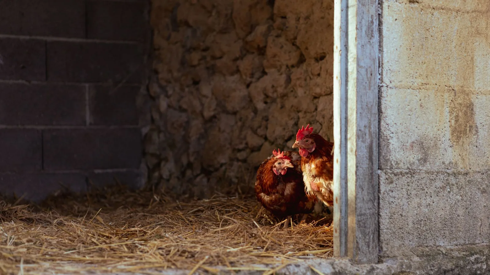
The clinical presentation of HPAI in poultry is highly variable, depending on viral strain, host species, age, and immune status. In susceptible chickens and turkeys, peracute death with minimal premonitory signs is common [5, 6]. Morbidity and mortality rates can approach 100% in naive flocks. Clinical signs, when observed, include severe depression, ruffled feathers, inappetence, cyanosis of the comb and wattles, edema of the head and neck, and respiratory distress characterized by gasping and coughing [6]. Neurological signs, including torticollis, ataxia, and paralysis, result from viral invasion of the central nervous system [25]. Diarrhea and a dramatic drop in egg production are frequently reported in laying flocks [6].
Clinical Signs in Waterfowl and Wild Birds
Ducks and geese often exhibit subclinical or mild infection, serving as efficient shedders without overt disease [5, 56]. However, some HPAI strains, particularly H5N1 clade 2.3.4.4b, have caused significant mortality in wild bird populations, including swans, geese, and raptors [3, 4]. Neurological signs, including disorientation and inability to fly, have been observed in affected wild birds. Mortality events in colonial nesting seabirds have been documented, raising conservation concerns [3].
Pathogenesis and Pathology
HPAI viruses replicate systemically, targeting endothelial cells, parenchymal cells of multiple organs, and lymphocytes [25]. The multibasic cleavage site in HA allows for activation by ubiquitous furin-like proteases, enabling replication in many tissues. Gross pathological findings include hemorrhages and necrosis in multiple organs, including the pancreas, heart, spleen, and kidneys [6]. Pancreatic necrosis is a characteristic lesion in some species. Microscopically, severe necrotizing inflammation and vasculitis are observed. The host innate immune response, including ZBP1/DAI-dependent cell death pathways, contributes to tissue damage and pathogenesis [25]. Gene expression meta-analysis has identified novel pathways involved in the host response to avian influenza virus infection, including dysregulation of immune signaling and metabolic processes [26].
Host Range Expansion and Mammalian Infections
The host range of HPAI H5N1 clade 2.3.4.4b has expanded dramatically, with documented infections in a broad range of mammalian species, including dairy cattle, foxes, seals, and domestic cats [27, 4]. The detection of H5N1 in dairy cattle represents a novel and concerning development, indicating adaptation to mammalian hosts [27]. The global panzootic in mammals underscores the pandemic potential of these viruses [4]. The PB2 mutation triad (I147T, K339T, A588T) has been associated with enhanced replication in mammalian cells and increased zoonotic risk [8]. Mechanistic models of cross-species transmission have identified critical gaps in understanding the factors that drive spillover events [1].
Diagnostic Approaches
Molecular Detection
Real-time RT-PCR targeting the matrix (M) gene or specific HA subtypes is the gold standard for HPAI diagnosis [22]. High-throughput sequencing enables comprehensive genomic characterization, facilitating phylogenetic analysis and molecular epidemiology [22, 51]. Sequencing of the HA cleavage site is essential for pathotyping. Point-of-care molecular diagnostics are being developed for rapid field deployment.
Serological Detection
Serological assays, including hemagglutination inhibition (HI) and enzyme-linked immunosorbent assay (ELISA), detect antibodies against influenza A virus [6]. These methods are useful for surveillance and for confirming infection in recovered or vaccinated populations. The detection of antibodies in unvaccinated flocks indicates prior exposure.
Differential Diagnosis
Clinical signs of HPAI can resemble those of other avian diseases. Differential diagnoses include Newcastle disease (velogenic viscerotropic), infectious coryza (caused by Avibacterium paragallinarum), fowl cholera (caused by Pasteurella multocida), and acute bacterial septicemias [6]. Laboratory confirmation is essential for definitive diagnosis.
Transmission Pathway Decision Framework
graph TD
 A[Wild Bird Reservoir] -->|Migratory Movements| B[Introduction to Poultry Region]
 B --> C{Direct Contact?}
 C -->|Yes| D[Infected Flock]
 C -->|No| E{Indirect Contact?}
 E -->|Fomites/Equipment| D
 E -->|Human Movement| D
 E -->|Aerosol| D
 D --> F[Viral Shedding]
 F --> G[Environmental Contamination]
 G --> H[Local Spread to Adjacent Flocks]
 G --> I[Long-Distance Spread via Live Bird Movements]
 I --> J[New Geographic Region]
 J --> B
 D --> K[Clinical Disease/Mortality]
 K --> L[Surveillance Detection]
 L --> M[Biosecurity Response]
 M --> N[Depopulation & Quarantine]
 N --> O[Disease Elimination]
Conclusion
The transmission of HPAI is a multifactorial process involving wild bird reservoirs, domestic poultry production systems, human activities, and environmental persistence. The emergence of clade 2.3.4.4b viruses has highlighted the capacity for rapid intercontinental spread and host range expansion. Effective control requires a comprehensive approach integrating robust biosecurity, active surveillance, rapid molecular diagnostics, and targeted movement restrictions. Continued research into the molecular determinants of transmission, host-pathogen interactions, and the ecology of wild bird reservoirs is essential for mitigating the impact of HPAI on animal health and food security.
References
[1] Wang M, Laison EKE, Philippsen T, et al. Mechanistic modelling of highly pathogenic avian influenza: A scoping review revealing critical gaps in cross-species transmission models. PLoS One. 2026. https://pubmed.ncbi.nlm.nih.gov/42060628/
[2] Ma J, Wang YH, Qiu YB, et al. A global dataset of spatiotemporal co-occurrence patterns of avian influenza virus-associated migratory birds. Sci Data. 2026. https://pubmed.ncbi.nlm.nih.gov/41634014/
[3] Steinfurth A, Lynton-Jenkins JG, Cleeland J, et al. Investigating high pathogenicity avian influenza virus incursions to remote islands: detection of H5N1 on Gough Island in the South Atlantic Ocean. Emerg Microbes Infect. 2026. https://pubmed.ncbi.nlm.nih.gov/41632631/
[4] Peacock TP, Moncla L, Dudas G, et al. The global H5N1 influenza panzootic in mammals. Nature. 2025. https://pubmed.ncbi.nlm.nih.gov/39317240/
[5] Alkie TN, Embury-Hyatt C, Signore AV, et al. Comparative pathogenicity of three A(H5N1) clade 2.3.4.4b HPAI viruses in blue-winged teal and transmission to domestic poultry. mSphere. 2025. https://pubmed.ncbi.nlm.nih.gov/40401902/
[6] EFSA Panel on Animal Health and Welfare (AHAW), More S, Bicout D, et al. Avian influenza. EFSA J. 2017. https://pubmed.ncbi.nlm.nih.gov/32625288/
[7] Yamaji R, Saad MD, Davis CT, et al. Pandemic potential of highly pathogenic avian influenza clade 2.3.4.4 A(H5) viruses. Rev Med Virol. 2020. https://pubmed.ncbi.nlm.nih.gov/32135031/
[8] Son SE, An SH, Lee CY, et al. Evolutionary trajectories and zoonotic potential of a PB2 mutation triad (I147T, K339T, and A588T) in avian influenza viruses. Vet Res. 2025. https://pubmed.ncbi.nlm.nih.gov/41361825/
[9] Youk SS, Leyson CM, Seibert BA, et al. Mutations in PB1, NP, HA, and NA Contribute to Increased Virus Fitness of H5N2 Highly Pathogenic Avian Influenza Virus Clade 2.3.4.4 in Chickens. J Virol. 2021. https://pubmed.ncbi.nlm.nih.gov/33268526/
[10] Li X, Li A, Qu F, et al. Evaluation of global distribution, genetic evolution, and mammalian infectivity and pathogenicity of H13 and H16 avian influenza viruses. Emerg Microbes Infect. 2025. https://pubmed.ncbi.nlm.nih.gov/40130325/
[11] Marandino A, Tomás G, Panzera Y, et al. Converging Transmission Routes of the Highly Pathogenic Avian Influenza H5N1 Clade 2.3.4.4b Virus in Uruguay: Phylogeographic Insights into Its Spread Across South America. Pathogens. 2025. https://pubmed.ncbi.nlm.nih.gov/40872303/
[12] Kovács L, Domaföldi G, Bertram PC, et al. Biosecurity Implications, Transmission Routes and Modes of Economically Important Diseases in Domestic Fowl and Turkey. Vet Sci. 2025. https://pubmed.ncbi.nlm.nih.gov/40284893/
[13] Tang H, Fournié G, Li J, et al. Analysis of the movement of live broilers in Guangxi, China and implications for avian influenza control. Transbound Emerg Dis. 2022. https://pubmed.ncbi.nlm.nih.gov/34693647/
[14] Willgert K, Meyer A, Tung DX, et al. Transmission of highly pathogenic avian influenza in the nomadic free-grazing duck production system in Viet Nam. Sci Rep. 2020. https://pubmed.ncbi.nlm.nih.gov/32439997/
[15] Guinat C, Durand B, Vergne T, et al. Role of Live-Duck Movement Networks in Transmission of Avian Influenza, France, 2016-2017. Emerg Infect Dis. 2020. https://pubmed.ncbi.nlm.nih.gov/32091357/
[16] Zeng X, Liu M, Zhang H, et al. Avian influenza H9N2 virus isolated from air samples in LPMs in Jiangxi, China. Virol J. 2017. https://pubmed.ncbi.nlm.nih.gov/28738865/
[17] Houston DD, Azeem S, Lundy CW, et al. Evaluating the role of wild songbirds or rodents in spreading avian influenza virus across an agricultural landscape. PeerJ. 2017. https://pubmed.ncbi.nlm.nih.gov/29255648/
[18] Cardona C, Wileman B, Malladi S, et al. The Risk of Highly Pathogenic Influenza A Virus Transmission to Turkey Hen Flocks Through Artificial Insemination. Avian Dis. 2021. https://pubmed.ncbi.nlm.nih.gov/34412462/
[19] Malladi S, Weaver JT, Lopez KM, et al. Surveillance and Sequestration Strategies to Reduce the Likelihood of Transporting Highly Pathogenic Avian Influenza Virus Contaminated Layer Manure. Avian Dis. 2021. https://pubmed.ncbi.nlm.nih.gov/34412451/
[20] Dunning J, Firth JA, Ward AI. The spread of highly pathogenic avian influenza virus is a social network problem. PLoS Pathog. 2025. https://pubmed.ncbi.nlm.nih.gov/40622959/
[21] Singh M, Toribio JA, Scott AB, et al. Assessing the probability of introduction and spread of avian influenza (AI) virus in commercial Australian poultry operations using an expert opinion elicitation. PLoS One. 2018. https://pubmed.ncbi.nlm.nih.gov/29494696/
[22] Howden K, French SK, Racicot M, et al. Applying Field and Genomic Epidemiology Methods to Investigate Transmission Networks of Highly Pathogenic Avian Influenza A (H5N1) in Domestic Poultry in British Columbia, Canada (2022-2023). Transbound Emerg Dis. 2025. https://pubmed.ncbi.nlm.nih.gov/40697881/
[23] Al-Eitan LN, Almahdawi DL, Khair IY. Phylogenetic Analysis and Spread of HPAI H5N1 in Middle Eastern Countries Based on Hemagglutinin and Neuraminidase Gene Sequences. Viruses. 2025. https://pubmed.ncbi.nlm.nih.gov/40431745/
[24] Ssematimba A, Malladi S, Bonney PJ, et al. Estimating the time of Highly Pathogenic Avian Influenza virus introduction into United States poultry flocks during the 2022/24 epizootic. PLoS One. 2024. https://pubmed.ncbi.nlm.nih.gov/39671367/
[25] Thomas PG, Shubina M, Balachandran S. ZBP1/DAI-Dependent Cell Death Pathways in Influenza A Virus Immunity and Pathogenesis. Curr Top Microbiol Immunol. 2023. https://pubmed.ncbi.nlm.nih.gov/31970498/
[26] Bharathi I U, Rani S, Patil SS, et al. Gene expression meta-analysis identifies novel pathways of the avian influenza virus disease. J Biomol Struct Dyn. 2026. https://pubmed.ncbi.nlm.nih.gov/39600177/
[27] Butt SL, Nooruzzaman M, Covaleda LM, et al. Hot topic: Influenza A H5N1 virus exhibits a broad host range, including dairy cows. JDS Commun. 2024. https://pubmed.ncbi.nlm.nih.gov/39429893/
[28] European Food Safety Authority (EFSA), Alvarez J, Bortolami A, et al. Risk posed by the HPAI virus H5N1, Eurasian lineage goose/Guangdong clade 2.3.4.4b. genotype B3.13, currently circulating in the US. EFSA J. 2025. https://pubmed.ncbi.nlm.nih.gov/40611987/
[29] Higgins D, Looker J, Sunnucks R, et al. Introducing a framework for within-host dynamics and mutations modelling of H5N1 influenza infection in humans. J R Soc Interface. 2025. https://pubmed.ncbi.nlm.nih.gov/40592463/
[30] Nidra FY, Monir MB, Dewan SMR. Avian Influenza A (H5N1) Outbreak 2024 in Cambodia: Worries Over the Possible Spread of the Virus to Other Asian Nations and the Strategic Outlook for its Control. Environ Health Insights. 2024. https://pubmed.ncbi.nlm.nih.gov/38585332/
[31] Navarro-Lopez R, Xu W, Gomez-Romero N, et al. Phylogenetic Inference of the 2022 Highly Pathogenic H7N3 Avian Influenza Outbreak in Northern Mexico. Pathogens. 2022. https://pubmed.ncbi.nlm.nih.gov/36365034/
[32] McDuie F, Matchett EL, Prosser DJ, et al. Pathways for avian influenza virus spread: GPS reveals wild waterfowl in commercial livestock facilities and connectivity with the natural wetland landscape. Transbound Emerg Dis. 2022. https://pubmed.ncbi.nlm.nih.gov/34974641/
[33] Yeo JY, Gan SK. Peering into Avian Influenza A(H5N8) for a Framework towards Pandemic Preparedness. Viruses. 2021. https://pubmed.ncbi.nlm.nih.gov/34835082/
[34] Nandi A, Allen LJS. Probability of a zoonotic spillover with seasonal variation. Infect Dis Model. 2021. https://pubmed.ncbi.nlm.nih.gov/33688600/
[35] Zhang J, Chen Y, Shan N, et al. Genetic diversity, phylogeography, and evolutionary dynamics of highly pathogenic avian influenza A (H5N6) viruses. Virus Evol. 2020. https://pubmed.ncbi.nlm.nih.gov/33324491/
[36] Yousefinaghani S, Dara RA, Poljak Z, et al. A decision support framework for prediction of avian influenza. Sci Rep. 2020. https://pubmed.ncbi.nlm.nih.gov/33149144/
[37] Nguyen NM, Sung HW, Yun KJ, et al. Genetic Characterization of a Novel North American-Origin Avian Influenza A (H6N5) Virus Isolated from Bean Goose of South Korea in 2018. Viruses. 2020. https://pubmed.ncbi.nlm.nih.gov/32709116/
[38] Arbi M, Souiai O, Rego N, et al. Historical origins and zoonotic potential of avian influenza virus H9N2 in Tunisia revealed by Bayesian analysis and molecular characterization. Arch Virol. 2020. https://pubmed.ncbi.nlm.nih.gov/32335769/
[39] Samir M, Vidal RO, Abdallah F, et al. Organ-specific small non-coding RNA responses in domestic (Sudani) ducks experimentally infected with highly pathogenic avian influenza virus (H5N1). RNA Biol. 2020. https://pubmed.ncbi.nlm.nih.gov/31538530/
[40] Ssematimba A, St Charles KM, Bonney PJ, et al. Analysis of geographic location and pathways for influenza A virus infection of commercial upland game bird and conventional poultry farms in the United States of America. BMC Vet Res. 2019. https://pubmed.ncbi.nlm.nih.gov/31088548/
[41] Gilbert M, Prosser DJ, Zhang G, et al. Could Changes in the Agricultural Landscape of Northeastern China Have Influenced the Long-Distance Transmission of Highly Pathogenic Avian Influenza H5Nx Viruses? Front Vet Sci. 2017. https://pubmed.ncbi.nlm.nih.gov/29312966/
[42] Wells SJ, Kromm MM, VanBeusekom ET, et al. Epidemiologic Investigation of Highly Pathogenic H5N2 Avian Influenza Among Upper Midwest U.S. Turkey Farms, 2015. Avian Dis. 2017. https://pubmed.ncbi.nlm.nih.gov/28665726/
Disclaimer: This article is for educational and informational purposes only. It is not intended to substitute for professional veterinary advice, diagnosis, treatment, or regulatory guidance. Always consult a licensed veterinarian or qualified specialist regarding animal health, disease diagnosis, and therapeutic decisions.