Tissue Definition in Biology: 4 Main Types, Structure & Functions
By Dr. Zubair Khalid, DVM, MS, PhD ·
Key Takeaways
- A tissue is a fundamental level of biological organization, comprising similar cells originating from the same embryological development, working collaboratively to perform a specific function, and often supported by an extracellular matrix.
- The four primary vertebrate tissue types are epithelial (forming protective barriers and linings), connective (providing support, binding, and transport, e.g., bone, blood), muscle (enabling movement via contractile cells), and nervous (transmitting electrical signals through neurons and glial cells).
- Histology, the study of tissues, is critical for clinical diagnosis; pathologists analyze tissue biopsies using stains like Hematoxylin and Eosin (H&E) to identify cellular and matrix abnormalities indicative of disease, such as cancer or inflammation.
- Regenerative medicine and drug development rely on a deep understanding of tissue structure and function; for instance, engineering artificial skin requires knowledge of epithelial-connective tissue interactions, and drug toxicity is often assessed by observing liver tissue architecture changes.
- Advanced techniques such as immunohistochemistry, confocal microscopy, and single-cell RNA sequencing are revolutionizing tissue analysis by enabling visualization at molecular resolution and detailed study of cellular communication and organization.
In the study of life, the term “tissue” sits at a critical junction between the microscopic world of cells and the visible complexity of organs. For students, researchers, and healthcare professionals, understanding exactly what a tissue is forms the backbone of histology, pathology, and regenerative medicine. This guide breaks down the biology of tissues in a clear, practical way, covering their definition, classification, and real-world importance.
What Is a Tissue in Biology?
A tissue is a group of similar cells that work together to perform a specific function. These cells share the same origin (embryological development) and are often surrounded by an extracellular matrix that provides structural and biochemical support. Tissues represent the second level of biological organization, sitting above cells and below organs.
In multicellular organisms, cells do not act in isolation. They organize into tissues to carry out tasks such as contracting muscles, lining the gut, transmitting signals, or providing structural support. The study of tissues, known as histology, relies on microscopic examination of thin tissue sections stained with dyes to reveal cellular details.
The four fundamental tissue types in vertebrates are epithelial, connective, muscle, and nervous tissues. Each type has unique structural features and functions, and they combine in various ways to form every organ in the body.
The Four Primary Tissue Types
To make the concept of tissue definition biology more actionable, here is a summary table that compares the four main categories:
| Tissue Type | Structure | Primary Function | Common Examples |
|---|---|---|---|
| Epithelial | Sheets of tightly packed cells with little extracellular matrix; may be simple (single layer) or stratified (multiple layers) | Protection, secretion, absorption, filtration | Skin epidermis, lining of stomach, kidney tubules |
| Connective | Cells scattered within an abundant extracellular matrix (fibers and ground substance) | Support, binding, protection, insulation, transport | Bone, blood, cartilage, adipose tissue |
| Muscle | Long, contractile cells (myocytes) filled with actin and myosin filaments | Movement (voluntary or involuntary) | Skeletal muscle, cardiac muscle, smooth muscle |
| Nervous | Specialized neurons and supporting glial cells | Transmission of electrical impulses, integration of signals | Brain, spinal cord, peripheral nerves |
Each tissue type has subtypes. For example, epithelial tissue can be squamous, cuboidal, or columnar; connective tissue ranges from loose areolar tissue to dense fibrous tissue and specialized forms like blood. Understanding these variations is essential for interpreting tissue biopsies or designing tissue engineering scaffolds.
Why Understanding Tissue Definition Matters
A solid grasp of tissue biology is not just academic. It has direct applications in medicine, biotechnology, and research.
In clinical diagnosis, pathologists examine tissue biopsies to detect abnormalities such as cancer, inflammation, or fibrosis. Knowing the normal histological appearance of each tissue type allows them to spot changes in cell shape, arrangement, or matrix composition.
In regenerative medicine, scientists engineer tissues from stem cells or biomaterials. For instance, creating artificial skin for burn victims requires a deep understanding of epithelial and connective tissue interactions. Similarly, cartilage repair relies on mimicking the properties of native connective tissue.
In pharmacology, drug toxicity is often assessed by observing changes in tissue architecture. Liver tissue, for example, is routinely examined in preclinical studies to evaluate potential damage from new compounds.
In evolutionary biology, comparing tissue types across species reveals how multicellularity evolved and how different organisms solve similar functional challenges.
How Tissues Are Studied
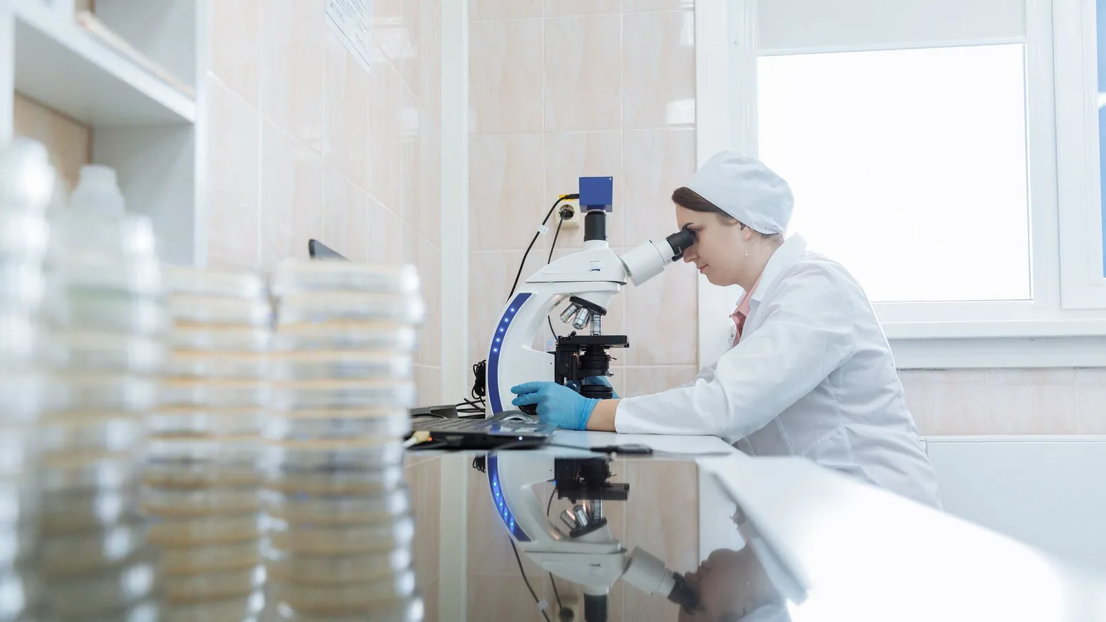
Tissue analysis begins with proper specimen handling. A small piece of tissue is fixed (usually in formalin) to preserve its structure, then embedded in paraffin wax, sliced into thin sections (5 to 10 micrometers thick) with a microtome, mounted on a glass slide, and stained. Hematoxylin and eosin (H&E) is the most common stain: hematoxylin colors nuclei blue, and eosin colors cytoplasm and extracellular matrix pink.
Special stains can highlight specific components. For example, Masson’s trichrome stains collagen fibers blue, useful in studying fibrosis. Immunohistochemistry uses antibodies to detect specific proteins, such as cancer markers.
In research labs, advanced techniques like confocal microscopy, single-cell RNA sequencing, and tissue clearing allow scientists to visualize tissues in three dimensions and at molecular resolution. These methods are redefining what we know about tissue organization and cellular communication.
The definition of a tissue in biology remains the same, but our ability to study it continues to expand, opening new doors for diagnosis and therapy.
--
Written by Zubair Khalid, DVM, MS, PhD, a molecular biologist and computational researcher sharing practical insights in bioinformatics and biotechnology.