Tympanic Ear: Eardrum, Ear Bones, and How Hearing Works
By Dr. Zubair Khalid, DVM, MS, PhD ·
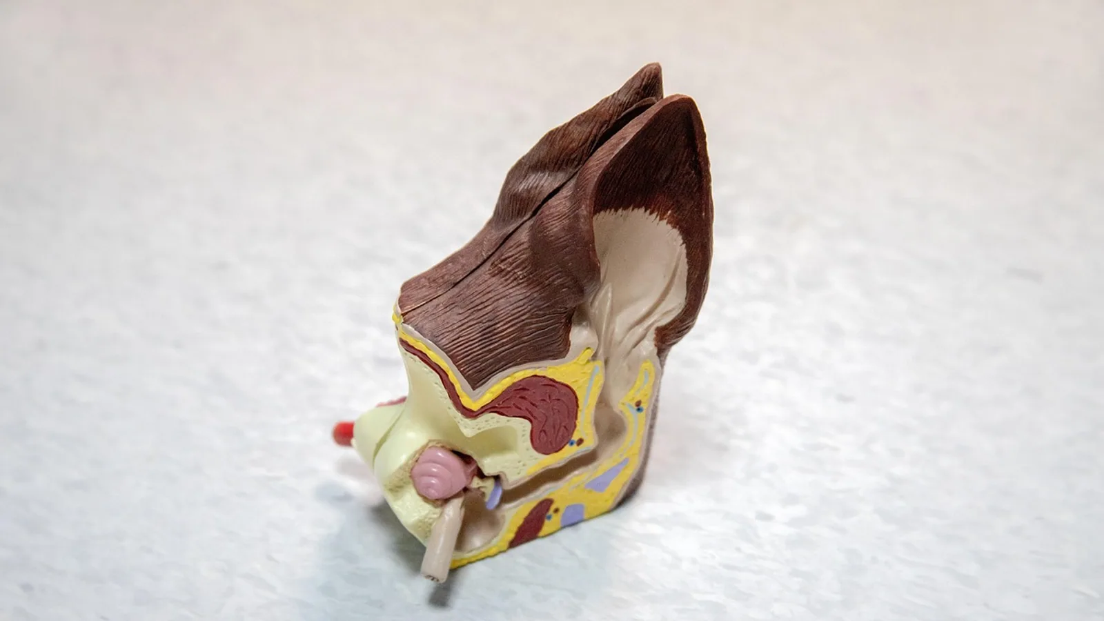
The tympanic ear is the sound-receiving system built around a tympanic membrane (eardrum) that vibrates when airborne sound strikes it and passes that motion through a chain of small bones to the fluid-filled inner ear. In mammals, that chain is the three auditory ossicles: the malleus, the incus, and the stapes.
This matters because air and fluid have very different mechanical properties. Sound travels easily through air but reflects off a fluid surface unless something boosts the pressure first. The tympanic ear solves that problem. It gathers sound over a relatively large membrane, concentrates the force onto a small piston at the oval window, and in doing so recovers most of the energy that would otherwise be lost. Without this middle-ear transformer, airborne hearing would be roughly 20 to 30 decibels weaker, which is the difference between a normal conversation and a whisper you can barely detect.
What the Tympanic Ear Actually Is
A tympanic ear has three functional parts in series:
- A tympanic membrane that catches pressure waves in air.
- A middle ear space containing one or more ossicles that transmit and amplify vibration.
- An oval window, a membrane-covered opening into the fluid of the inner ear.
The term "tympanic ear" distinguishes this arrangement from other vertebrate hearing strategies. Some animals detect vibration through bone conduction or skin, and some rely on a single bone without a true eardrum. The tympanic ear is the version that most mammals, birds, and reptiles use.
The outer ear, where present, funnels sound toward the membrane. In humans the external ear includes the auricle (pinna), the external auditory canal, and the outer surface of the tympanic membrane [1]. The canal shapes the incoming spectrum and helps with localization before any bone moves.
The Eardrum and Tympanic Membrane


The eardrum and tympanic membrane are the same structure. "Eardrum" is the everyday name, and "tympanic membrane" is the anatomical one. It is a thin, roughly cone-shaped sheet of tissue stretched across the end of the ear canal.
Two features make it a good sound receiver. First, it is thin and compliant, so even weak pressure changes move it. Second, it is coupled to the ossicular chain across most of its surface, which lets it drive the bones efficiently rather than flexing in place.
The membrane is not uniform. The pars tensa, the taut lower portion, is the main vibrating region. The pars flaccida, the smaller upper portion, is looser. Clinicians care about this distinction because retraction of the membrane, where it pulls inward toward the middle ear, is a common finding and can progress to more serious disease in some patients [1]. Perforations of the pars flaccida also appear in congenital middle ear malformations [2].
Why the Membrane's Shape Matters
A cone, not a flat disk, is the useful geometry. When sound arrives, the cone's curved profile means different parts of the membrane move with slightly different timing. This produces a lever-like effect inside the membrane itself and helps the manubrium, the handle of the malleus attached to the membrane, move as a unit. The result is a more efficient transfer of energy into the ossicular chain than a simple flat sheet would give.
The Three Ear Bones: Malleus, Incus, and Stapes
The ear bones are the smallest bones in the body and the only ones that do not bear weight. They form a mobile chain suspended in the middle ear cavity by ligaments and two tiny muscles.
Malleus (Hammer)
The malleus is the first bone of the chain. Its handle, the manubrium, is embedded in the tympanic membrane, and its head articulates with the incus. When the eardrum moves inward, the malleus rotates about an axis and pushes the incus.
Incus (Anvil)
The incus sits between the malleus and the stapes. Its body receives the malleus, and its long process reaches down to the stapes head. The incus is the most commonly damaged ossicle in chronic middle ear disease, and its long process is a frequent site of erosion in cholesteatoma [3]. When surgeons reconstruct a damaged chain, the incus is often the bone they reshape or replace.
Stapes (Stirrup)
The stapes is the smallest bone in the human body, at roughly 3 mm long. It has a head, two crura (legs), and a flat footplate that sits in the oval window. The footplate is held in place by the annular ligament, a ring of tissue that seals the window and allows the footplate to piston in and out.
The stapes is the final mover. Every vibration that reaches the inner ear passes through its footplate. Because the footplate is small, the force collected over the much larger eardrum is concentrated onto a tiny area, which is the core of the amplification.
How Hearing Works, Step by Step
Step 1: Sound Arrives and Moves the Eardrum
Sound is a pressure wave. When it reaches the tympanic membrane, higher pressure pushes the membrane inward and lower pressure lets it spring back. The membrane follows the waveform of the sound, oscillating at the same frequency.
Step 2: The Ossicular Chain Transmits the Motion
The malleus, incus, and stapes move as a linked unit. The malleus rotates, the incus follows, and the stapes pistons at the oval window. The chain acts as a lever system, and the lever ratio adds a modest mechanical advantage on top of the area ratio.
Step 3: Impedance Matching Amplifies the Signal
This is the central trick of the middle ear. Air has low acoustic impedance (it offers little resistance to sound), and the fluid of the inner ear has high impedance. When a wave traveling in a low-impedance medium hits a high-impedance one, most of the energy reflects instead of entering. The middle ear reduces that reflection.
Two mechanisms do the work:
- Area ratio. The effective vibrating area of the tympanic membrane is much larger than the area of the stapes footplate. Force collected over a large area, delivered to a small area, produces higher pressure. This is the same principle as pressing a thumbtack: the same push over a tiny point creates far more pressure.
- Lever ratio. The ossicular chain is arranged so the malleus arm is longer than the incus arm, giving a small additional force advantage.
Together these effects raise the pressure delivered to the inner ear fluid enough to overcome most of the impedance mismatch [4]. Without them, a large fraction of incoming sound energy would bounce off the oval window and never reach the sensory cells.
Step 4: The Oval Window Drives Inner Ear Fluid
The stapes footplate pushes into the oval window, displacing fluid in the cochlea. Because fluid is nearly incompressible, a second flexible window, the round window, bulges outward to accommodate the movement. This in-and-out motion sets up a traveling wave along the cochlear partition.
Step 5: Hair Cells Convert Motion to Nerve Signals
Inside the cochlea, the traveling wave bends hair cells at a position that depends on frequency. High frequencies peak near the base and low frequencies near the apex. Hair cells convert that mechanical bending into electrical signals, which travel along the auditory nerve to the brainstem and auditory cortex, where they are interpreted as sound.
Summary Table: Key Facts About the Tympanic Ear
| Structure | Everyday name | Function | Key detail |
|---|---|---|---|
| Tympanic membrane | Eardrum | Receives airborne sound and vibrates | Pars tensa does most of the vibrating |
| Malleus | Hammer | First ossicle, attached to the eardrum | Manubrium embedded in the membrane |
| Incus | Anvil | Middle ossicle linking malleus to stapes | Long process commonly eroded in disease |
| Stapes | Stirrup | Final ossicle, drives the oval window | Smallest bone in the body, about 3 mm |
| Oval window | (no common name) | Entry point into inner ear fluid | Sealed by the annular ligament |
| Round window | (no common name) | Pressure relief for cochlear fluid | Moves opposite to the stapes footplate |
Comparative Anatomy: Mammals, Birds, and Reptiles
The number of ossicles is the clearest difference between groups. Mammals have three. Birds and reptiles have one.
In birds, the bony element of the middle ear is the columella. Its outer end joins a cartilaginous extracolumella, which connects to the tympanic membrane, and its inner end forms a footplate that sits in the oval window, held by the stapedial (columellar) annular ligament [5]. The columella is a single bone doing the job that three bones do in mammals.
Reptiles follow the same single-bone plan. The middle ear evolved independently at least four times across tetrapods, and the tympanic membrane arose at the interface between the mandibular and hyoid arches [6]. That shared developmental origin explains why the same basic parts keep reappearing in different lineages even when the final anatomy differs.
The evolutionary story behind the mammalian three-bone chain is that two of the bones, the malleus and incus, were originally jaw joint bones in the ancestors of mammals. As the jaw joint reorganized, those bones were repurposed for hearing and moved into the middle ear. Birds and reptiles kept the older arrangement, so their jaw joint and their single middle ear bone reflect a different solution to the same problem.
Comparative Table: Tympanic Membrane, Ossicle Number, and Middle Ear Structure
| Group | Tympanic membrane | Ossicles | Middle ear structure |
|---|---|---|---|
| Mammals | Present | Three (malleus, incus, stapes) | Three-bone chain with lever and area-ratio amplification |
| Birds | Present | One (columella) | Single bone plus cartilaginous extracolumella, footplate in oval window [5] |
| Reptiles | Present in most groups | One (columella) | Single bone, footplate in oval window, no mammalian-style chain |
The functional consequence is that birds and reptiles still achieve impedance matching, but with a simpler mechanical arrangement. Their middle ear relies more on the geometry of the columella and the annular ligament than on a multi-bone lever system. In diving birds, the annular ligament is proportionally larger than in terrestrial relatives, and narrower ligaments appear in species specialized for high-frequency hearing [5]. That kind of variation shows how finely the single-bone system can be tuned to ecology.
Primate middle ears add another layer of variation. The spatial relationships among the ossicles and the epitympanic sinuses of the middle ear change through development, and the middle ear cavity and ossicular chain follow different growth trajectories across species [7]. This matters for comparative anatomy because the same three bones can sit in quite different cavities depending on the animal.
How the Tympanic Ear Is Studied and Observed
Researchers and clinicians examine the tympanic ear in several ways, and each method reveals a different layer.
Anatomical Imaging
Micro-computed tomography (micro-CT) resolves the ossicles and middle ear cavity in fine detail. Combining micro-CT with diffusible iodine-based contrast-enhanced CT lets researchers visualize soft structures alongside bone, which is how the developmental changes in primate middle ears were mapped [7]. High-resolution synchrotron X-ray microtomography goes further and quantifies mineral density, porosity, and vascular canal density inside individual ossicles [3].
Mechanical Measurement
Laser Doppler vibrometry measures how much a structure moves in response to sound. It has been used to test 3D-printed middle ear models, comparing their middle ear transfer functions against cadaver temporal bones [8]. This kind of measurement turns an anatomical model into a functional one.
Physical and Computational Models
Because cadaver temporal bones are scarce and variable, researchers build artificial middle ears. One approach uses photopolymer resin for rigid parts and silicone or hot-melt glue for the tympanic membrane, joints, and ligaments, with different silicone hardness values to test how ligament stiffness affects sound transmission [8]. These models support both prosthesis development and surgical training.
Clinical Observation
Otoscopy lets a clinician see the tympanic membrane directly. A dark mass behind the membrane, for example, can indicate an abnormally positioned jugular bulb, which in one reported case protruded into the middle ear and contacted the stapes [9]. Imaging then confirms what the otoscope suggested.
Clinical and Comparative Relevance
The tympanic ear is not just a textbook diagram. Its mechanics explain a large share of conductive hearing loss, the type caused by a problem in the outer or middle ear rather than the sensory cells.
When the ossicular chain is disrupted, sound cannot reach the oval window efficiently. Chronic otitis media with cholesteatoma can erode ossicles and alter their internal microarchitecture, reducing mineral density and increasing porosity, especially around vascular canals [3]. That internal damage matters because surgeons sometimes use the patient's own reshaped ossicles as grafts, and a weakened ossicle may not hold up.
Ossiculoplasty, the reconstruction of the ossicular chain, is built directly on middle ear mechanics. The goal is to restore a conduit that alleviates the impedance mismatch between air and fluid [4]. Surgeons choose among partial and total replacement prostheses depending on which parts of the chain survive.
Congenital malformations add another dimension. Middle ear malformations account for roughly 0.5% to 3% of conductive hearing loss cases, and they can involve the oval window, the ossicular chain, or the facial nerve's course through the middle ear [2]. One reported case involved agenesis of the long process of the incus together with a facial nerve canal defect [2]. These are rare, but they show how tightly the middle ear's parts are packed together.
Vascular anomalies can also intrude. An aberrant internal carotid artery coursing through the middle ear occurs in about 1% of the population and often causes no specific symptoms, which makes it easy to miss until imaging reveals it [10]. Recognizing it before surgery prevents serious bleeding.
Comparative anatomy has practical value too. The single columella of birds and reptiles is a natural experiment in how far a simpler system can go. Studying the columellar annular ligament across bird species shows that ligament geometry tracks ecology, with larger ligaments in diving birds and narrower ones in high-frequency specialists [5]. That is a reminder that "simpler" does not mean "less capable."
Common Mistakes and Limitations
Confusing the eardrum with the ossicles. The eardrum and the ear bones are separate structures that work in series. A perforated eardrum and a damaged ossicular chain are different problems with different treatments.
Assuming the stapes is the only bone that matters. All three ossicles are needed for normal amplification. Losing the incus, for instance, breaks the chain even if the stapes is intact.
Thinking the middle ear creates energy. It does not. It redistributes force over area and through leverage. Total energy is not increased, and some is always lost.
Overlooking the round window. The round window is not a minor detail. Without it, the incompressible cochlear fluid could not move, and the stapes would be pushing against a rigid wall.
Treating the three-bone plan as universal. Mammals have three ossicles. Birds and reptiles have one. Generalizing from human anatomy to all vertebrates leads to errors in comparative work.
Assuming a visible abnormality explains the hearing loss. A high-riding jugular bulb or a retracted membrane may be incidental. Correlating anatomy with function requires proper testing, and individual cases need a veterinarian or physician for diagnosis and management.
Forgetting developmental timing. The middle ear cavity and ossicles do not grow at the same rate, and their spatial relationships shift through ontogeny [7]. Adult proportions are not a reliable guide to juvenile anatomy.
Quick Review
- The tympanic ear is a three-part system: eardrum, ossicles, and oval window.
- The eardrum and tympanic membrane are the same structure.
- Mammals have three ear bones: malleus, incus, and stapes.
- The stapes is the smallest bone in the human body, about 3 mm long.
- Impedance matching works mainly through the area ratio between the eardrum and the stapes footplate, with a smaller contribution from the ossicular lever ratio.
- Birds and reptiles have a single middle ear bone, the columella.
- The round window relieves pressure so cochlear fluid can move.
Frequently Asked Questions
What is the tympanic ear?
The tympanic ear is the hearing system built around a tympanic membrane that catches airborne sound and transmits its vibration through middle ear ossicles to the oval window of the inner ear.
Are the eardrum and tympanic membrane the same thing?
Yes. Eardrum is the common term and tympanic membrane is the anatomical term for the same thin, cone-shaped tissue at the end of the ear canal.
What are the three ear bones and what does each do?
The malleus (hammer) attaches to the eardrum, the incus (anvil) links the malleus to the stapes, and the stapes (stirrup) drives the oval window.
Which is the smallest bone in the human body?
The stapes, the third ossicle, is the smallest bone in the human body at roughly 3 mm long.
How does impedance matching amplify sound?
The eardrum's large effective area collects force and delivers it to the much smaller stapes footplate, raising pressure. A lever ratio in the ossicular chain adds a smaller boost.
Do birds and reptiles have three ear bones like mammals?
No. Birds and reptiles have a single middle ear bone called the columella, while mammals have the three-ossicle chain.
Related Articles
- Membrane Fouling in Protein Ultrafiltration
- Cell Membrane Function Biology
- Ultrafiltration for Protein Concentration
- Selecting a Host System for Membrane Protein Expression
- Membrane-Based Protein Enrichment
- Molecular Dynamics Simulations of Membrane-Bound Viral Glycoproteins
Sources
- Contemporary management of tympanic membrane retraction.
- Middle ear malformations combined with cerebrospinal fluid otorrhea: A case report.
- Spatial quantification of alterations in ossicular microarchitecture related to middle ear disease.
- Middle Ear Mechanics: The Science of Ossiculoplasty.
- Adaptation in the Avian Middle Ear-The Columellar Annular Ligament in Aquatic and Diving Birds.
- Evolution of the Jaw Joint and Middle Ear Morphologies in the Lineage Towards Birds.
- "Ontogenetic Scaling of the Primate Middle Ear".
- Acoustically functional 3D-printed middle ear model for prosthesis development and otosurgical training.
- Extremely High-Riding Jugular Bulb With Conductive Hearing Loss Presenting as a Mass Posterior to the Tympanic Membrane.
- Silent obstruction: The hidden role of aberrant internal carotid artery in middle ear pathology.