Cytoplasm Role: Functions in Cell Biology
By Dr. Zubair Khalid, DVM, MS, PhD ·
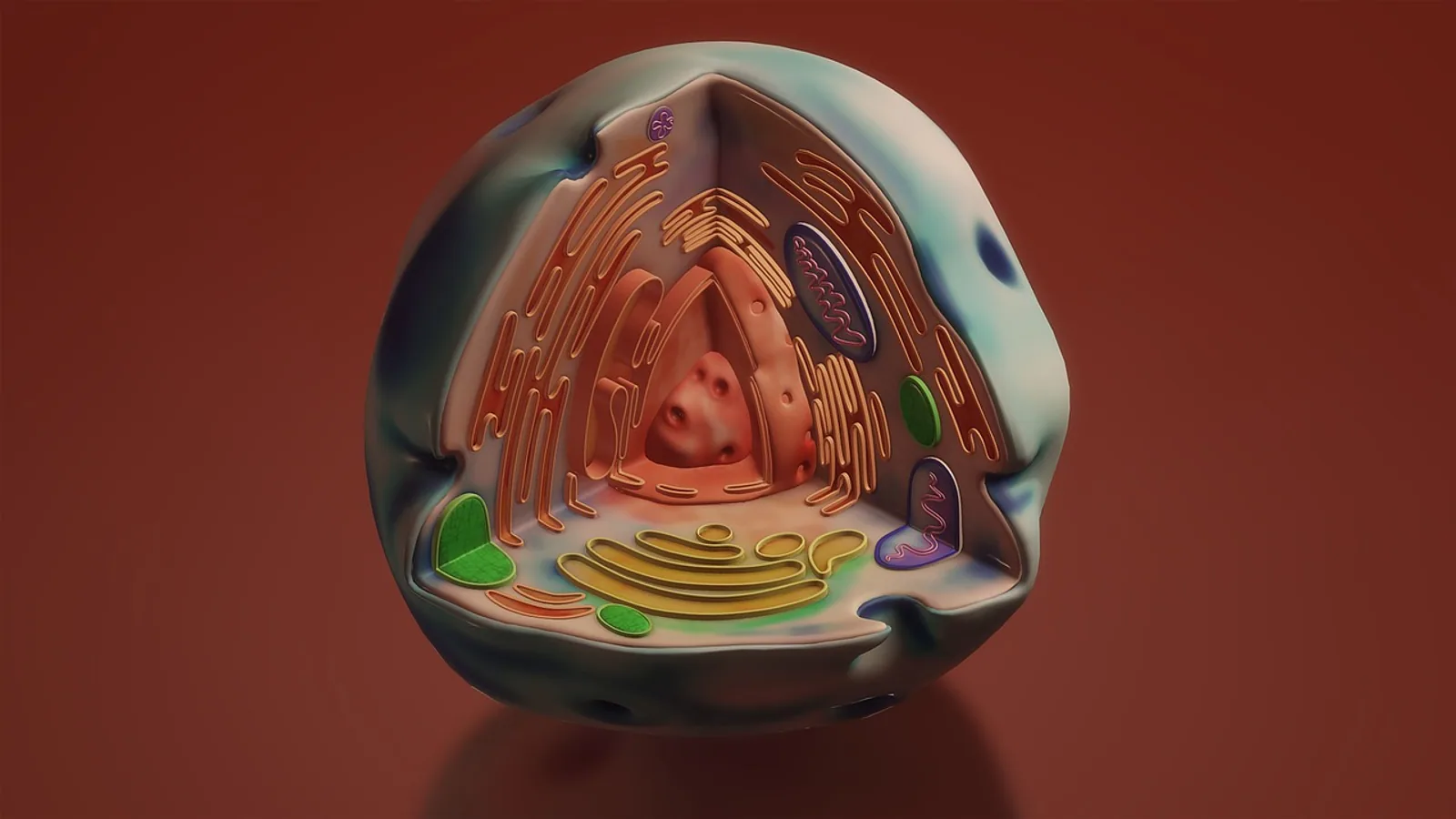
Cytoplasm is everything inside the plasma membrane of a cell except the nucleus: the fluid cytosol plus the organelles, cytoskeletal filaments, and stored inclusions suspended in it. Its role is to host and organize the chemistry of life, from glycolysis and protein synthesis to the directed movement of cargo and the relay of signals from the cell surface inward.
That definition sounds simple, but the cytoplasm carries most of the cell's day-to-day workload. It is where enzymes meet their substrates, where ribosomes build proteins, where molecular motors haul vesicles along tracks, and where signals are converted into responses. Understanding the cytoplasm and its functions explains why cell shape, metabolism, and communication are so tightly linked.
Cytosol vs. Cytoplasm: Getting the Terms Right


The most common point of confusion is the difference between cytoplasm and cytosol. They are not synonyms.
- Cytoplasm is the entire contents of the cell outside the nucleus. It includes the cytosol, all membrane-bound organelles (except the nucleus), the cytoskeleton, and non-membrane-bound structures such as ribosomes and biomolecular condensates.
- Cytosol is only the aqueous, gel-like fluid fraction. It is the liquid in which the organelles and filaments are suspended.
A useful mental model is a bowl of soup. The broth is the cytosol. The broth plus the vegetables, noodles, and everything else in the bowl is the cytoplasm.
This distinction matters because many statements in cell biology are technically about one or the other. Glycolysis happens in the cytosol. Mitochondria, which sit in the cytoplasm, are not part of the cytosol. The National Human Genome Research Institute defines cytoplasm as the gel-like substance inside a cell that surrounds the nucleus and holds the organelles [1].
Why the Cytoplasm Is Not One Uniform Compartment
Textbooks often draw the cytoplasm as a pale, even background. Real cytoplasm is heterogeneous. It is crowded with macromolecules, and that crowding changes the behavior of the water inside it. Cells are densely packed with proteins, nucleic acids, and metabolites, and this crowding disrupts the normal hydrogen-bond networks of water, altering protein folding, enzymatic activity, and the formation of membraneless condensates [2].
The cytoplasm also varies from place to place within a single cell. Regions near the plasma membrane differ in composition from regions near the nucleus. Local pH, ion concentrations, and metabolite levels can shift across short distances. A metabolic enzyme can behave differently depending on which compartment it occupies. One study compared the glycolytic enzyme glyceraldehyde 3-phosphate dehydrogenase (GAPDH) in the cytoplasm and nucleus of Xenopus laevis oocytes and found that the enzyme's activity profile differed between the two compartments, with nuclear GAPDH showing up to 2.5-fold higher peak velocity and non-Michaelis-Menten kinetics, while cytoplasmic GAPDH followed Michaelis-Menten behavior [3]. Same enzyme, same cell, different environment, different catalytic behavior.
Summary Table: Cytoplasm Function Categories
| Function category | Example processes | Key molecules and structures |
|---|---|---|
| Metabolism | Glycolysis, fatty acid synthesis, pentose phosphate pathway, amino acid metabolism | Soluble enzymes (GAPDH, GOT1), NAD+/NADH, ATP, metabolites |
| Protein synthesis and folding | Translation, post-translational modification, chaperone-assisted folding | Ribosomes, tRNA, mRNA, chaperones, aminoacyl-tRNA synthetases |
| Intracellular transport | Vesicle trafficking, organelle positioning, receptor recycling, motor-driven cargo movement | Microtubules, actin filaments, kinesin, dynein, myosin, IFT proteins, Hook adaptors |
| Structural support | Cell shape, mechanical integrity, anchoring of organelles, cortical stiffness | Actin cortex, intermediate filaments, microtubule networks |
| Signal transduction | Receptor signaling, second messenger cascades, signal relay to the nucleus | G proteins, kinases, phosphatases, calcium ions, cAMP |
| Storage and inclusion | Glycogen granules, lipid droplets, pigment granules, secretory vesicles | Glycogen, triglycerides, melanin, stored secretory products |
| Cytoplasmic streaming | Bulk mixing of contents, nutrient distribution in large plant cells | Actin-myosin networks, myosin XI motors |
Metabolic Reactions in the Cytoplasm
The cytoplasm is the cell's main metabolic floor. Most soluble enzymes reside in the cytosol, and many core pathways run there.
Glycolysis
Glycolysis converts glucose into pyruvate through a ten-step pathway, and every step occurs in the cytosol. The pathway produces ATP and NADH, and it operates in nearly every cell type. Because glycolytic enzymes are soluble and not membrane-bound, they depend on the cytoplasmic environment for their activity.
That dependence is real and measurable. In cells lacking the mitochondrial transporter CITRIN, the cytosolic ratio of reduced to oxidized NAD (NADH:NAD+) rises, and glycolysis drops. Normalizing that ratio with nicotinamide riboside restored glycolysis and fatty acid oxidation, showing how sensitive cytoplasmic metabolism is to the local redox state [4].
The Malate-Aspartate Shuttle and Cytosolic Redox
Cytosolic and mitochondrial metabolism are linked by shuttle systems. The malate-aspartate shuttle moves reducing equivalents from the cytosol into mitochondria, and it depends on two forms of aspartate aminotransferase: GOT1 in the cytosol and GOT2 in mitochondria [5]. When GOT1 is lost in rod photoreceptors, NADH accumulates in the cytosol, creating reductive stress that drives photoreceptor degeneration [5]. This is a clear example of why the cytoplasm's redox balance matters for cell survival, not just for metabolism.
Fatty Acid Metabolism and Cytosolic Carbon Flow
Fatty acid oxidation happens in mitochondria, but the carbon products do not stay there. In proliferating cells, a significant portion of carbon derived from fatty acid oxidation exits the canonical TCA cycle as citrate and is converted to malate in the cytosol [6]. This means the cytoplasm is not just a passive recipient of mitochondrial output. It actively processes and redirects carbon toward biosynthetic reactions.
Protein Synthesis
Translation occurs in the cytosol. Ribosomes, transfer RNA (tRNA), and most of the machinery for protein synthesis are located there. Messenger RNA (mRNA) is exported from the nucleus and translated by cytosolic ribosomes, either free in the cytosol or attached to the endoplasmic reticulum.
The cytoplasm also handles protein folding and quality control. Chaperone proteins in the cytosol help newly synthesized polypeptides fold correctly, and misfolded proteins are targeted for degradation.
Intracellular Transport Along Cytoskeletal Tracks
The cytoplasm is not a well-mixed bag. Cargo moves along defined routes, and the cytoskeleton provides the roads.
Microtubules and actin filaments form a dynamic scaffold that ensures membrane-bound compartments are sorted, moved, and distributed correctly [7]. Motor proteins carry cargo along these tracks. Kinesin motors generally move cargo toward the cell periphery along microtubules, while dynein moves cargo toward the cell center. Myosin motors move cargo along actin filaments.
Motor Proteins and Adaptors
The specificity of transport comes from adaptor proteins that link cargo to motors. The intraflagellar transport (IFT) system, originally identified for its role in building cilia, is now recognized as a broader trafficking coordinator. In T lymphocytes, IFT20 works with other IFT components to target recycling vesicles within the endosomal system, delivering receptors and signaling molecules to the immune synapse [7].
Adaptor proteins such as Hook1 link specific organelles to motors. In the filamentous fungus Podospora anserina, the kinesin-3 motor KIN2 and the HOOK1 adaptor are required for early endosome and peroxisome motility, and for positioning mitochondria and endoplasmic reticulum at sites of polarized growth [8]. Without them, organelle distribution breaks down and hyphal growth fails.
Transport in Specialized Cells
Transport through the cytoplasm is not limited to the cell body. In myelinating oligodendrocytes, microtubule-dependent organelle transport occurs within the cytoplasmic spaces of the myelin sheath itself. Peroxisome movement in these spaces is modulated by neuronal electrical activity, and loss of the motor KIF21B or the protein CNP leads to organelle stasis and secondary axon pathology [9]. This continuous cytoplasmic network, which the authors call TRAM (transport route across myelin), enables metabolites and organelles to move between the oligodendrocyte soma and the periaxonal space [9].
Structural Support and Cell Shape
The cytoplasm provides mechanical support through the cytoskeleton, a network of protein filaments that gives cells their shape and organizes their interior.
The actin cortex, a dense mesh of actin filaments just beneath the plasma membrane, controls cell surface tension and shape changes. Intermediate filaments provide tensile strength. Microtubules resist compression and serve as tracks for transport.
Organelle positioning depends on this structural framework. Mitochondria, endoplasmic reticulum, vacuoles, and peroxisomes are not randomly scattered. They occupy specific regions, and that positioning depends on cytoskeletal integrity and motor activity [8]. When transport fails, organelle organization fails with it, and cell function follows.
The physical properties of the cytoplasm itself also contribute. The crowded, gel-like nature of the cytosol restricts the movement of large molecules and creates local microenvironments that influence reaction rates and molecular interactions [2].
Signal Transduction and the Cytoplasm
Signals from outside the cell must cross the plasma membrane and travel through the cytoplasm to reach their targets. The plasma membrane physically and functionally connects the cytoplasm to the extracellular matrix, creating a microenvironment that supports metabolic demands and maintains cellular homeostasis [10].
When a receptor on the plasma membrane binds a ligand, the signal is relayed inward through cytoplasmic signaling proteins. G proteins, kinases, phosphatases, and second messengers such as calcium ions and cyclic AMP carry the message. Many of these molecules are soluble and diffuse through the cytosol. Others are anchored to membranes or scaffolded onto cytoskeletal elements, which keeps signaling localized and specific.
The cytoplasm also integrates stress signals. When cells lose homeostasis, a stress response is triggered with three sequential components: sensors detect the damage, a signaling cascade transmits the alarm, and effectors execute the adaptive action [10]. One such mechanism, the integrated stress response, adjusts protein biosynthetic rates to redirect resources and restore homeostasis [10]. Much of this coordination happens in the cytoplasm.
Cytoplasmic Streaming in Plants
Large plant cells face a transport problem. Diffusion is too slow to move nutrients and organelles across a cell that may be hundreds of micrometers long. Plants solve this with cytoplasmic streaming, also called cyclosis.
Cytoplasmic streaming is the directed, active movement of the cytoplasm driven by actin-myosin networks. In plant cells, myosin XI motors move along actin filaments, dragging organelles and fluid with them. This bulk flow mixes the cell contents and distributes nutrients, metabolites, and signaling molecules throughout the cell.
Streaming is especially important in cells with large central vacuoles, where the cytoplasm is confined to a thin layer near the plasma membrane. Without streaming, that thin layer would be poorly mixed, and organelles at one end of the cell would have limited access to resources produced at the other end.
How Cytoplasm Functions Are Studied
Researchers observe cytoplasm function through several methods.
Live imaging with fluorescent markers tracks organelle movement and cytoskeletal dynamics in real time. In oligodendrocytes, live imaging revealed microtubule-dependent organelle transport within myelin sheaths and showed that peroxisome movement responds to neuronal activity [9].
Immunofluorescence uses antibodies to visualize specific proteins and their locations. A detailed protocol exists for studying cargo transport mediated by cytoskeleton-associated molecular complexes, with a focus on the IFT system and receptor recycling [7].
Whole compartment activity profiling isolates intact nuclei and cytoplasms and measures enzyme behavior in near-physiological conditions. This approach revealed that GAPDH behaves differently in the cytoplasm and nucleus [3].
Stable isotope tracing follows the fate of labeled carbons through metabolic pathways. Using 13C-labeled fatty acids, researchers showed that fatty acid-derived carbon exits the TCA cycle as citrate and is converted to malate in the cytosol of proliferating cells [6].
Spatial organellomics combines automated segmentation with machine learning to classify cell states from multi-organelle signatures. This approach has been used to map cell-state diversity in liver and pancreas and to link organelle remodeling to metabolic adaptation during nutritional stress [11].
Clinical and Comparative Relevance
Cytoplasmic dysfunction appears in many diseases.
In Sjögren's syndrome, salivary gland epithelial cells undergo structural changes that lead to loss of cell polarity and disrupted barrier function. These changes are sensed by subcellular organelles as a condition of stress, triggering adaptive responses that attempt to restore homeostasis [10].
In retinal disease, loss of the cytosolic enzyme GOT1 causes NADH accumulation and photoreceptor degeneration. NADH oxidation, whether by metabolic or genetic means, prolongs photoreceptor survival, implicating reductive stress as a driver of cell death [5].
In liver disease, the presence of anti-LC1 antibodies (liver cytosol antibody type 1) is associated with an increased risk of decompensated cirrhosis or liver failure. Patients positive for anti-LC1 with viral hepatitis had a 2.25-fold increased risk compared with patients with other diagnoses [12]. This antibody targets a cytoplasmic antigen, showing that immune responses to cytoplasmic components can have clinical consequences.
In aging and metabolic stress, mitochondrial quality control and cytoplasmic redox balance are tightly linked. A study of D-galactose-induced kidney aging in rats found that mitochondrial impairment, oxidative stress, and altered mitophagy-related proteins contributed to renal dysfunction, and that nano-encapsulated chrysin partially restored mitochondrial function and structure [13]. The cytoplasm is where many of these stress signals converge.
Common Mistakes and Limitations
Confusing cytoplasm with cytosol. Cytoplasm includes the cytosol plus organelles and inclusions. Cytosol is only the fluid.
Treating the cytoplasm as a single well-mixed compartment. The cytoplasm is heterogeneous. Local composition, crowding, and redox state vary, and these differences affect enzyme behavior and signaling [2][3].
Assuming all cytoplasmic processes are freely diffusible. Many molecules are anchored, scaffolded, or transported along cytoskeletal tracks. Diffusion alone does not explain how cargo reaches its destination [7][8].
Overlooking the role of water structure. The crowded environment of the cytoplasm alters water's hydrogen-bond networks, which affects protein folding, enzymatic function, and phase separation [2]. The cytoplasm is not just a solvent.
Forgetting that transport requires energy and specificity. Motor proteins consume ATP, and adaptor proteins determine which cargo goes where. Transport failure leads to organelle mispositioning and cell dysfunction [9][8].
Assuming findings from one cell type apply to all. Cytoplasmic organization differs between cell types. What is true for a hepatocyte may not be true for a neuron or a plant cell.
Individual cases of disease related to cytoplasmic dysfunction require clinical evaluation. A veterinarian or physician should assess any specific health concern.
Quick Review
- Cytoplasm = cytosol + organelles + inclusions. Cytosol = the fluid alone.
- Glycolysis, protein synthesis, and most soluble enzyme activity occur in the cytosol.
- The cytoplasm is crowded and heterogeneous, not uniform. Crowding affects water structure, enzyme behavior, and condensate formation.
- Intracellular transport depends on microtubules, actin filaments, motor proteins, and adaptor proteins such as IFT components and Hook1.
- The cytoskeleton provides structural support and positions organelles.
- Signal transduction relays external signals through the cytoplasm to internal targets.
- Cytoplasmic streaming in plants uses actin-myosin networks to mix contents and distribute nutrients.
Frequently Asked Questions
What is the main function of the cytoplasm?
The cytoplasm hosts most of the cell's metabolic reactions, supports protein synthesis, enables intracellular transport, provides structural support, and relays signals from the cell surface to internal targets.
Is cytoplasm the same as cytosol?
No. Cytosol is the aqueous fluid fraction. Cytoplasm includes the cytosol plus all organelles, the cytoskeleton, and stored inclusions outside the nucleus.
Where does glycolysis occur?
Glycolysis occurs in the cytosol. All ten enzymatic steps of the pathway take place there, and the pathway depends on the local redox state of the cytoplasm.
How do organelles move through the cytoplasm?
Organelles move along microtubules and actin filaments using motor proteins such as kinesin, dynein, and myosin. Adaptor proteins link specific cargo to specific motors.
What is cytoplasmic streaming?
Cytoplasmic streaming is the active, directed movement of cytoplasm driven by actin-myosin networks. It mixes cell contents and distributes nutrients, especially in large plant cells.
Why is the cytoplasm described as heterogeneous?
The cytoplasm varies in composition, crowding, pH, and redox state across different regions of the cell. These local differences affect enzyme activity, signaling, and molecular interactions.
Related Articles
- Cell Membrane Function Biology
- Cell Membrane Cholesterol: Its Role in Membrane Fluidity and Function
- Buffer Solutions: Types, Purpose, and Applications in Biology
- Red Blood Cell Biology: Structure, Function, and Measurement Context
- The Role of Chromatin Remodelers in Cell Differentiation: A Review of SWI/SNF and ISWI Functions in Lineage Commitment
- Anticodon Sequence: Definition, Function, and Role in Translation
- Centriole Function: Roles in Cell Division
Sources
- Cytoplasm
- Water Under Molecular Crowding: From Femtosecond H-Bond Networks Dynamics to Biological Function.
- Biochemical evidence that the whole compartment activity behavior of GAPDH differs between the cytoplasm and nucleus.
- Nicotinamide riboside rescues dysregulated glycolysis and fatty acid β-oxidation in a human hepatic cell model of citrin deficiency.
- Redox imbalance dictates dependence on GOT1 versus GOT2 for rod photoreceptor health during aging and stress.
- (13)C stable isotope tracing reveals distinct fatty acid oxidation pathways in proliferative versus oxidative cells.
- Immunofluorescence-Based Study of Cargo Transport Mediated by Cytoskeleton-Associated Molecular Complexes.
- Kinesin-3 and the cargo adaptor Hook1 regulate early endosome and peroxisome motility and are required for mitochondrial and endoplasmic reticulum spatial organization during polarized fungal cell growth in Podospora anserina.
- An activity-modulated transport route across myelin (TRAM) for motor-driven organelle transfer in oligodendrocytes.
- Cellular stress leading to epithelial cell dysfunction in Sjögren's syndrome.
- Multi-organelle signatures map cell-state diversity and metabolic adaptation in tissues.
- Viral hepatitis is associated with increased risk of decompensated cirrhosis or liver failure in patients positive for liver cytosol antibody type 1.
- Chrysin preserves renal structure and mitochondrial quality control in D-galactose-induced kidney aging: enhanced efficacy via nano-encapsulation.