Are Bats Blind? Echolocation and Vision Explained
By Dr. Zubair Khalid, DVM, MS, PhD ·
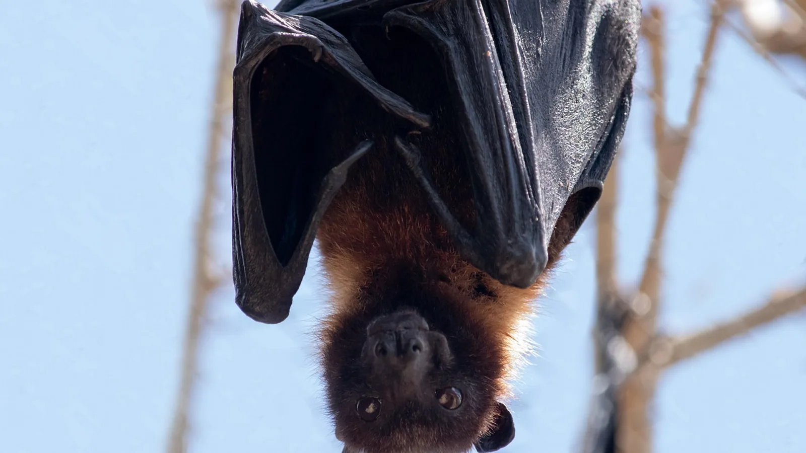
No, bats are not blind. Every bat species studied to date has functional eyes with a retina, a lens, and a working optic nerve, and most bats use vision alongside echolocation. The myth that bats are blind likely comes from the fact that many insect-eating bats have small eyes and hunt at night using sound, so people assumed the eyes were vestigial. They are not.
The real story is a division of labor. Bat eyesight handles long-range navigation, horizon detection, and spotting predators. Echolocation, a biological sonar system, handles the fine-scale work of tracking and catching prey in the dark. Some bats lean heavily on one system. Others use both. A few, like the Old World fruit bats, barely echolocate at all and rely on large, well-developed eyes.
This guide explains what bats actually see, how echolocation works, how the two systems interact, and where the "blind as a bat" idea came from. It also covers the two major bat groups, the microchiropterans and megachiropterans, because their eyes and ears are built very differently.
The Short Answer: Bats Are Not Blind
Bats are mammals in the order Chiroptera, and like other mammals they have two eyes with rod and cone photoreceptors. Rods handle dim-light vision. Cones handle brighter-light vision and, in many species, color. Every bat retina examined so far contains both cell types, even in species that echolocate constantly.
A study of the greater horseshoe bat (Rhinolophus ferrumequinum), a microbat with a sophisticated constant-frequency echolocation system, found a strongly rod-dominated retina. Rods made up 97.49% of photoreceptors and cones 2.51%, with a mean rod-to-cone ratio of about 39 to 1 [1]. That sounds like a weak eye until you remember what it is built for. A rod-heavy retina is a low-light retina. It captures photons efficiently at dusk and after dark, which is exactly when this bat flies.
The same retina also contained cone cells, and the presence of cones means the eye retains a photopic (brighter-light) capability alongside its scotopic (dim-light) one [1]. The bat eye is not a degenerate leftover. It is a specialized nocturnal camera.
Why People Think Bats Are Blind
The myth has several roots.
First, most bats are nocturnal and many are small. A little brown bat or a pipistrelle has eyes that look like shiny beads, not the large forward-facing eyes of an owl. Small eyes read as "poor vision" to a human observer, but eye size alone does not determine visual capability. Retinal structure, photoreceptor density, and neural processing matter more.
Second, echolocation is spectacular and easy to demonstrate. A bat can thread through a room full of wires in total darkness. That performance is so impressive that it overshadows the visual system, and early naturalists assumed the eyes must be useless.
Third, the phrase "blind as a bat" entered common language long before anyone studied bat retinas under a microscope. It is a figure of speech, not a finding.
Fourth, some bats really do have reduced visual systems. The David's myotis (Myotis davidii), an echolocating insectivore, has dim-light but not daylight-adapted vision, and five visual protein genes have degenerated into pseudogenes in its lineage [2]. But reduced is not absent. Even this species retains a working dim-light visual system, and its close relatives fly and forage successfully with it.
The Two Great Bat Groups
Bat biologists traditionally split Chiroptera into two informal groups based on anatomy and sensory strategy. Modern genetics has rearranged the family tree, but the sensory contrast still holds and it is the fastest way to understand bat vision.
Microchiropterans: Echolocation First
Microchiropterans are the smaller bats, and nearly all of them use laryngeal echolocation. That means they produce ultrasonic calls with their larynx and analyze the returning echoes. Their eyes are often small relative to body size, and their retinas are rod-dominated for dim light [1][3].
This does not mean their eyes are decorative. The big brown bat (Eptesicus fuscus) navigates complex obstacle courses using vision and echolocation together, or with echolocation alone when visual cues are removed [4]. When researchers impaired the bats' vestibular system with heavy water and also removed visual cues, performance dropped sharply. With vision available, the bats compensated. That is direct evidence that the visual channel is doing real work during flight [4].
Megachiropterans: Vision First
Megachiropterans are the Old World fruit bats, family Pteropodidae. They have large eyes, a well-developed visual system, and no laryngeal echolocation. They orient primarily by vision and olfaction [5]. The one exception is the genus Rousettus, the cave-roosting fruit bats, which evolved a clicking form of echolocation that is produced with the tongue rather than the larynx. That is a separate invention, not the ancestral bat system.
The megachiropteran eye is a serious optical instrument. In the African straw-colored fruit bat (Eidolon helvum), the ratio of mean corneal diameter to mean axial eye diameter was 0.58, meaning the cornea is large relative to the eye [6]. A large cornea gathers more light. The retina also has an undulating structure with reflective material, a tapetum lucidum, that acts like a series of tiny mirrors. Each parabolic surface of the choroidal papillae reflects light onto the adjacent surface, re-sensitizing photoreceptors in the region. This boosts dim-light sensitivity and helps explain why fruit bats can navigate and forage visually at night [6].
Comparison Table
| Feature | Microchiropterans (most insect-eating bats) | Megachiropterans (Old World fruit bats, Pteropodidae) |
|---|---|---|
| Echolocation type | Laryngeal (voice box) echolocation | None, except Rousettus, which clicks with the tongue |
| Eye size | Often small relative to body, varies by species | Large |
| Retina | Strongly rod-dominated, cones present | Rod-dominated with specialized reflective structures |
| Primary sensory mode | Echolocation for prey capture, vision for navigation | Vision and olfaction |
| Typical diet | Insects, some blood, frogs, fish, nectar | Fruit, nectar, pollen |
| Vision role | Long-range orientation, predator detection, some prey tasks | Main orientation and foraging sense |
| Ultraviolet sensitivity | Present in some species, lost in others | Present in tree-roosting species, lost in cave-roosting species |
How Bat Eyes Work
Bat eyes follow the standard mammalian blueprint. Light enters through the cornea, passes through the pupil and lens, and lands on the retina at the back of the eye. The retina converts photons into electrical signals, and the optic nerve carries those signals to the brain.
Rods, Cones, and the Dim-Light Trade-Off
Rods are the night-vision cells. They are extremely sensitive to light but do not distinguish color well. Cones need more light but support sharper vision and, in animals with multiple cone types, color discrimination.
Across bats, retinas are rod-heavy. The greater horseshoe bat's retina is about 97.5% rods [1]. That ratio is a design choice. A retina packed with rods can detect very faint light, which is what a bat needs when it flies at dusk or after dark. The cost is reduced color vision and lower acuity in bright light.
Cones are still present, and they are not vestigial. Cone photoreceptors were found in both the Egyptian fruit bat (Rousettus aegyptiacus) and the insectivorous Kuhl's pipistrelle (Pipistrellus kuhlii), despite rods dominating in both [7]. Cones give bats a photopic channel for the brighter edges of twilight.
The Neural Side of Bat Vision
Photoreceptors are only the front end. The brain has to process what the eye collects, and bat brains appear tuned for sensitivity over speed.
Researchers recorded activity in the superior colliculus, a midbrain visual center, of the echolocating bat Phyllostomus discolor. Response latencies were in the normal mammalian range, but the neural spatial sampling areas were unusually large compared with other mammals [3]. Large sampling areas mean each neuron pools light from a wider patch of retina, which boosts sensitivity in dim conditions at the cost of fine detail. The authors concluded that echolocating bats likely use spatial summation, not longer integration times, to see better in low light [3].
That is a meaningful design trade. A bat does not need to read fine print. It needs to detect a tree line, a water surface, or a predator silhouette at a distance, and it needs to do so with very few photons.
A Retina Built for Both Worlds
The presence of AII amacrine cells in bat retinas is another sign that the eye handles both rod and cone signals. AII amacrine cells are interneurons that route rod signals into the cone pathway, letting a rod-heavy retina use the same output wiring for both dim and brighter light. These cells have been identified in the greater horseshoe bat retina using parvalbumin immunoreactivity, and their existence suggests the bat retina processes both rod-driven and cone-driven signals [8].
Cholinergic amacrine cells, another key retinal element, have also been mapped in the horseshoe bat retina, with an average total density of 788 cells per square millimeter [9]. The bat retina is not a stripped-down sensor. It has the full complement of retinal circuitry.
How Echolocation Works
Echolocation is active sensing. The bat emits a sound, the sound bounces off objects, and the returning echo carries information about distance, size, shape, and movement. The bat's auditory system then reconstructs a spatial image from those echoes.
Call Design
Most bats use frequency-modulated (FM) calls, which sweep rapidly through a range of frequencies. FM calls give excellent distance resolution, which is why they are common in bats that hunt insects in cluttered spaces. Some bats, including horseshoe bats and Old World leaf-nosed bats, use constant-frequency (CF) calls with a long steady tone, often with a short FM sweep at the end. CF calls are exquisitely sensitive to the Doppler shift caused by a moving insect, which helps the bat detect fluttering prey against background clutter [10].
Reading the Echo
The bat's brain compares the outgoing call with the returning echo. Time delay gives distance. Loudness gives size and reflectivity. Frequency shifts give relative motion. The auditory system of echolocating bats is highly specialized, and comparative genomics has found strong signatures of adaptation in auditory perception genes across rhinolophoid bat lineages [10].
Echolocation Is Not Perfect
Echolocation has limits. It works poorly in noise, it has a maximum useful range, and it costs energy. When researchers exposed Daubenton's bats (Myotis daubentonii) to broadband noise while they landed on a target, the bats were almost three times more likely to need multiple attempts compared with silent controls, and they showed a Lombard response of 0.18 dB of call increase per dB of noise [11]. Echolocation is powerful, but it is not magic, and bats adjust their behavior when it degrades.
Vision and Echolocation Working Together
The most useful way to think about bat senses is as a layered system. Vision and echolocation cover different ranges and different tasks.
Long-Range Navigation: Vision's Job
Vision is good at long range and at detecting large, stable features. A bat leaving a roost at dusk can use the dim outline of a tree line, a river, or a hillside to orient. Echolocation is less efficient at very long range because echoes weaken with distance and the bat must keep calling.
Echolocating bats can support biosonar with vision for far-range operations such as homing [3]. This is the core division of labor. Vision answers "where am I and where is the big stuff," while echolocation answers "what is directly in front of me right now."
Prey Capture: Echolocation's Job
Catching a flying insect requires millimeter-scale precision and millisecond-scale timing. Echolocation delivers that. The bat emits a rapid series of calls as it closes in, updating the target position on each echo, and the final capture happens in a fraction of a second.
The Overlap
The systems are not strictly separated. In the big brown bat, complex maneuvers through fixed and rotating obstacle arrays were carried out using vision and echolocation together, or with echolocation alone [4]. The bat can run on either channel, but it performs best when both are available.
Some bats add a third sense. Substrate gleaners, such as the pallid bat, hunt surface-dwelling prey using a mixture of echolocation, prey-generated sounds, and vision [12]. For these species, echolocation is often limited to general orientation and obstacle avoidance, while passive listening and vision do the hunting [12]. This is a reminder that "bat" covers a huge range of foraging strategies, and the sensory mix shifts with the ecology.
Do Bats See Color?
This is where careful language matters. Bats have cone photoreceptors, and cone cells are the prerequisite for color vision [13]. But having cones does not automatically mean a bat sees the world in rich color, and the evidence does not support strong claims about bat color perception.
What the evidence does support is ultraviolet sensitivity in some species. Two phyllostomid microbats, the nectar-feeding Glossophaga soricina and the fruit-eating Carollia perspicillata, have a substantial cone population expressing two cone opsins, a shortwave-sensitive (S) opsin and a longwave-sensitive (L) opsin [13]. The S opsin sequence suggests ultraviolet tuning, and corneal electroretinogram recordings confirmed elevated sensitivity to UV light [13]. The authors concluded that bats have the prerequisite for daylight vision, dichromatic color vision, and UV vision [13].
UV sensitivity is not universal across bats. A separate study tested four species: the insectivores Hipposideros armiger (constant-frequency echolocator) and Scotophilus kuhlii (frequency-modulated echolocator), and the fruit bats Rousettus leschenaultii (cave-roosting) and Cynopterus sphinx (tree-roosting). After UV stimulation, the visual cortex stained more distinctly in S. kuhlii and C. sphinx, while H. armiger and R. leschenaultii showed no such increase [14]. The pattern suggests that constant-frequency echolocators and cave-roosting fruit bats have lost UV vision, while frequency-modulated bats and tree-roosting fruit bats retain it [14].
The proposed explanation is sensory trade-off plus roosting ecology. Bats that rely heavily on a narrow-band echolocation system may have relaxed selection on short-wavelength vision. Bats that roost in the open, where UV light is available, may keep it [14].
The Sensory Trade-Off Hypothesis
The idea that bats trade vision for hearing has been tested at the molecular level.
Researchers assayed rhodopsin, the dim-light visual pigment, from bat species with different echolocation abilities. Spectral tuning was similar across bats, but the rate at which the light-activated state decayed was significantly slower in a non-echolocating bat than in species using distinct echolocation strategies [15]. Slower decay means the pigment stays active longer, which is consistent with a visual system tuned for dim light. The rates were also remarkably slower than in other mammals, suggesting adaptation to low-light environments [15].
The same study found that evolutionary constraint on the rhodopsin gene increased as Chiroptera diversified, even though no clear shifts were tied to specific echolocation abilities [15]. The picture is not a simple "echolocation replaces vision" story. It is a story of both systems being tuned to the bat's particular niche.
Genomic work points the same way. In David's myotis, an echolocating bat with dim-light vision, five visual protein genes have become pseudogenes, and genome-wide codon usage analysis found enrichment for both vision-related and hearing-related genes [2]. The black flying fox (Pteropus alecto), which has highly developed day vision and does not echolocate, did not show the same pattern [2]. Each species invests in the senses its lifestyle demands.
Common Mistakes and Limitations
Mistake 1: Assuming all bats are the same. "Bat" describes more than 1,400 species with wildly different sensory strategies. A horseshoe bat, a fruit bat, and a pallid bat have different eyes, different ears, and different hunting methods. Any blanket statement about bat vision will be wrong for some group.
Mistake 2: Treating echolocation and vision as competitors. They are complementary. Vision handles long-range orientation and predator detection. Echolocation handles fine-scale prey capture. In the big brown bat, removing visual cues alone did not break flight, but removing visual cues and vestibular input together caused major performance drops [4]. The systems back each other up.
Mistake 3: Reading "rod-dominated" as "poor vision." A rod-dominated retina is a low-light specialist retina. That is an adaptation, not a defect. The greater horseshoe bat's 97.5% rod retina [1] is built for the light levels it actually flies in.
Mistake 4: Claiming bats have excellent color vision. The evidence supports cone-based UV sensitivity in some species and the anatomical prerequisite for dichromatic color vision [13], but it does not support broad claims about how colorful the bat world looks. Overstating this is a real error in popular writing.
Mistake 5: Forgetting that echolocation has limits. Noise degrades it. Daubenton's bats needed almost three times as many attempts to land on a target under broadband noise, and they increased call loudness in response [11]. Echolocation is robust but not invulnerable.
Limitation: species coverage. Retinal anatomy has been described in detail for only a fraction of bat species. The horseshoe bat, the fruit bats, and a handful of phyllostomids are well studied. Many species have never had their photoreceptors counted. Conclusions drawn from one species should not be extended to all bats without evidence.
Limitation: laboratory versus field. Many vision and echolocation experiments use trained bats in controlled rooms. Field conditions include rain, clutter, competing bats, and predators, and the sensory weighting may differ. This is an active area of research.
Limitation: individual variation. Age, health, and experience affect sensory performance in any animal. A specific bat's behavior is not fully predicted by its species average. If you keep bats or work with them in a rehabilitation setting, individual assessment by a qualified veterinarian is the right approach.
Practical Implications for Bat Watchers and Keepers
If you watch bats at dusk, you are watching both senses at work. The bat that appears at your porch light is likely using vision to find the lit area and echolocation to catch the insects drawn to it. That is why some bats hunt around streetlights and why light pollution can change bat foraging behavior.
If you are involved in bat rehabilitation or captive care, remember that light matters. A rod-dominated retina needs time to adapt to changing light levels, and sudden bright light can be aversive. Enclosures that mimic natural twilight cycles are more appropriate than constant bright lighting. Ultraviolet output from some artificial lamps may also be visible to bats that retain UV sensitivity [13], so lamp choice is worth considering.
For researchers and students, the practical lesson is to measure both channels. A study that manipulates only echolocation, or only vision, will miss the interaction. The big brown bat experiments showed that the two systems compensate for each other [4], and the Daubenton's bat experiments showed that bats do not simply switch to vision when echolocation is masked [11]. They adjust their echolocation instead.
What Is Still Uncertain
Several questions remain open.
How do bats weight vision and echolocation in real time? Laboratory work shows both are available, but the moment-to-moment arbitration is not fully mapped.
How widespread is UV vision? It has been confirmed in some phyllostomids and inferred in some fruit bats, but the full distribution across Chiroptera is unknown [14][13].
What does the bat brain do with color signals? Cones are present, and dichromatic vision is plausible, but behavioral color discrimination tests are rare.
How do sensory trade-offs shape genomes over time? The rhodopsin and opsin gene patterns suggest real trade-offs [15][2][16], but the causal direction and the role of ecology are still being worked out.
How do bats handle noisy urban environments? The Lombard response and increased landing attempts under noise [11] suggest costs, but long-term population effects are not established.
Frequently Asked Questions
Are bats blind?
No. All bats have functional eyes, and every bat retina studied contains both rods and cones. The "blind as a bat" phrase is a myth.
Can bats see?
Yes. Bats use vision for long-range navigation, orientation, and predator detection, and many species combine vision with echolocation during flight.
Are bats really blind or do they just have bad eyesight?
They are not blind, and their eyesight is not simply "bad." Bat eyes are specialized for dim light, with rod-dominated retinas that trade fine detail for sensitivity.
Do all bats use echolocation?
No. Most microbats use laryngeal echolocation, but Old World fruit bats (Pteropodidae) generally do not, with the exception of Rousettus, which clicks with its tongue.
How do bats see in the dark?
They use rod-heavy retinas, large corneas in some species, reflective tapetum structures in fruit bats, and neural pooling that increases sensitivity at the cost of detail.
Do bats see color?
Bats have cone photoreceptors, which are the prerequisite for color vision, and some species are sensitive to ultraviolet light. Strong claims about bat color perception are not well supported.
Which bat group has better eyesight?
Megachiropterans, the Old World fruit bats, have larger eyes and a stronger reliance on vision. Microchiropterans have smaller eyes and lean more on echolocation, though their vision is still functional.
Why do people say bats are blind?
Because many bats are small, nocturnal, and famous for echolocation, people assumed their eyes were useless. The saying predates any scientific study of bat retinas.
Related Articles
- Can Cats See in the Dark? Feline Vision Explained
- Computer Vision for Livestock Health and Welfare Observation
- Single-Blind vs. Double-Blind vs. Open Peer Review
- Double-Blind Studies: Design and Interpretation
- Why Is My Cat suddenly going blind? Hypertensive Retinal Detachment and Uveitis
- Cat Eyes: Understanding Feline Vision, Common Eye Problems & Care Tips
Sources
- The photoreceptor populations in the retina of the greater horseshoe bat Rhinolophus ferrumequinum.
- Sensory rewiring in an echolocator: genome-wide modification of retinogenic and auditory genes in the bat Myotis davidii.
- The Neural Basis of Dim-Light Vision in Echolocating Bats.
- Interaction of vestibular, echolocation, and visual modalities guiding flight by the big brown bat, Eptesicus fuscus.
- A comparative analysis of brain size in relation to foraging ecology and phylogeny in the Chiroptera.
- Ocular morphology of the fruit bat, Eidolon helvum, and the optical role of the choroidal papillae in the megachiropteran eye: a novel insight.
- Retinal characterization in the eyes of two bats endemic in the Egyptian fauna, the Egyptian fruit bat (Rousettus aegyptiacus) and insectivorous bat (Pipistrellus kuhlii), using the light microscope and transmission electron microscope.
- AII amacrine cells in the inner nuclear layer of bat retina: identification by parvalbumin immunoreactivity.
- Immunocytochemical localization of cholinergic amacrine cells in the bat retina.
- The Genomes of Two Bat Species with Long Constant Frequency Echolocation Calls.
- Daubenton's bats maintain stereotypical echolocation behaviour and a lombard response during target interception in light.
- Adaptations for Substrate Gleaning in Bats: The Pallid Bat as a Case Study.
- Bat eyes have ultraviolet-sensitive cone photoreceptors.
- Immunohistochemical evidence of cone-based ultraviolet vision in divergent bat species and implications for its evolution.
- Functional Shifts in Bat Dim-Light Visual Pigment Are Associated with Differing Echolocation Abilities and Reveal Molecular Adaptation to Photic-Limited Environments.
- Parallel and convergent evolution of the dim-light vision gene RH1 in bats (Order: Chiroptera).