Peptide Mass and Fragmentation Calculators Explained
By Dr. Zubair Khalid, DVM, MS, PhD ·

By the end of this guide you will be able to calculate the mass of any peptide from its sequence, convert that mass into the m/z values a mass spectrometer reports at each charge state, predict the b and y fragment ions from a tandem mass spectrometry experiment, and run a tryptic digest prediction on paper or in Python. You need three things on hand: a peptide sequence in single-letter amino acid code, a web browser for the online tools, and optionally Python 3.8 or later with the pyteomics package installed using pip install pyteomics.
A peptide mass calculator is one of the most frequently used entries in the broader family of biology calculator tools. Purpose-built computational tools of this kind play an established role in molecular biology research, letting investigators generate quantitative predictions before and after an experiment [1]. This guide explains what peptide mass and fragmentation calculators compute, the exact constants behind every result, a step-by-step workflow, and the common mistakes that produce a wrong number.
What a Peptide Mass Calculator Does
A peptide mass calculator converts an amino acid sequence into predicted masses. You supply the sequence, the tool sums standardized residue masses, adds the mass of one water molecule for the two termini, and reports a molecular weight in daltons (Da), the standard unit of molecular mass. Most tools also convert the neutral mass into m/z, the mass-to-charge ratio an instrument actually measures, and many extend to digest predictions and fragment ion lists.
The need comes from scale. The human genome contains about 20,000 protein-coding sequences [2], and no laboratory can characterize that many proteins without predictions. The standard workflow therefore runs from sequence to expected mass: the sequence comes from DNA sequencing or prior peptide sequencing, and the calculator predicts the mass a spectrometer should report. Agreement between the predicted and observed mass supports the identity, purity, or successful modification of the molecule.
One assumption matters from the start. Calculators treat the chain as a linear polymer, which is correct for mass even when the real molecule is folded, bound to a partner, or assembled into a larger structure. Bacteria, for example, build surface polymers called pili from protein subunits, and each subunit still carries a mass fixed by its linear sequence [3]. Shape changes biology, not mass, so the calculator ignores shape on purpose.
The Constants Every Calculation Depends On
Three constants drive every result.
Residue masses. A residue is an amino acid incorporated in a chain, and its mass equals the free amino acid minus one water molecule. Glycine shows the difference: free glycine has a monoisotopic mass of 75.03203 Da, while the glycine residue contributes 57.02146 Da. The finished peptide gains one H2O (18.010565 Da) back at its termini, so the neutral mass is the sum of residue masses plus water.
The proton. Instruments measure ions, not neutral molecules. Adding one proton (1.007276 Da) gives the singly charged ion, and for a charge state z the m/z is (M + z × 1.007276) / z.
Isotopes. The monoisotopic mass assumes the lightest stable isotope of every element, mostly carbon-12. The average mass weights all natural isotopes and runs roughly 0.06 percent higher for typical peptides. The peptide PEPTIDE has a monoisotopic mass of 799.3599 Da and an average mass of 799.8328 Da, a gap of 0.47 Da that a high-resolution instrument resolves easily.
The standard monoisotopic residue masses used by essentially every tool fit in one dictionary:
MONO = {
    "G": 57.02146,  "A": 71.03711,  "S": 87.03203,  "P": 97.05276,
    "V": 99.06841,  "T": 101.04768, "C": 103.00919, "I": 113.08406,
    "L": 113.08406, "N": 114.04293, "D": 115.02694, "Q": 128.05858,
    "K": 128.09496, "E": 129.04259, "M": 131.04049, "H": 137.05891,
    "F": 147.06841, "R": 156.10111, "Y": 163.06333, "W": 186.07931,
}
H2O = 18.010565    # one water for the two termini
PROTON = 1.007276  # charge carrier
This table collects the formulas you will use most often.
| Quantity | Formula or constant (monoisotopic) |
|---|---|
| Neutral peptide mass | sum of residue masses + 18.01056 Da |
| Singly protonated ion, [M+H]+ | (M + 1.00728) / 1 |
| Doubly protonated ion, [M+2H]2+ | (M + 2 × 1.00728) / 2 |
| b ion, charge 1 | sum of N-terminal residues + 1.00728 |
| y ion, charge 1 | sum of C-terminal residues + 19.01784 |
| a ion | b ion - 27.99491 (CO loss) |
| Water neutral loss | -18.01056 |
| Ammonia neutral loss | -17.02655 |
| Carbon-13 isotope spacing | +1.00335 per carbon |
Peptide Mass Calculator Step by Step
Work through the steps on the test peptide PEPTIDE, then repeat them on your own sequence.
Step 1: Fix the sequence and its chemical state
Write the sequence in single-letter code and decide its state: free or blocked termini, reduced or alkylated cysteines, and any known modifications. PEPTIDE is simple, with free N- and C-termini and no cysteine. A sequence containing cysteine is not simple, because alkylation changes the mass, so record the chemical state before you calculate anything.
Step 2: Choose monoisotopic or average
Use monoisotopic masses for peptides measured on high-resolution instruments, where individual isotope peaks are resolved. Use average masses for large peptides and proteins on low-resolution instruments, where the isotope envelope merges into one broad peak. For peptides up to a few kilodaltons, monoisotopic is the default choice.
Step 3: Add modification masses
Modifications shift the mass by fixed amounts. These are the shifts you will apply most often.
| Modification | Target | Mass shift (Da, monoisotopic) |
|---|---|---|
| Carbamidomethylation | Cysteine | +57.02146 |
| Oxidation | Methionine | +15.99491 |
| Phosphorylation | Serine, threonine, tyrosine | +79.96633 |
| Acetylation | N-terminus or lysine | +42.01057 |
| Deamidation | Asparagine, glutamine | +0.98402 |
| C-terminal amidation | C-terminus | -0.98402 |
Worked example: the peptide ACDE with carbamidomethylated cysteine has a neutral mass of 71.03711 + (103.00919 + 57.02146) + 115.02694 + 129.04259 + 18.01056 = 493.1479 Da, and its [M+H]+ appears at 494.1551 m/z.
Step 4: Run a web calculator
Open PeptideMass on ExPASy, the bioinformatics portal maintained by the SIB Swiss Institute of Bioinformatics. Paste the sequence, set the mass type to monoisotopic, and submit. For PEPTIDE you should get these values:
Sequence:          PEPTIDE
Monoisotopic mass: 799.3599 Da
Average mass:      799.8328 Da
PeptideMass also performs digestion. Select trypsin and the tool cuts after lysine and arginine, except before proline, then lists the mass of every fragment at your chosen number of missed cleavages.
Step 5: Reproduce the number in Python
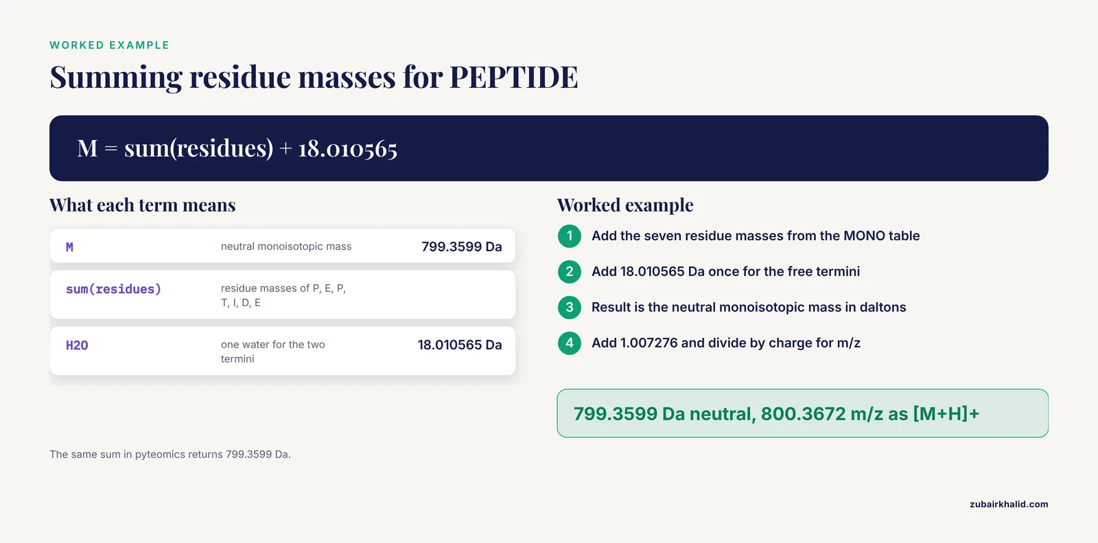
The same calculation in code makes the arithmetic transparent and lets you batch-process sequences.
def peptide_mass(seq, charge=1):
    seq = seq.upper().replace(" ", "")
    unknown = set(seq) - set(MONO)
    if unknown:
        raise ValueError(f"Unknown residue code(s): {', '.join(sorted(unknown))}")
    neutral = sum(MONO[a] for a in seq) + H2O
    return neutral, (neutral + charge * PROTON) / charge

neutral, mz = peptide_mass("PEPTIDE")
print(f"Neutral monoisotopic mass: {neutral:.4f} Da")
print(f"[M+H]+: {mz:.4f} m/z")
Expected output:
Neutral monoisotopic mass: 799.3599 Da
[M+H]+: 800.3672 m/z
The pyteomics library gives the same answer from a maintained mass table:
from pyteomics.mass import calculate_mass

m = calculate_mass("PEPTIDE")
mh = calculate_mass("PEPTIDE", charge=1)
print(f"Neutral: {m:.4f} Da")
print(f"[M+H]+:  {mh:.4f} m/z")
Neutral: 799.3599 Da
[M+H]+: 800.3672 m/zStep 6: Convert to charge states
Electrospray ionization usually produces a series of charge states rather than one ion. For PEPTIDE, the singly protonated ion appears at 800.3672 m/z, the doubly protonated ion at 400.6872 m/z, and the triply protonated ion at 267.4606 m/z. Isotope spacing identifies the charge in an unknown spectrum: 1.00335 divided by the charge gives the spacing between consecutive isotope peaks, so a spacing of about 0.5017 indicates z = 2.
Step 7: Validate with a calibrant peptide
Run the same calculation on angiotensin II (DRVYIHPF), a peptide used in instrument calibration mixtures. Its monoisotopic [M+H]+ is 1046.5418 m/z. If your script or web tool returns that number, your constants and logic are correct, and any later mismatch points to the input rather than the method.
Fragmentation Calculators: Predicting b and y Ions
A fragmentation calculator predicts where a peptide breaks. Tandem mass spectrometry (MS/MS) isolates one peptide and fragments it, and collision-based methods such as collision-induced dissociation (CID) and higher-energy collisional dissociation (HCD) cleave the amide bonds along the backbone. Each cleavage produces two complementary pieces: b ions from the N-terminal side and y ions from the C-terminal side. Matching a ladder of b and y ions to predicted masses is how peptide sequences get confirmed.
The formulas appear in the constants table above. A b ion of charge 1 is the sum of the N-terminal residue masses plus one proton, and a y ion of charge 1 is the sum of the C-terminal residue masses plus water and a proton (19.01784 combined). The following script generates both series:
def by_ions(seq):
    seq = seq.upper()
    n = len(seq)
    b = [sum(MONO[a] for a in seq[:i]) + PROTON for i in range(1, n)]
    y = [sum(MONO[a] for a in seq[n - i:]) + H2O + PROTON for i in range(1, n)]
    return b, y

b, y = by_ions("PEPTIDE")
for i in range(len(b)):
    print(f"b{i+1}: {b[i]:8.4f}   y{i+1}: {y[i]:8.4f}")
Expected output:
b1:  98.0600   y1: 148.0604
b2: 227.1026   y2: 263.0874
b3: 324.1554   y3: 376.1714
b4: 425.2031   y4: 477.2191
b5: 538.2871   y5: 574.2719
b6: 653.3141   y6: 703.3145
Three features make these lists useful.
Complementary pairs. For every cut position, b_i plus y_(n-i) equals the precursor mass plus two protons, 801.3745 for PEPTIDE. Any fragment list that violates this identity contains an error.
Neutral losses. Ions containing serine, threonine, aspartate, or glutamate often lose water (-18.01056), and ions containing lysine, arginine, glutamine, or asparagine often lose ammonia (-17.02655). Phosphopeptides prominently lose phosphoric acid (-97.9769). Fragment calculators list these satellite peaks because search engines must match them.
Immonium markers. Low-mass fragments near m/z 70 flag proline and near m/z 136 flag tyrosine, giving quick residue calls before you read the full ladder.
Two extensions cover the rest of real spectra. a ions equal b ions minus CO (-27.99491) and appear alongside b ions in many spectra. Electron-based fragmentation methods such as electron-transfer dissociation (ETD) and electron-capture dissociation (ECD) cleave the backbone differently and produce c and z ions, and the same calculators that list b and y ions list those series as well.
Choosing a Bioinformatics Calculator for Peptide Work
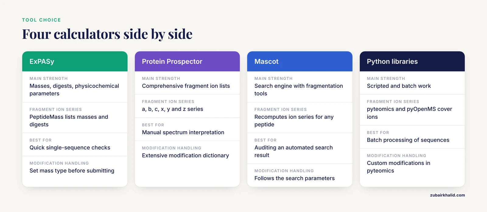
Several mature tools cover the same math with different depth.
ExPASy (SIB). The suite works as a broad biology calculator collection. PeptideMass handles masses and digests, ProtParam adds physicochemical parameters such as isoelectric point and extinction coefficient, FindPept matches observed masses to sequences, and PeptideCutter maps protease cleavage sites.
Protein Prospector (UCSF). MS-Product generates comprehensive fragment ion lists across the a, b, c, x, y, and z series with an extensive modification dictionary, which makes it a standard fragmentation calculator for manual spectrum interpretation.
Mascot (Matrix Science). The search engine includes fragmentation tools that recompute ion series for any candidate peptide, useful when you audit an automated search result.
Python libraries. pyteomics.mass, pyOpenMS, and Biopython cover scripted and batch work. pyteomics computes monoisotopic masses with custom modifications, while Biopython's ProtParam module reports average masses.
Tool choice matters less than consistency. Confirm three settings in any bioinformatics calculator before you trust a number: the mass type (monoisotopic or average), the modification dictionary, and the enzyme specificity rules for digest predictions.
Peptide Mass Calculator Tips
These peptide mass calculator tips prevent most wasted hours.
- Run every important sequence through two independent tools. Agreement between ExPASy and your own script rules out a broken constant table.
- Keep a written list of your lab's fixed modifications, such as carbamidomethylation at +57.02146, and apply it before every calculation rather than after.
- Calculate the full charge-state series, not just [M+H]+, because electrospray spectra of peptides frequently contain 2+ and 3+ ions.
- Use isotope spacing to assign charge before you compare any observed peak to a predicted mass.
- Allow one or two missed cleavages in digest predictions, since incomplete digestion is the norm in real samples.
- Record the mass type next to every number in your notebook. A mass without "monoisotopic" or "average" attached is ambiguous.
- Account for purification tags. A recombinant peptide carrying the common 6xHis tag gains six histidine residues, about 822.35 Da [4].
- Save the exact input string and settings alongside the output, because a prediction you cannot reproduce is not a prediction.
Common Mistakes and Limitations
Most wrong numbers trace back to a short list of peptide mass calculator mistakes.
- Summing free amino acid masses. Free amino acid tables already include the terminal water, so adding another 18.01056 double-counts it. Use residue masses plus one water, or free amino acid masses minus one water.
- Mixing monoisotopic and average values. Comparing a monoisotopic prediction to an average-mass reference creates an error of roughly 0.06 percent that grows with peptide size. Match the mass type to the instrument and to whatever you compare against.
- Dropping fixed modifications. An alkylated cysteine without its +57.02146 shift is wrong by more than most instruments' mass accuracy. Define the chemical state first, as in Step 1.
- Comparing m/z to neutral mass. A doubly charged ion of a 1,000 Da peptide appears near m/z 501, not 1,000. Apply the charge formula every time.
- Ignoring terminal processing. N-terminal acetylation adds +42.01057, signal peptide removal changes the sequence itself, and C-terminal amidation subtracts 0.98402. The peptide substance P (RPKPQQFFGLM) carries a C-terminal amide, so its [M+H]+ of 1347.7354 only appears when that shift is applied.
- Trusting ambiguous codes. X, B, Z, and J stand for sets of residues. Most tools reject them, and a tool that silently substitutes an average value gives a number with hidden uncertainty. Isoleucine and leucine are the exception, because they are isobaric at 113.08406 and interchangeable for mass purposes.
- Over-reading digest predictions. Trypsin cuts after K and R except before P, but real digests also contain missed cleavages, semi-tryptic ends, and enzyme variability. A digest list is a search space, not a sample inventory.
Limitations. Calculators predict positions, not intensities, so they cannot tell you which fragment will be strong. They assume a single covalent chain, so disulfide-linked peptides need a correction of -2.01565 per bond from the reduced sum, and noncovalent complexes need separate treatment. Salt adducts shift ions, with the sodium adduct [M+Na]+ appearing 21.98194 above [M+H]+. None of these are errors in the calculator, they are chemistry the calculator expects you to declare.
How to Check Your Results
Four checks catch nearly every error.
First, compute a calibrant. Angiotensin II should return 1046.5418 m/z for [M+H]+, and any deviation means a constant or a setting is wrong.
Second, test internal consistency. In a fragment list, every b_i plus y_(n-i) must equal the precursor mass plus 2.01455 Da.
Third, compute the same peptide in a second tool. Two independent implementations agreeing on 799.3599 Da for PEPTIDE is stronger evidence than one.
Fourth, express agreement in parts per million (ppm). The relative error is (observed - theoretical) / theoretical × 10^6. At m/z 800, a 5 ppm tolerance corresponds to about 0.0040 Da, which is the window a high-resolution instrument actually delivers.
In engineered-protein work this checking step carries extra weight. Recombinant DNA technology now produces designed structural proteins with control at the level of individual monomers [5], and the predicted mass of the designed construct is the first quantitative check that the expressed product matches the plan.
Where Peptide Calculations Fit in Real Projects
Peptide mass and fragmentation calculators sit at the start of several standard workflows.
In protein engineering and materials design, predicted masses verify expressed constructs before expensive characterization begins [5]. In teaching laboratories, project-based courses that move from gene manipulation through expression, purification, protein quantification, and western blotting [6] rely on predicted masses to interpret sodium dodecyl sulfate polyacrylamide gel electrophoresis (SDS-PAGE) bands and assay results, and semester-long courses built around purifying and analyzing a fusion protein follow the same pattern [7]. In applied proteomics, mass prediction underpins the identification step in settings from clinical biomarker research to aquaculture breeding and disease-resistance programs that now include proteomics among their molecular tools [8].
Mass is also the bridge to structure. Cryo-EM, NMR, and molecular dynamics now delineate the molecular properties of protein complexes at high spatial and temporal resolution [9], and small-angle X-ray scattering coupled with crystallography quantifies conformational ensembles of assemblies in solution [10]. Those methods answer shape and dynamics questions, but every one of them starts from a construct whose composition the mass calculator confirmed first.
Frequently Asked Questions
What is the difference between monoisotopic and average peptide mass?
The monoisotopic mass uses only the lightest stable isotope of each element, while the average mass weights all naturally occurring isotopes. High-resolution instruments resolve individual isotope peaks, so monoisotopic values fit peptides, and average values fit large peptides and proteins on low-resolution instruments.
Why does my calculated peptide mass not match the observed m/z?
The usual causes are a charge state mismatch, a missing modification such as carbamidomethylation, or comparing m/z directly to neutral mass. Apply the charge formula and confirm the peptide's chemical state before you suspect the calculator.
Do I need to add water when I sum residue masses?
Yes. Residue masses exclude the termini, so the neutral peptide mass is the sum of residues plus one water molecule, 18.010565 Da in monoisotopic terms.
How do I calculate the m/z of a doubly charged peptide?
Divide the mass plus two protons by two, using (M + 2 × 1.007276) / 2. The same pattern extends to any charge state z.
What are b and y ions in a fragmentation calculator?
b ions are N-terminal fragments and y ions are C-terminal fragments formed when collision energy breaks the peptide backbone. A fragmentation calculator lists their m/z values so you can match them against a tandem mass spectrum.
Which modification masses matter most for a standard tryptic digest?
Carbamidomethylation of cysteine at +57.02146 Da as a fixed modification and oxidation of methionine at +15.99491 Da as a variable modification cover most standard digest setups.
Can a peptide mass calculator handle X, B, Z, or J in a sequence?
Most calculators reject these codes or silently substitute average values, because they represent ambiguous residues. Replace them with concrete letters or use an ambiguity-aware tool before trusting the output.
Are web peptide mass calculators accurate enough for publication?
Yes, when the mass type, constants, and modifications are set correctly, because mainstream tools draw on the same standard isotope masses. Confirm the result with a calibrant peptide and a second tool, and state the mass type in your methods.
Sources
- emDNA - A Tool for Modeling Protein-decorated DNA Loops and Minicircles at the Base-pair Step Level
- The RNA Revolution in the Central Molecular Biology Dogma Evolution
- Structural and Molecular Biology of a Protein-Polymerizing Nanomachine for Pilus Biogenesis.
- A simple and sensitive SYBR Gold-based assay to quantify DNA-protein interactions
- Molecular design of protein-based materials - state of the art, opportunities and challenges at the interface between materials engineering and synthetic biology
- Development of an integrated and project-based laboratory course in upper-level biochemistry and molecular biology
- Demonstrating core molecular biology principles using GST-GFP in a semester-long laboratory course
- Applications of Molecular Biology and Genomic Technologies in Modern Aquaculture: Current Advances and Future Perspectives
- Editorial: Recent advances in molecular properties of DNA-protein interactions, chromatin and their biological roles
- X-ray scattering reveals disordered linkers and dynamic interfaces in complexes and mechanisms for DNA double-strand break repair impacting cell and cancer biology