Molecular Weight Calculators for DNA, RNA, and Peptides
By Dr. Zubair Khalid, DVM, MS, PhD ·
A molecular weight calculator for DNA, RNA, and peptides converts a sequence into its molecular mass by adding the standard masses of its building blocks and subtracting the small mass lost each time two of them are joined. The result, reported in daltons (Da) or kilodaltons (kDa), is the number behind molar conversions, dosing calculations, and sanity checks on every gel and purification you run.
Molecular weight is the mass of one mole of a molecule, and one dalton equals one gram per mole, so a 50 kDa protein has a molar mass of 50,000 grams per mole. A calculator automates the arithmetic. You paste in a nucleotide or amino acid sequence, and the tool returns the mass along with related outputs such as the elemental formula, the theoretical isoelectric point (pI, the pH at which the molecule carries no net charge), and microgram-to-picomole conversions. These tools sit inside the broader family of biology calculator software, and sequence-based mass calculation is a standard fixture of every bioinformatics calculator package.
The arithmetic is simple. The interpretation is where accuracy is won or lost. A 69-amino-acid scorpion toxin was calculated at 7,721.77 g/mol from its sequence, and the value accompanied the sequence into GenBank, the public sequence database [1]. A 434-amino-acid parasite protein was run through the widely used ProtParam tool and reported at 49.1 kDa [2]. A DNA repair protein with a calculated mass of 31 kDa, meanwhile, migrated on SDS-PAGE (sodium dodecyl sulfate polyacrylamide gel electrophoresis, which separates proteins by chain length) at 40 to 45 kDa and behaved as a roughly 92 kDa species in gel filtration [3]. Calculated and apparent molecular weights are different measurements, and this guide explains both, along with the formulas, the worked examples, and the traps.
What a Molecular Weight Calculator for DNA, RNA, and Peptides Means
Strictly speaking, molecular weight is a dimensionless ratio and molar mass carries units of grams per mole, but laboratory usage treats the two as the same number. When a paper reports a 32.7 kDa protein, it means one mole of those molecules weighs 32,700 grams, and one molecule weighs 32,700 daltons, where the dalton is defined relative to one twelfth of the mass of a carbon-12 atom.
A molecular weight calculator for DNA, RNA, and peptides is any tool that performs this summation from sequence data. The minimum input is a string of letters: A, C, G, and T for DNA, A, C, G, and U for RNA, and the one-letter or three-letter amino acid code for proteins. The minimum output is a single mass. Useful tools add the elemental formula, the theoretical pI, the ultraviolet absorption coefficient at 280 nm for proteins (the extinction coefficient, estimated from tryptophan, tyrosine, and cystine content), and conversion utilities between mass and moles.
The sequences come from everywhere. Cloned complementary DNA (cDNA, a DNA copy of an RNA transcript) yields an open reading frame (ORF, the stretch between start and stop codons that encodes protein) whose base length predicts protein length at three bases per residue. A study of a thioredoxin-like protein in rotifers cloned a full-length cDNA of 1,111 bp, identified an ORF of 878 bp encoding 292 amino acids, and calculated a molecular weight of 32.7 kDa and a pI of 4.97 for the deduced peptide [4]. Transcriptome sequencing (the full set of expressed sequences) feeds calculators as well, as with the scorpion venom toxin above [1], and model-organism databases supply ready-made sequences, as in the analysis of the Toxoplasma gondii RNA-binding protein DDX39, where the sequence retrieved from the ToxoDB database was pushed through ProtParam and a panel of prediction tools [2].
The Chemistry Behind the Numbers
DNA
DNA is a chain of nucleotides joined by phosphodiester bonds (the linkages between adjacent nucleotides), and each bond-forming condensation releases one molecule of water. Calculators sum the masses of the nucleotide units and apply a small correction at the ends.
For everyday planning, two numbers carry most of the load. One base pair of double-stranded DNA averages about 660 Da, so a 1,000 bp fragment weighs roughly 660 kDa, and one microgram of it corresponds to about 1.5 picomoles, roughly 9 × 10^11 molecules. Single-stranded DNA is estimated near 330 Da per nucleotide by the same convention. These quick values are deliberately coarse and often assume sodium-salt forms, so expect the exact calculator output to differ by a few percent.
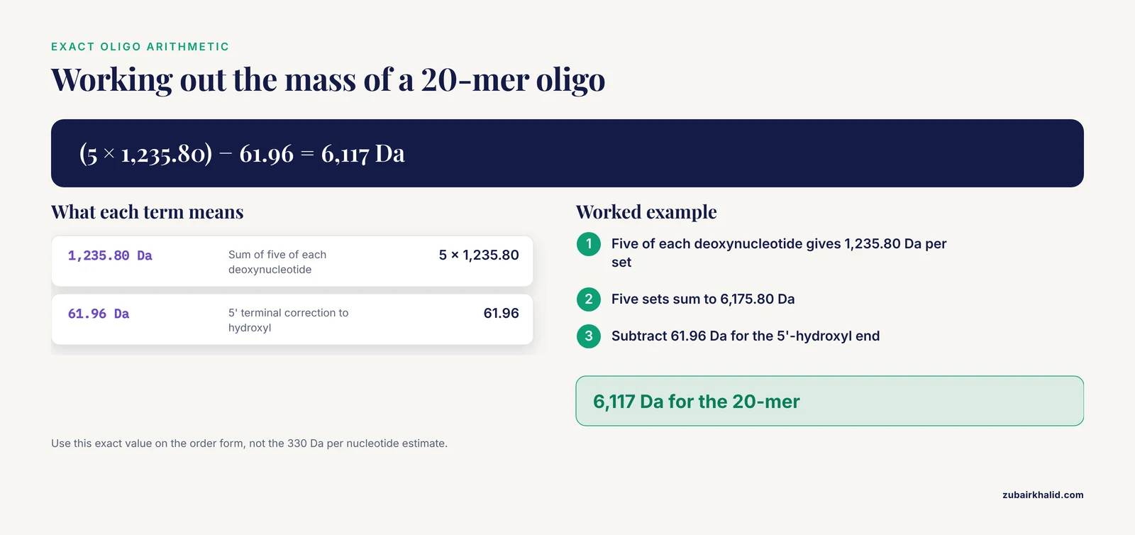
Exact oligonucleotide calculators work base by base. Standard average masses for internal residues are 313.21 Da for dA, 289.18 Da for dC, 329.21 Da for dG, and 304.20 Da for dT. The calculator subtracts 61.96 Da from the total to convert the 5' terminal residue from its internal phosphorylated form to a 5'-hydroxyl end. A 20-mer containing five of each deoxynucleotide therefore comes to (5 × 1,235.80) - 61.96 = 6,117 Da. That exact value, not the rule of thumb, belongs in your lab notebook and on your order form.
RNA
RNA uses ribose, which carries one more oxygen than the deoxyribose in DNA, and uracil replaces thymine. Standard internal residue masses are 329.21 Da for rA, 305.18 Da for rC, 345.21 Da for rG, and 306.17 Da for rU, which lands between roughly 320 and 340 Da per nucleotide depending on composition. The traditional quick estimate of 340 Da per nucleotide sits at the high end of that range.
Two real-world features push an RNA's true mass above the bare sequence value. Most eukaryotic messenger RNAs carry a 7-methylguanosine cap, a modified guanine added at the 5' end, and most carry a poly(A) tail, a run of adenosine residues at the 3' end whose variable length adds about 329 Da per adenosine. A calculator that handles RNA properly lets you specify a cap and a tail length rather than assuming a bare transcript.
Peptides and proteins
Peptide calculators sum the masses of the free amino acids and subtract 18.015 Da for each peptide bond formed, which works out to n minus one water losses for a chain of n residues. A useful anchor is that the occurrence-weighted average residue mass in proteins is close to 110 Da, so a 300-residue protein lands near 33 kDa, and the published examples in this article follow that pattern.
Composition matters as much as length. Protamine, the antimicrobial protein from salmon sperm, has a molecular weight of approximately 5 kDa, yet 60 to 70 percent of its residues are arginine, giving the molecule a strong positive charge that drives its binding to bacterial membranes and its inhibition of bacterial translation [5]. Two peptides with identical masses can behave in opposite ways if one is arginine-rich and the other is not.
Standard corrections a good calculator offers include disulfide bonds, which remove 2.016 Da per bond as two hydrogens are lost, N-terminal acetylation, which adds 42.04 Da, and C-terminal amidation, which removes 0.98 Da. Signal peptide removal is the correction beginners forget most often, and it gets its own entry in the mistakes section below.
Average and monoisotopic mass
Every element has an average atomic mass that reflects its natural isotope mixture (carbon at 12.011, for example) and a monoisotopic mass for its most abundant isotope (carbon-12 at exactly 12.000). Calculators built for molecular biology and protein chemistry default to average masses, which is what you want for SDS-PAGE comparisons and molar conversions. Mass spectrometrists working with small peptides switch to monoisotopic masses, because a small ion resolves into separate isotope peaks and the monoisotopic peak serves as the reference. For a 50 kDa protein the isotope envelope blurs into one broad peak, and the average mass is the meaningful number.
| Molecule | Calculator input | Quick planning estimate | What the calculator actually computes | Corrections to look for |
|---|---|---|---|---|
| Double-stranded DNA | Sequence or length in bp | 660 Da per bp (1 kb ≈ 660 kDa) | Exact mass from base composition, plus μg-to-pmol conversions | Linear versus circular ends, sodium salt basis |
| Single-stranded DNA oligo | Sequence | About 330 Da per nt | Sum of internal residue masses (dA 313.21, dC 289.18, dG 329.21, dT 304.20) minus 61.96 for a 5'-hydroxyl end | 5' phosphate (adds about 80 Da), 3' labels |
| Single-stranded RNA | Sequence | 320 to 340 Da per nt | Sum of ribonucleotide residue masses with terminal correction | 5' cap, poly(A) tail length, modified bases |
| Peptide or protein | Sequence | About 110 Da per residue | Sum of free amino acid masses minus 18.015 Da per peptide bond | Disulfides, acetylation, amidation, signal peptide removal |
Worked Examples from the Research Literature
Published papers show the calculator in routine use, and each example teaches a slightly different lesson.
The scorpion toxin meuNa10 came out of a venom gland transcriptome from the scorpion Mesobuthus eupeus. The sequence encodes 69 amino acids, and physicochemical software calculated a molecular weight of 7,721.77 g/mol with a pI of 8.7. The researchers noted eight cysteine residues stabilizing a cysteine-stabilized alpha/beta fold, deposited the sequence in GenBank, and used the calculated properties to frame the toxin as a stable, hydrophilic three-finger protein [1]. The precision of the reported value, down to hundredths of a dalton, is the signature of a sequence-based calculation rather than a gel estimate.
The Toxoplasma gondii protein TgDDX39 shows the database-to-calculator pipeline. Its 434-amino-acid sequence from ToxoDB was analyzed with ProtParam, which returned a molecular weight of 49.1 kDa, a theoretical pI of 5.55, and the elemental formula C2173H3458N598O661S18, alongside predictions of unstable, hydrophilic character, no transmembrane regions, and 27 phosphorylation sites [2]. The molecular weight anchored a profile used to judge the protein's suitability as a vaccine antigen.
The rotifer protein BpTXNL shows the cloning-to-calculator path. Its 878 bp ORF encodes 292 amino acids, and the calculated mass of 32.7 kDa with a pI of 4.97 accompanied the characterization of a conserved thioredoxin domain with its redox-active CGPC motif [4].
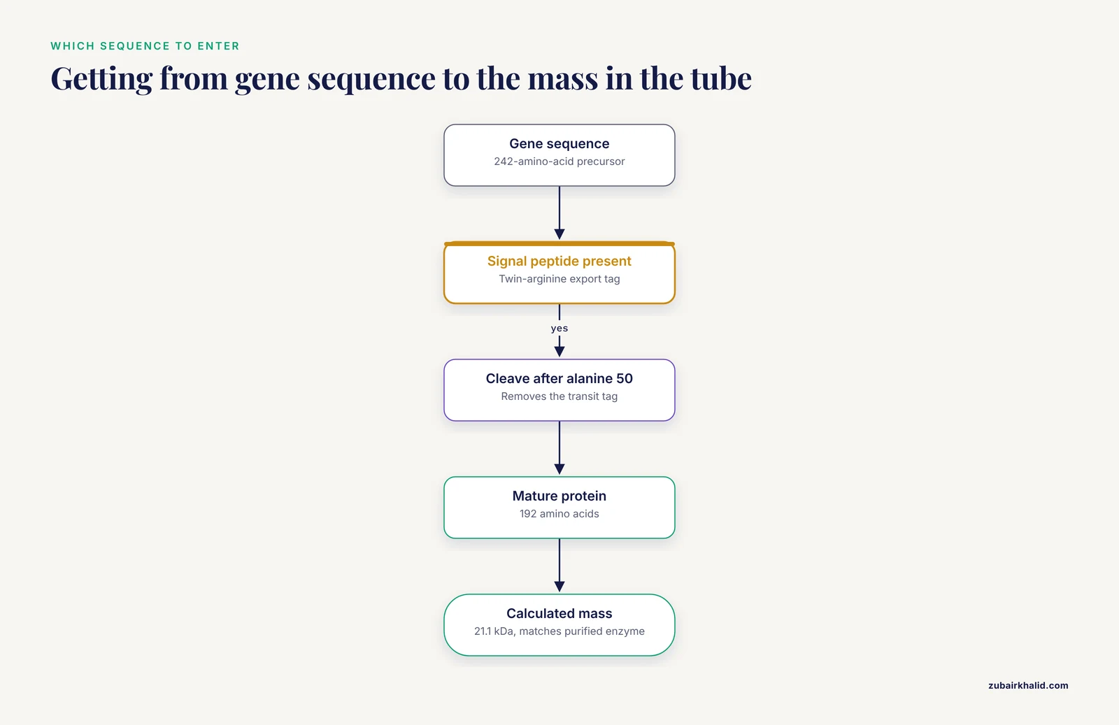
The bacterial xylanase XynCvir from Streptomyces viridodiastaticus MS9 (a xylan-degrading enzyme from a soil bacterium) shows why the sequence you enter must match the molecule in the tube. The gene encodes a 242-amino-acid precursor carrying a twin-arginine translocation signal peptide, a bacterial export tag. Cleavage between alanines 50 and 51 releases a mature protein of 192 amino acids with a calculated mass of 21.1 kDa, which matches the approximately 21 kDa enzyme purified from culture [6].
Older enzymology used subunit molecular weights as the primary identity of a complex. Purified RNA polymerase II from Acanthamoeba castellanii resolved into nine polypeptides reported at 185,000, 140,000, 70,000, 35,000, 21,000, 21,000, 19,000, 18,500, and 16,200 Da, and differences in the largest subunit distinguished two polymerase forms [7]. In an era before genome sequences were routine, those numbers were the enzyme's identity card.
How to Interpret the Output: Calculated Versus Apparent Molecular Weight
A calculated molecular weight is a property of the sequence. An apparent molecular weight is a property of the sequence plus its modifications plus the physics of the measurement. Confusing the two is the most common interpretation error, and the literature supplies a vivid demonstration.
The DNA repair protein XPA has a calculated mass of 31 kDa, yet it ran on SDS-PAGE as multiple bands from about 40 to 45 kDa and behaved as an approximately 92 kDa species in gel filtration chromatography (a separation method that sorts proteins by hydrodynamic size, the space a molecule sweeps through solution). Neither discrepancy reflected post-translational modifications, since mass spectrometry found none and all nine cysteines in the active protein were reduced. Treatment with iodoacetamide, a cysteine-blocking reagent, collapsed the pattern to a single 42 kDa band, and intramolecular crosslinking, which constrains the protein's floppy regions, produced a roughly 32 kDa monomer much closer to the calculated value [3]. The explanation is intrinsic disorder. Unstructured regions migrate anomalously in SDS-PAGE and inflate the hydrodynamic radius that gel filtration measures.
Each method answers a different question. SDS-PAGE reports migration calibrated against standards, so glycosylation, disorder, and extreme charge shift the answer away from mass. Gel filtration reports shape as well as size, so an elongated protein appears larger than its mass. Mass spectrometry measures mass itself and speaks the calculator's language. Molecular assemblies add another layer, which is why a study of pathogenic adenoviruses described host cofactors RuvBL1 and RuvBL2 being redirected into cytoplasmic high molecular weight complexes as a behavioral description rather than a mass measurement [8].
Nucleic acids carry their own gaps between calculated and apparent values. Supercoiled plasmid DNA, in which the closed circular helix is twisted upon itself, has exactly the mass the sequence predicts, but its migration on agarose gels depends on topology, so size estimation requires linear standards. RNA populations add heterogeneity. Northern blots (a method that sizes RNA transcripts by hybridization) of guinea pig lipoprotein lipase detected transcripts of 3.8, 3.3, and 2.1 kb from a single gene, with a fourth 4.5 kb species in adipocytes and heart [9]. Alternative splicing and polyadenylation mean one calculator run on one transcript never describes the whole RNA population.
Why Molecular Weight Matters in Real Experiments
Molar arithmetic is the daily bread of the bench. Ligation, PCR setup, transfection, and dosing all convert between mass and moles, and the conversion factor is the molecular weight. One microgram of a 660 kDa kilobase-scale DNA fragment is about 1.5 picomoles, while one microgram of a 5 kDa peptide is 200 picomoles, a difference of more than 100-fold in molecule count at identical mass.
Delivery experiments make the same point at the nanoscale. When calcium phosphate nanoparticles were tested as carriers for molecules of different sizes, the low-molecular-weight peptide analogue enalaprilat loaded by coprecipitation (trapping during particle formation) at 250 ± 10 to 340 ± 30 μg per mg of particles, the enzyme superoxide dismutase 1 loaded at only 6.6 ± 2 μg per mg, and DNA reached up to 88 μg per mg by sorption (surface binding) [10]. Comparing those loading figures in molar terms requires each cargo's molecular weight, and the loading differences track both cargo size and charge.
Antisense oligonucleotide (ASO, a short synthetic DNA-like strand that binds a target RNA) programs run on the same arithmetic. Adipose-directed ASOs targeting the transcriptional repressor ZFP423, dosed weekly in mice, induced thermogenic beiging of white fat (a shift of white fat cells toward energy-burning brown-like cells) and, when combined with the GLP-1 receptor agonist semaglutide, produced greater fat loss than either agent alone while preserving lean mass [11]. Every step from synthesis scale to dose in milligrams per kilogram passes through the oligo's calculated mass.
Conjugate design needs both halves of the mass at once. A recent mRNA quantification strategy built peptide-DNA chimera probes through a click reaction (a copper-catalyzed coupling chemistry), then used protease cleavage of the peptide segment to release fragments that report target binding in protein nanopores (nanoscale channels that detect single molecules) [12]. The stoichiometry of such a construct, and the identity of every cleaved fragment, comes straight from sequence-based mass calculations.
Choosing and Using a Bioinformatics Calculator
A capable tool handles all three molecule classes, offers both average and monoisotopic masses, accepts modified bases and residues, and converts between mass and moles in both directions. Batch mode matters once your input is a library rather than a single sequence.
ProtParam is the reference example for proteins. It takes a plain amino acid sequence and returns molecular weight, theoretical pI, elemental formula, extinction coefficient, instability and aliphatic indices, and grand average hydropathicity. The TgDDX39 analysis shows how one tool sits inside a larger pipeline, with ProtParam for physicochemical properties, SignalP 5.0 for signal peptides, TMHMM 2.0 for transmembrane regions, NetPhos 3.1 for phosphorylation sites, and Phyre2 for structural models [2].
Treat the molecular weight as the first output in a chain rather than the last. Structural bioinformaticians build full-length enzyme models from domain pieces and validate them before docking and dynamics, as in the in-silico reconstruction of yeast topoisomerase I (the enzyme that relieves torsional stress in DNA) and its modeled interface with the RNA polymerase III subunit Rpc82 [13]. Atomistic simulations with free energy calculations, increasingly paired with AI-guided enhanced sampling, then test how these molecular machines move during catalysis [14]. Even DNA behavior beyond mass is sequence-dependent, with mechanical models showing that the APOE and COL4A1 genes form bend-prone regions at different sequence positions under weak torsional stress [15].
Verification closes the loop. Calculated values predict, and analytical methods confirm. Mass spectrometry measures true mass, SDS-PAGE gives a fast apparent size, and gel filtration reports hydrodynamic behavior, with the XPA study showing exactly how far the three can diverge for a disordered protein [3].
Common Mistakes and Limitations
- Entering the precursor sequence instead of the mature one. Signal peptides and pro-peptides ride along in the gene sequence but not in the final molecule. The guinea pig lipoprotein lipase sequence begins with a 17-residue leader peptide ahead of the 448-residue ORF [9], and the bacterial xylanase above only matched its purified mass once the 50-residue transit tag was removed [6]. Calculate the sequence that exists in your sample.
- Forgetting the water correction. Summing free amino acid or nucleotide monomer masses without subtracting water for each bond overestimates mass by 18.015 Da per peptide bond and inflates oligonucleotide totals.
- Mixing monoisotopic and average masses. A monoisotopic value pasted into a protocol written for average masses creates small errors that compound in molar conversions.
- Ignoring terminal chemistry. A 5' phosphate adds about 80 Da to an oligo, a 5' cap adds more to an RNA, and N-terminal acetylation adds 42.04 Da to many natural peptides.
- Counting the stop codon. A stop codon ends translation and contributes no residue, so it must be removed before a protein calculation.
- Trusting SDS-PAGE as a mass measurement. As the XPA case showed, disorder and charge can shift apparent size far above the true mass [3].
- Assuming all DNA is linear. Circular plasmids have no ends, so terminal corrections do not apply, and supercoiling changes migration without changing mass.
- Overlooking sequence errors. A single base insertion shifts the reading frame, changes every downstream residue, and can move the calculated mass by kilodaltons.
- Expecting the mass to predict behavior. Molecular weight is one property among many. Charge, shape, disorder, and binding partners govern how a molecule actually behaves, and individual experimental cases call for expert judgment rather than a single number.
What Remains Uncertain
Calculators are exact about arithmetic and silent about biology, and several gaps remain open. Predicting how far a disordered protein will drift from its calculated mass on a given gel is not yet possible from sequence alone. Post-translational modification prediction covers common cases but misses many rare or cell-type-specific ones. Mass itself says nothing about mechanism, which is why the field increasingly turns to atomistic simulation and free energy calculations to understand how nucleic acid processing enzymes actually work [14]. Sequence-based numbers start projects. Experiments finish them.
Frequently Asked Questions
What does a molecular weight calculator for DNA, RNA, and peptides mean?
It is a tool that converts a nucleotide or amino acid sequence into molecular mass by summing standard building-block masses and correcting for the bonds that join them. The result is reported in daltons or kilodaltons and feeds molar conversions and dosing calculations.
How accurate is a calculated molecular weight?
For an unmodified sequence the arithmetic is essentially exact. Real molecules carry modifications, processing steps, and structural quirks the sequence alone does not show, so treat the calculated value as a prediction that analytical methods confirm.
How do I convert micrograms to picomoles using a molecular weight?
Multiply the mass in micrograms by one million and divide by the molecular weight in daltons. One microgram of a 1 kb DNA fragment, at roughly 660,000 Da, equals about 1.5 picomoles.
Should I include the stop codon when calculating a protein's molecular weight?
No. The stop codon terminates translation and adds no amino acid, so remove it and calculate only the encoded residues.
Why does my peptide run at a different size on SDS-PAGE than the calculator predicted?
SDS-PAGE measures migration, not mass. Extreme charge, disulfide bonding, glycosylation, and unstructured regions all shift apparent size away from the calculated value.
What is the difference between monoisotopic and average mass?
Average mass uses the natural isotope mixture of each element, while monoisotopic mass uses only the most abundant isotope. Use average masses for routine bench work and monoisotopic masses for high-resolution mass spectrometry of small peptides.
What is the molecular weight of one base pair of DNA?
One base pair of double-stranded DNA averages about 660 Da, so a 1,000 bp fragment weighs roughly 660 kDa. Exact calculators compute the value from the actual base composition.
Can a bioinformatics calculator work from an amino acid sequence alone?
Yes. Protein calculators need only the amino acid sequence, and most return the elemental formula, theoretical pI, and estimated 280 nm absorption coefficient from the same input.
Sources
- Identification, Characterization, and Modeling of a Bioinsecticide Protein Isolated from Scorpion Venom gland: A Three-Finger Protein.
- [[Bioinformatics analysis of the RNA binding protein DDX39 of Toxoplasma gondii].](https://pubmed.ncbi.nlm.nih.gov/37926470/)
- Aberrant mobility phenomena of the DNA repair protein XPA.
- Molecular cloning and characterization of a thioredoxin-like protein gene in rotifer Brachionus plicatilis.
- "Rich arginine and strong positive charge" antimicrobial protein protamine: From its action on cell membranes to inhibition of bacterial vital functions.
- Molecular and Biochemical Characterization of Xylanase Produced by Streptomyces viridodiastaticus MS9, a Newly Isolated Soil Bacterium.
- DNA-dependent RNA polymerase II from Acanthamoeba castellanii. Comparison of the catalytic properties and subunit architectures of the trophozoite and cyst enzymes.
- Molecular insights into type I interferon suppression and enhanced pathogenicity by species B human adenoviruses B7 and B14.
- Molecular cloning and sequence analysis of cDNA encoding lipoprotein lipase of guinea pig.
- Calcium Phosphate Nanoparticles as Carriers of Low and High Molecular Weight Compounds.
- An RNA thermogenic therapy to preserve lean mass and enhance metabolic health during GLP-1 weight loss.
- Protease-hydrolysis-driven approach towards the quantification of cellular mRNA after drug treatment in protein nanopores.
- Exploring molecular interactions between yeast topoisomerase I and RNA polymerase III subunit, Rpc82 using an in-silico approach.
- Atomistic Simulations Decode the Mechanisms of DNA and RNA Processing Enzymes: Function through Motion.
- Sequence-dependent genesis of similar collective states in DNA molecules under low external torque.