Molarity and Dilution Calculators for Molecular Biology
By Dr. Zubair Khalid, DVM, MS, PhD ·
By the end of this guide you will be able to turn any reagent label into a solution of the exact concentration your protocol asks for. You will calculate a stock from powder, resuspend a quantified primer, dilute that stock to a working concentration, plan a serial dilution for a standard curve, and check every number for errors before it reaches the bench.
You need three things on hand:
- A molarity dilution calculator. The short Python script in this article does the job, and any reputable web tool or spreadsheet works the same way.
- The molecular weight of your reagent, printed on the bottle or on its certificate of analysis.
- Your target concentration and final volume, taken from the protocol you are following.
Everything else is arithmetic you can verify by hand, and verifying it is the point.
What a Molarity Dilution Calculator Does
Molarity expresses concentration as moles of solute per liter of solution, abbreviated M. A mole is 6.022 × 10^23 particles, a count known as Avogadro's number. Molecular weight (MW) connects mass to moles. It is the mass in grams of one mole of a substance, so Tris base, with a molecular weight of 121.14 g/mol, contains 121.14 g in every mole.
Concentration prefixes scale in thousands: 1 M equals 1,000 mM, which equals 1,000,000 µM, which equals 10^9 nM. Molecular biology lives between roughly 1 nM and 1 M, and most concentration errors live at the boundaries between those prefixes.
A stock solution is the concentrated version you store. A working solution is the dilute version you make fresh for today's experiment. Molarity and dilution calculators for molecular biology all run on the same two relationships:
- The definition of molarity: M = moles ÷ liters, which expands to M = mass ÷ (MW × volume in liters).
- The dilution equation: C1 × V1 = C2 × V2, which states that the moles of solute before dilution equal the moles after, because dilution adds solvent and not solute.
Hand any three of the four quantities in either equation to a calculator and it solves for the fourth.
Among biology calculator tools, this one is the workhorse of daily bench work. Sequence and structure tools sit further along the same spectrum. A bioinformatics calculator translates a DNA sequence into a restriction digest or a reverse complement, and at the research frontier, atomistic simulations now compute the free energies that DNA and RNA processing enzymes surmount during catalysis, resolving molecular motions that static structures alone cannot show [1]. The habit that transfers across the whole spectrum is identical: check the units before you trust the number.
The Formulas Behind Every Molarity Dilution Calculator
Pick one unit per quantity and convert before you calculate. The table collects the six relationships a full-featured calculator uses.
| Goal | Formula | Worked example |
|---|---|---|
| - | - | - |
| Molarity from a weighed mass | M = mass ÷ (MW × volume in L) | 60.57 g Tris base (MW 121.14 g/mol) in 0.5 L gives 1.0 M |
| Mass for a target molarity | mass = M × MW × volume in L | 1.0 M Tris in 500 mL needs 60.57 g |
| Stock volume for a dilution | V1 = (C2 × V2) ÷ C1 | 100 µM stock into 10 µM at 1 mL total needs 100 µL stock |
| Dilution factor | DF = C1 ÷ C2 | 100 µM to 10 µM is a 1:10 dilution |
| Serial dilution endpoint | Cn = C0 ÷ DF^n | Four 1:10 steps from 100 µM end at 0.01 µM |
| Molecules per aliquot | N = M × volume in L × 6.022 × 10^23 | 1 fM in 1 µL holds about 602 molecules |
One caution the table cannot carry: the dilution equation assumes that volumes add. For dilute aqueous solutions they do, well within the tolerance of a micropipette. For concentrated or viscous stocks such as glycerol or 10 M sodium hydroxide, combined volumes can shift slightly, so prepare those to the mark in a volumetric flask rather than by adding exact volumes.
Step-by-Step: Using a Molarity Dilution Calculator
The walkthrough below uses a small Python script you can paste into any interpreter, notebook, or Colab session. Spreadsheet users can follow the same steps, since each function is a one-cell formula. In a spreadsheet, molarity is =mass/(MW*vol_L) and a dilution is =(C2*V2)/C1.
AVOGADRO = 6.022e23

def molarity(mass_g, mw, volume_l):
    """Mol per liter from a weighed mass."""
    return mass_g / (mw * volume_l)

def mass_needed(target_molarity, mw, volume_l):
    """Grams required for a target molarity and volume."""
    return target_molarity * mw * volume_l

def stock_volume(c_stock, c_final, v_final):
    """Stock volume needed. Use one unit for both concentrations and one unit for both volumes."""
    return (c_final * v_final) / c_stock

def serial_dilution(c_start, dilution_factor, n_steps):
    """Concentration after each round of a fixed dilution factor."""
    return [c_start / dilution_factor ** i for i in range(n_steps + 1)]

def molecule_count(molarity_m, volume_l):
    """Solute molecules in an aliquot."""
    return molarity_m * volume_l * AVOGADRO
Step 1: Gather the Inputs
Read three values off your materials before you touch the keyboard. The molecular weight comes from the bottle label or certificate of analysis, never from memory. The target concentration and final volume come from the protocol. The diluent identity comes from the protocol too, because water, buffer, and growth media are not interchangeable for sensitive samples. This walkthrough prepares 500 mL of 1.0 M Tris base from powder, then works through a 42 nmol primer tube, a 10 µM working solution, and a four-step serial dilution.
Step 2: Calculate the Mass for Your Stock
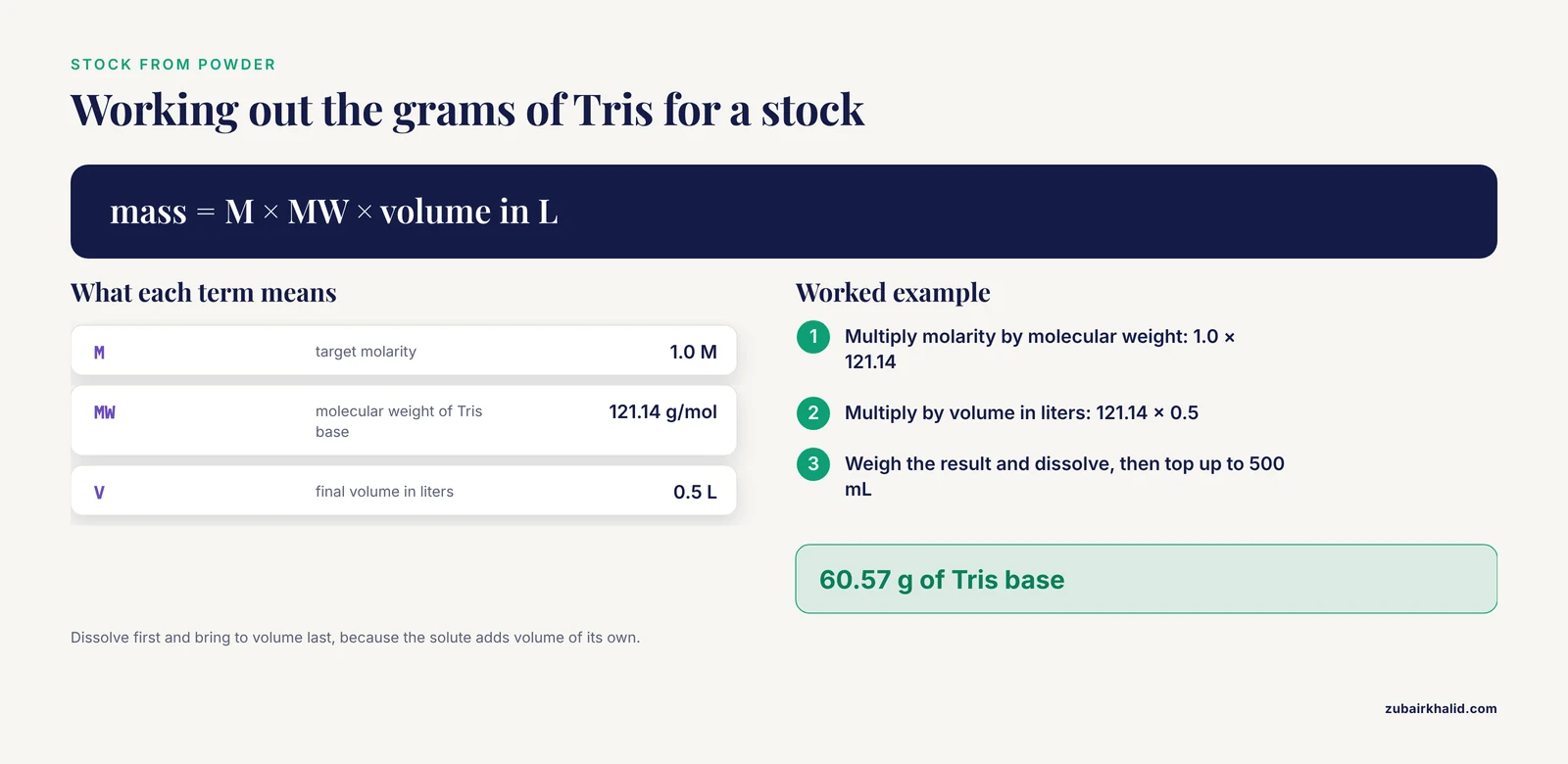
Ask the calculator for the mass behind 500 mL of 1.0 M Tris base:
>>> mass_needed(1.0, 121.14, 0.5)
60.57
The expected output is 60.57, in grams. Weigh the Tris, dissolve it in about 400 mL of water, titrate with hydrochloric acid to the target pH if your protocol calls for Tris-HCl, then bring the volume to exactly 500 mL in a graduated cylinder or volumetric flask. Dissolve first and top up last, because the solute adds volume of its own.
Step 3: Resuspend a Quantified Sample
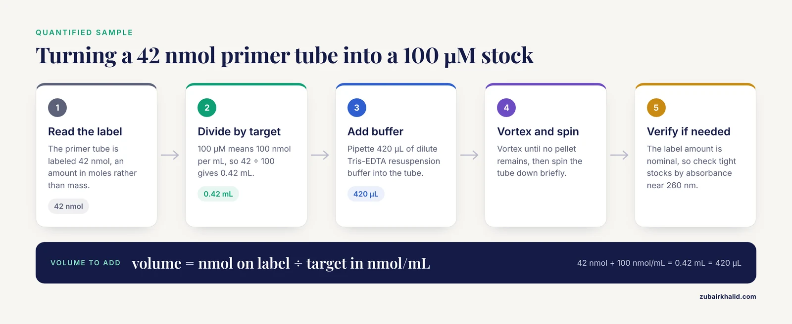
Oligonucleotides and many proteins arrive quantified in moles rather than mass. A primer tube labeled 42 nmol needs this much buffer to become a 100 µM stock:
100 µM is 100 nmol per mL, so the volume is 42 nmol ÷ 100 nmol/mL = 0.42 mL, or 420 µL.
>>> 42 / 100
0.42
Add 420 µL of resuspension buffer, typically a dilute Tris-EDTA solution that protects DNA from degradation, vortex until no pellet remains, and spin the tube down briefly. The label amount is nominal rather than measured, so labs that need tight concentrations verify the stock by ultraviolet absorbance near 260 nm, where nucleic acids absorb strongly, before trusting it.
Step 4: Calculate the Working Dilution
Dilute the 100 µM primer stock to a 10 µM working solution with a 1 mL final volume:
>>> stock_volume(100, 10, 1000)
100.0
The expected output is 100.0, in microliters, because the volumes were entered in µL. Pipette 100 µL of stock into 900 µL of diluent. Record both absolute volumes in your notebook, not only the 1:10 ratio. The ratio is the dilution factor, and the absolute volumes are what your hands actually do.
Step 5: Plan a Serial Dilution
A standard curve needs a ladder of concentrations, and a serial dilution builds one by repeatedly diluting the previous tube. Four 1:10 steps from the 10 µM working solution:
>>> serial_dilution(10, 10, 4)
[10, 1.0, 0.1, 0.01, 0.001]
The list reads 10, 1, 0.1, 0.01, and 0.001 µM. Each tube receives 1 part of the previous tube plus 9 parts of diluent. Prepare enough volume at each step to cover pipette dead volume, and change tips between transfers so the ladder does not drift upward.
Step 6: Check the Result
Two checks catch most errors in under a minute.
First, back-calculate. You planned 100 µL of 100 µM stock into 900 µL of diluent, so the final concentration is (100 µL × 100 µM) ÷ 1000 µL = 10 µM. The number you pipetted toward must come back out of the arithmetic.
Second, count the molecules. This check tells you whether the target concentration is physically meaningful at your working volume:
>>> molecule_count(1e-15, 1e-6)   # 1 fM in 1 µL
602.2
>>> molecule_count(1e-18, 1e-6)   # 1 aM in 1 µL
0.6022
A 1 fM solution in a 1 µL aliquot holds about 602 molecules, enough for ordinary statistics to behave. A 1 aM solution holds fewer than one molecule per aliquot on average, which means many aliquots contain none. Any protocol that asks for bulk concentrations below the femtomolar range deserves a second look at what it actually expects you to pipette.
Molarity Dilution Calculator Tips from Real Experiments
Tip 1: Treat molarity as an experimental variable
Magnesium chloride concentration is one of the classic variables in PCR optimization because it drives primer-template binding and polymerase activity. An optimization study of an HIV reverse transcription PCR (RT-PCR) assay, which first copies viral RNA into complementary DNA (cDNA) before amplification, tested MgCl2 at 1.25, 1.5, and 5.0 mM while also diluting the cDNA template from 10^-1 to 10^-10. Optimal amplification came from 1.25 mM MgCl2 combined with cDNA dilutions of 10^-1 to 10^-2 [2]. The entire experimental design consisted of one molarity series and one dilution series, which is exactly the kind of plan a calculator lets you lay out in minutes.
Tip 2: Sometimes the right molarity is very low
Conventional DNA gels run in high-ionic-strength buffers, but that is a convention rather than a law. One study separated double-stranded DNA, single-stranded DNA, and RNA in agarose gels run in low-molarity conductive media, using 1 mM alkali metal solutions for small fragments and 5 to 10 mM media for RNA and larger fragments, at least 15-fold faster than standard runs because the dilute media generate less Joule heating, the heat produced as electric current passes through the buffer [3]. Preparing such media puts your dilution calculator at the millimolar edge, where relative errors get large. A 0.5 mM absolute error is invisible in a 1 M stock and catastrophic in a 1 mM medium, so weigh carefully and verify with a second calculation or a conductivity measurement when the target is dilute.
Tip 3: Convert mass concentrations to molarity before comparing studies
Papers in pharmacology and microbiology often report concentrations in µg/mL, micrograms of compound per milliliter, which cannot be compared across compounds of different molecular weight until you convert. A benzimidazole anticancer study reported IC50 values, the concentrations inhibiting a measured response by 50 percent, of 10.83 µM in a liver cancer cell line and 11.55 µM in a breast cancer cell line for compound 3b [4]. An anti-Toxoplasma study, working against the parasite Toxoplasma gondii, reported complete killing of tissue cysts by compound 9c at 100 µg/mL and above [5]. Converting that 100 µg/mL to molar units takes one division. A compound with a molecular weight of 300 g/mol at 100 µg/mL is 333 µM. Without the conversion, the two studies cannot be placed on the same concentration scale.
Tip 4: Dilution changes more than concentration
The respiratory syncytial virus (RSV) fusion glycoprotein offers a sharp lesson. A soluble form of the protein was purified on a nickel column through its histidine tag and eluted in 50 mM phosphate buffer containing 500 mM NaCl and 250 mM imidazole, the small molecule used to displace tagged proteins from the column. In that buffer it stayed in its native pretriggered state. Dialysis, the gradual exchange of small solutes across a semipermeable membrane, against a 10 mM buffer triggered the protein into its extended form, and triggering efficiency tracked the drop in buffer molarity [6]. The endpoint concentration looked reasonable on paper. The path to it changed the molecule. When a dilution crosses a large ionic-strength gap, step down through intermediate buffers instead of plunging the sample into water.
Tip 5: The route between concentrations matters as much as the endpoint
Osmotic stress makes the same point for cells. When the glycerol cryoprotectant, a solute that protects cells during freezing and thawing, was removed from fresh equine spermatozoa (sperm cells), rapid one-step dilution into isosmotic media, media matching the cells' internal osmolality or dissolved-particle concentration, damaged motility and viability significantly more than fixed-molarity or fixed-volume step-wise dilution [7]. The final glycerol concentration was the same in every case. The step-wise route gave the cells time to exchange water and glycerol without osmotic shock. Plan intermediate steps whenever you dilute cells, embryos, or any osmotically sensitive material, and have the calculator compute each step rather than only the endpoints.
Tip 6: Effective concentration can exceed bulk concentration
Bulk molarity is not the only concentration that matters. In chemical biology, molecular templates assemble reaction partners so their local concentrations at the point of reaction far exceed their bulk concentrations, which is how proximity-driven reactions reach high rates and selectivity under highly dilute conditions [8]. If an assay works at a bulk concentration that seems too low to explain its rate, the calculator is not wrong and neither is the experiment. Molecular recognition can concentrate reactants locally, and that is a mechanism rather than an arithmetic error.
Common Mistakes and Limitations
The most common mistakes with a molarity dilution calculator fall into a short list. Each item names the mistake, the symptom, and the fix.
- Prefix slips. Confusing mM with µM produces a 1000-fold error. The symptom is an absurd answer, such as 0.06 g of Tris for a 500 mL, 1 M stock instead of 60.57 g. Convert every input to base units (moles, liters, grams) before computing, then convert the answer back for the bench.
- Units typed into numeric fields. In code, pasting a value with its unit as text, such as "100" carried along with the word µM, produces TypeError: unsupported operand type(s) for /: 'str' and 'float'. A stock concentration entered as zero produces ZeroDivisionError: division by zero. Strip the unit, enter the number alone, and keep the unit in the column header or variable name.
- Wrong molecular weight. Salt forms and hydration states change the number. Tris base has a molecular weight of 121.14 g/mol while Tris hydrochloride is 157.60 g/mol. Calcium chloride is 110.98 g/mol anhydrous and 147.01 g/mol as the dihydrate. Picking the wrong form shifts your concentration by roughly 30 percent. Read the exact compound name on your bottle, not the name in the protocol.
- Treating the diluent as interchangeable. Diluting a protein or a cell suspension into plain water changes ionic strength, pH capacity, and osmolality at once. The RSV fusion protein example above shows a molecule that changed conformation purely because its buffer was dialyzed to a lower molarity [6]. Dilute into the same buffer, or step down through a buffer series.
- One-step dilution of osmotically sensitive samples. Cells exchange water across their membranes as their surroundings change. The equine sperm study above found one-step glycerol removal significantly more damaging than step-wise routes [7]. Build intermediate concentrations into the plan.
- Serial dilution error compounding and early rounding. Every transfer adds its own pipetting error, and per-step errors accumulate through the ladder rather than canceling. Rounding intermediate values adds a second source of drift, so carry at least four significant figures through the chain and round only the final answer. Larger intermediate volumes, calibrated pipettes, and positive-displacement tips for viscous solutions such as glycerol keep the drift small.
- Ignoring volume non-additivity. The dilution equation assumes the final volume equals the sum of the combined volumes. That holds for dilute aqueous solutions and breaks down at high solute fractions, so make concentrated or viscous solutions to the mark in a volumetric flask.
- Diluting past the last molecule. Every dilution series eventually reaches a concentration where the average aliquot holds less than one molecule of solute. A study of high-dilution bullfrog thyroid preparations on tadpole development worked at 1:10^26 by weight, a molarity beyond Avogadro's number, meaning the bulk solution contains essentially none of the original solute [9]. Whatever one makes of such preparations, a dilution calculator applied to them will honestly return a target with zero molecules per practical volume. Run the molecule count check from Step 6 whenever a protocol pushes into attomolar territory.
- Trusting the stock label. Nominal amounts on primer tubes and protein vials are estimates that drift with handling, hydration, and time. Where the concentration matters, verify the stock by absorbance or another direct measurement, then recalculate the dilution from the measured value.
A note on the limits of the tool itself. A molarity dilution calculator computes ideal-solution arithmetic. It does not know activity coefficients at high ionic strength, temperature effects on volume and pH, solubility ceilings, or whether your solute stays stable at the concentration you chose. It cannot check that the stock you started with matches its label. Treat the output as a theoretical target, verify concentrations by measurement where they matter, and adjust for the behavior of your specific reagent. Critical applications are validated empirically, not by formula alone.
Choosing and Checking a Biology Calculator
Features that separate a dependable calculator from a risky one:
- Unit switching on both input and output, covering M, mM, µM, and nM.
- A dilution mode that returns absolute volumes, not just a ratio.
- A serial dilution planner that lists every intermediate tube.
- A visible formula or worked example, so the math can be checked against this article.
- A molecular weight lookup is convenient, but never automatic. Confirm the value against your actual bottle, because the tool cannot know whether you hold the free base or the salt.
Three verification habits work with any tool. Back-calculate one result by hand. Run the same inputs through a second independent tool and compare. Dimensional-check the answer, confirming that grams, moles, and liters cancel into the unit you expect.
Sensible next steps: save the Python functions above as a personal snippet, record every calculation in your notebook alongside the molecular weight you used, and batch multi-reagent protocols in a spreadsheet with one row per reagent. The same unit-checked arithmetic scales upward through computational biology, where docking and binding free-energy calculations performed in silico, meaning in computer models, now guide experiments [10], and the discipline that keeps all of it honest is the one practiced throughout this article. Know your inputs, check your units, and verify the output.
Frequently Asked Questions
What does a molarity dilution calculator do?
It converts between mass, moles, molarity, and volume using the relationships M = mass ÷ (MW × liters) and C1V1 = C2V2. Give it any three of the four quantities and it returns the fourth with the units handled.
How do I calculate a dilution from a stock solution?
Multiply the target concentration by the target volume, then divide by the stock concentration. For 10 µM final in 1 mL from a 100 µM stock, that is (10 × 1000) ÷ 100 = 100 µL of stock plus 900 µL of diluent.
What is the difference between mM and µM?
A factor of 1000. One millimolar equals 1000 micromolar, so a 5 mM solution is 5000 µM. Most lab concentration errors are prefix slips at exactly this boundary.
How do I resuspend a lyophilized primer to a specific concentration?
Divide the amount on the label by the target concentration. A 42 nmol primer brought to 100 µM, which is 100 nmol per mL, needs 42 ÷ 100 = 0.42 mL, so add 420 µL of buffer.
How many molecules are in my aliquot?
Multiply the molarity by the volume in liters by Avogadro's number. A 1 fM solution in 1 µL holds about 600 molecules, and a 1 aM solution in 1 µL holds fewer than one.
Why did my diluted sample stop working even though the final concentration is correct?
Dilution changes ionic strength, osmolality, and buffering capacity, not just the solute concentration. Sensitive proteins and cells can change state or suffer osmotic shock on the way to a correct endpoint, so dilute into the same buffer and step down gradually.
What dilution factor should I use for a serial dilution?
Use the factor that keeps every tube inside the assay's working range, commonly 1:10 for broad screening and 1:2 or 1:3 for fine dose-response curves. After n steps, the concentration is the starting value divided by the factor raised to n.
Can a dilution calculator be wrong?
The arithmetic is reliable, but the assumptions are not always met. The tool does not know your hydrate state, the true concentration of your stock, volume non-additivity at high solute loads, or the biological response to the dilution path, so verify critical concentrations by measurement.
Sources
- Atomistic Simulations Decode the Mechanisms of DNA and RNA Processing Enzymes: Function through Motion.
- [[Optimization of HIV diagnosis using the RT-PCR (gag) method with various dilutions of cDNA and MgCl2 molarity].](https://pubmed.ncbi.nlm.nih.gov/10386040/)
- Ultra-fast high-resolution agarose electrophoresis of DNA and RNA using low-molarity conductive media.
- Synthesis and multilevel evaluation of benzimidazole-based anticancer compounds: DNA/serum albumin interaction, and in-depth theoretical insights.
- Imidazopyridine derivatives as promising anti-Toxoplasma gondii agents: in vitro studies, in vivo testing, and molecular modeling analysis.
- Soluble respiratory syncytial virus fusion protein in the fully cleaved, pretriggered state is triggered by exposure to low-molarity buffer.
- Step-wise dilution for removal of glycerol from fresh and cryopreserved equine spermatozoa.
- Toward Ideal Proximity-Driven Reactions in Chemical Biology.
- Homeopathically prepared dilution of Rana catesbeiana thyroid glands modifies its rate of metamorphosis.
- Exploring molecular interactions between yeast topoisomerase I and RNA polymerase III subunit, Rpc82 using an in-silico approach.